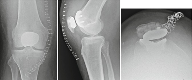
Fig. 1
Radiographs (anterior, lateral radiographs, skyline view) of the right knee showing a femoral and tibial cemented TKR (Emotion, Aesculap, B.Braun Medical AG, Sempach, Switzerland). A pseudo-patella baja was found, which was due to joint line elevation

Fig. 2
Radiographs (anteroposterior, lateral) of the right knee after PFJ. On the skyline view, obvious patella maltracking and a conflict of the metal-backed patella with the metallic trochlea were noted
These findings were confirmed by the surgical report at TKR, in which it was stated that the entire intra-articular joint space appeared black due to metal wear. On the removed implant, deep fissures were found in the trochlear and patellar component.
Questions
1.
What is your differential diagnosis now?
2.
What are your next steps in diagnostics?
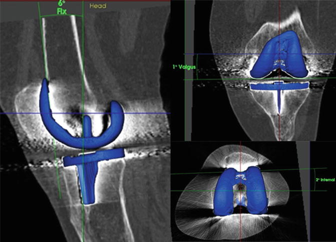
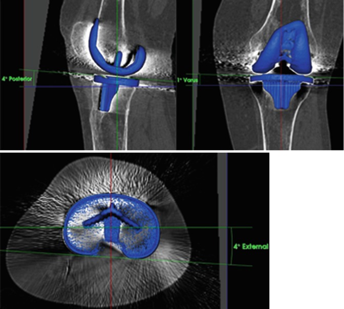
SPECT/CT was performed to accurately determine TKR component position and investigate loosening or infection (Figs. 3 and 4). SPECT/CT showed no signs of loosening or infection. Using a customized software (Orthoexpert v1.15©, OrthoImagingSolutions Ltd., London, UK), 3D-CT revealed an overall correctly positioned TKR (femoral component, 2° internally rotated, 6° flexed, and 0° varus; tibial component, 6° external rotated, 5° posterior, and 1° valgus).


Fig. 3
Measurements of the right femoral TKR component position using a customized and validated software

Fig. 4
Measurements of the right tibial TKR component position using a customized and validated software
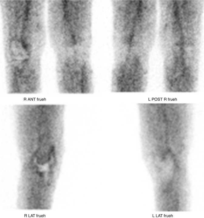
However, in three-phase bone scans, an extensive synovitis was observed, especially in the anterior knee compartment (Fig. 5).


Fig. 5


Soft tissue phase of 99mTc-HDP-SPECT images of the right knee showed a clear synovitis of the knee, in particular in the anterior compartment
Stay updated, free articles. Join our Telegram channel

Full access? Get Clinical Tree


