CHAPTER 80 Developmental and Functional Anatomy of the Lumbar Spine
INTRODUCTION
A thorough knowledge of human anatomy continues to be the cornerstone for all diagnosis and treatment planning. This awareness allows spinal physicians to rise above mediocrity and arm themselves for the complex issues involved in spine care. Unlike peripheral joints, spinal anatomy involves many joints interacting in conjunction to bring about complex, multiplanar motions. These motions have physiological restrictions and weaknesses based upon this anatomy. These weaknesses allow for the unfortunate consequences resulting in axial and radicular pain syndromes that spinal physicians know so well. This chapter will hopefully provide the reader with a basic understanding of both the developmental and functional anatomy of the lumbar spine.1
Since the lumbar and cervical vertebrae develop embryologically in a similar manner, it will only be reiterated here from Chapter 46. The aspects specifically pertaining to the cervical spine will be deleted. Specific differences concerning ossification centers and growth pattern will also be discussed.
EMBRYOLOGIC DEVELOPMENT
In order to give a more temporal flow to the embryologic development of the spine we will use the general staging scale devised originally by Streeter.2 This scale consists of 23 stages of growth that starts with fertilization and runs to the sixtieth day of growth.3 Each stage typically has duration of 2–3 days in length.4 Growth and differentiation of the neurospinal axis begins in embryonic stage 6. These cells typically originate from the primitive streak and node at the thirteenth to fifteenth day from fertilization. In the following stage the cells that will form the notochord travel rostrally from the primitive node. The notochordal cells’ growth cause abutment to the overlying ectoderm, which thickens and becomes the neural plate.5,6 This neural plate serves as the primordium for all nervous system evolution.7 This stage occurs at the 17–19 day mark (stage 8) and marks the first stage of nervous system development. The superior notochord promulgates growth of neuroectodermal structures, while the inferior notochord stimulates growth of mesodermal structures.8 In stage 9 the neural plate molds itself into a neural fold/groove.6 This stage is also notable as the first three somites are formed during this 19–21 day period. The somites represent paired, block-like masses of mesoderm lying alongside the neural tube. The human embryo develops 42–44 paired somites: 4 occipital, 8 cervical, 12 thoracic, 5 lumbar, 5 sacral, and 8–10 coccygeal somites. They consist of two cell collections; the ventromedially placed sclerotomal cells and dorsolaterally situated dermo-myotomes.9 The sclerotomal structures are responsible for the formation of the vertebral column, and the dermo-myotomal cells form the segmental skin and musculature.10 It is postulated that early vertebral segmentation occurs during this stage, most notably is the formation of the first two cervical vertebrae and cranial vertebral junction.6 These bony structures are formed by the first eight occipital somites. Spinal cord primordium is also present during this stage with continuing growth occurring through stage 12. In this stage the inferior neuropore closes and 21–29 somite pairs have formed. At this point vertebral differentiation has occurred through the lumbar segments. Stage 13 (28–32 days) marks completion of pontine and cervical flexures of the spine and continuing neurulation caudal to closure of the posterior neuropore.11 Concluding contouring of the caudal neural tube and adjacent vertebrae begins in this stage and progresses through gestation and into early infancy.6,11 Structures such as the conus medullaris, ventricular terminalis, and filum terminale originate from this caudal neural tube.12
Specific vertebral development occurs through three stages: membrane, cartilage, and bone formation.13 Membrane formation occurs as the notochord encounters the neural tube dorsally and the foregut ventrally. As growth progresses, cavities are formed between the notochordal and neural tube/foregut interface. Sclerotomic cells then infiltrate these cavities. Dorsal migration of sclerotomic cells also occurs during this period. These migrating cells settle in regions that ultimately will become the neural arches.13 This occurs through stages 10 through 12 of embryologic growth. Anterolateral movement of sclerotomal cells occurs in stage 13. These pockets provide primordium for future growth of the ribs. Stages 15 and 16 are characterized by fissuring and flexures of the notochord alongside vertebral body and intervertebral disc primordium.6
The period of cartilage formation in vertebral development takes place through embryonic stages 17 through 23. This chondrification begins in the vertebral bodies and subsequently migrates dorsally into the neural arches.6 During the latter part of stage 23 the dorsal aspect of the neural arches begins to deviate medially.
Bone formation starts in the early fetal stage of growth with ossification of the cervical and thoracic regions followed by lumbar and sacral vertebral segments. This final period continues into infancy and adolescence.14
Growth of spinal nerves begins with formation of the spinal cord. This initial differentiation involves clumping of neural crest cells in embryologic stage 12.15 Over the next three stages of development the neural crest cells form spinal ganglia that expand and move ventrally. The first ventral root fibers occur during stage 13. The primordium of the cervical and brachial plexuses also forms during this stage. Stage 14 is marked by complete development of all motor roots from C1 to S2 and growth of intersegmental anastomoses between these motor roots.16 Shortly after this stage the motor nerve roots move away from the spinal ganglia and toward their respective intervertebral foramen. Formation of the spinal nerve by mingling of motor and sensory root fibers occurs in stage 15. The roots enter the medial aspect of the foramen during stage 18, and progress completely into the foramen by stage 23. Other motor nerve developments worth noting are the following: the end of stage 14 marks the motor nerve fibers from the L2–4 segments moving a short distance into the leg.17 Creation of the brachial plexus begins in stage 16 and progresses through stage 23. At this point, the major motor subdivisions of the brachial plexus resemble those of the adult.15 Stage 16 also heralds formation of the lumbar plexus and the individual femoral, obturator, tibial, and peroneal nerves, and stage 17 marks development of the hypogastric, pudendal, genitofemoral, and ilioinguinal nerves.15
The sensory nerves develop in a similar manner as their neighboring motor fiber counterparts. As discussed above, the formation of sensory spinal ganglia anlage begins in stage 12 of embryologic development.18 Sensory root fibers grow out first centrally from the ganglia to make connections with the spinal cord in stage 14. In the following stage the fibers move peripherally to blend with the motor root fibers to form the spinal nerve. Entrance to the intervertebral foramen is gained by the end of embryologic stage 16, and complete access to the foramen by all sensory nerve roots is gained by stage 23.19
The autonomic system also begins its formation during the middle embryologic stages of development. Unlike the motor and sensory nerve roots, the autonomic nerve differentiation begins with primordium formation in the lower thoracic, esophageal, and upper abdominal regions rather than in the cephalocaudal growth of the vertebra and nerve roots.20 This occurs at embryologic stage 13. This primordium continues differentiation in the lower cervical to sacral region in stage 14. The first white communicantes fibers can be identified in the T2–9 level at this stage.15 Anlage of the autonomic pelvic plexus begins in stage 15.21 Stage 16 is marked by continued growth of the white rami communicantes through the L3 spinal level. The sympathetic chain also has a cephalad movement from the thoracic to upper cervical segments during this stage. The first gray rami communicantes begin to appear at the C8–T1 level around this time (37–42 days).22 Auerbach’s plexus begins differentiation in embryologic stage 17. Continued growth of gray rami communicantes occurs through stage 23 with formation of autonomic nerve fibers to the S5 spinal level.16
The development of the meninges originates from a thin cellular network that exists between the somites, notochord, and neural tube.6 Its early formation is termed the ‘meninx primitiva.’ It serves as a host site for the migration of cells and eventual development of the meninges. This meninx primitiva generation occurs in embryologic stage 15, and grows to surround the complete neural tube in stage 16. In the following stage, the denticulate ligaments begin to form. By stage 18 the spinal canal has started to take shape with cavitation of the meninx primitiva quickly following in embryologic stage 19. Evidence of pia mater structure formation also occurs during this time. It consists primarily of meninx primitiva cells with a small contribution from neural crest cells. Formation of the dura mater in the cervical and thoracic regions follows pia mater growth in stage 20. It consists of mesodermal cells from sclerotomal and meninx primitiva origins. It initiates growth anterior to the spinal cord with progression longitudinally and circumferentially around the cord. Dura mater growth reaches the lumbar region by stage 22, and completely surrounds the spinal cord end to end at stage 23. Constructs of the epidural space also begin during this last stage. Arachnoid mater development continues to be poorly understood at this time. It is believed to be the last of the meninges to form. This occurs after completion of the embryologic stages, i.e. post 60 days fertilization.
FETAL DEVELOPMENT
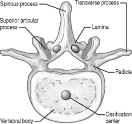
All major structures of the spine are clearly distinguishable by the end of the embryologic period of spinal growth. The next stage of development occurs during the fetal period. Its temporal boundaries include the termination of stage 23 of the embryologic period to birth. As discussed above, chondrification of the vertebra begins in the embryological period. It can be identified in the pedicles, lamina, and transverse processes of vertebrae prior to the fetal period. Early into the fetal period these chondrification centers enlarge and migrate into the posterior arches and vertebral bodies. Fusion of the posterior vertebral structures and dorsal growth of the spinous processes occur at the end of the first trimester (twelfth week).23 This period is also marked by the onset of ossification anteriorly in the body and posteriorly in the lamina of the vertebra (Fig. 80.1). These two bony growth centers progress in a dissimilar manner. Ossification of the vertebral body first occurs in the thoracic region and spreads bi-directionally towards the cervical and lumbar segments. The laminar bony growth takes place in a more typical cephalocaudal pattern from cervical to coccygeal segments.24 In addition, one sees a longitudinal growth of the spinal structures during this fetal period. This growth is demonstrated by measurement of the vertex–coccyx segment over this period. At 3 months, this segment measures about 10 cm long, and reaches to roughly 34 cm at birth.25
The intervertebral disc undergoes changes during this fetal period. The end of the embryologic stage and beginning of the fetal period see the notochord expand into the center of the disc. This cell ingrowth to the disc forms a strand-like appearance that is implanted into an amorphous mucoid substance called chorda reticulum.26 The embryologic cartilage surrounding the expanding notochordal cells begins to have collagen fibers formed within it around the tenth week of gestation.27 Interestingly, their growth patterns are identical to those in the adult.28 Their formation is well established by the sixth month of fetal growth. In addition, the neighboring anterior and posterior longitudinal ligaments begin to develop around the same time as the annular fibers. They grow from perivertebral mesenchymal cells.29 As the fetal period continues, the chorda reticulum enlarges and radially expands. During the terminal stage of this period the chorda reticular cells bordering the annular cartilage themselves begin a transition to fibrocartilage cells. These cells arrange themselves into patterns similar to those seen in the collagen fibers of the adult anulus fibrosus.26,30
POSTNATAL DEVELOPMENT
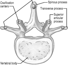
This final stage of development takes place from birth to adulthood. It is by far the longest of the three stages in duration. In contrast, this stage involves minimal plastic changes in the spinal tissues. This period primarily involves tissue maturation and longitudinal growth of the spine.25 The spinal maturation process involves continued ossification of the vertebrae. This is a continuation from its onset in the fetal period. As discussed above, the vertebrae typically have three primary centers of ossification: one in the ventral vertebral body and one in each of the lamina. The cervical and thoracic vertebrae typically have two secondary centers of ossification, whereas the lumbar spine is unique as it has two additional secondary centers of ossification. This gives the lumbar spine a total of seven ossification centers (Fig. 80.2).48 At birth, approximately 30% of the spine is ossified, primarily from these ossification centers. The articular processes, transverse processes, spinous processes, and the last few sacral and coccygeal segments still demonstrate a cartilaginous pattern at birth.31 Hyaline cartilage also still persists in the superior, inferior, and peripheral portions of the vertebral body. A striking feature at birth is that all pars interarticularis of the cervical, thoracic, and lumbar spine are ossified at birth. Failure of this pars ossification at birth has clinical implications for development of spondylolysis and spondylolisthesis in adulthood.32
The atlas and axis are unique as to their ossification centers number and placement. The reader is referred to the cervical spine anatomy chapter for a more detailed discussion regarding these two vertebrae. The remaining cervical, thoracic, and lumbar segments all develop in a more traditional pattern. As noted above, they have three main primary ossification centers: one located in the vertebral body, and one in each lamina (see Fig. 80.1). The vertebral body is almost entirely ossified by 6 years of age, and the neural arches approximate posteriorly at around 2–3 years of age.31 The epiphyseal plates of the vertebral bodies do not undergo ossification until completion of growth. This occurs around the twentieth year of life. This bony growth progresses in a radial pattern from a peripheral to central direction. However, this inward growth is arrested to leave a central cartilaginous portion that becomes the end-plate zone in the adult. The pedicles remain cartilaginous until the sixth year of life when their ossification process is complete and they fuse with their respective vertebral bodies.37 Secondary ossification sites occur at the transverse processes and spinous processes around the fifteenth to sixteenth year of life. As mentioned earlier, the lumbar vertebrae have two more secondary ossification sites than the thoracic and cervical vertebrae (see Fig. 80.2). These additional sites are found in the dorsal aspect of the bilateral superior articular processes.49 These areas usually fuse in the second to third decade of life. An accessory secondary ossification site may occur in the transverse process of the seventh cervical vertebrae.32 If this accessory center fails to fuse with the main secondary center in the transverse process, the accessory site may progress to formation of a cervical rib.33
The intervertebral disc also undergoes changes during the postnatal period. There is a progressive decline in the notochordal mesenchymal cells at birth. This is accompanied by a steady increase in the mucoid substance of the nucleus pulposus. A fibrocartilage capsule surrounds these diminishing notochordal cells and increasing mucoid substance. This change is typically evident by the fifth month of growth.38 By 4 years of age there are no active notochordal cells within the nucleus.39 After this point the nucleus pulposus halts any further growth. Future growth of the disc thereafter occurs through the anulus fibrosus. This growth involves primarily production of fibrous elements. As years advance there is ingrowth of fibrous tissue into the nucleus pulposus with resultant progressive loss of its liquid content.40 Intervertebral disc shape in the newborn has a relatively wedge-shaped appearance with the nucleus located posteriorly in the disc.41 The nucleus reverses position and is primarily anteriorly placed by 2 years of age.41 As the child assumes a more upright posture and gait, the nucleus takes on a more central position in the disc. This occurs around the fourth to the eighth year of life.30,41 A significant horizontal and longitudinal growth phase occurs in the disc after birth. This growth progresses late into adolescence.
A more generalized discussion on growth after birth and to adulthood will be summarized to demonstrate the developmental requirements of the spine. It is marked by two main growth spurts: one occurring after birth and the second during puberty.42 The early growth spurt takes place from birth through the age of 5 years. Almost half of this initial growth occurs through the first year of life.41 As mentioned above, at birth the sitting height measures around 35 cm. After the first year of growth this sitting height increases to about 47cm2. In the following 4 years of growth the sitting height increases from 47 cm to 62 cm. So by the time the normal infant reaches 5 years age they have increased their height 27 cm. Over the next 5 years the truncal growth slows dramatically in comparison to the prior 5 years. It increases about 10 cm over this period. The pubertal, or adolescent, phase marks the last stage of linear spinal growth. This commonly occurs at the tenth through eighteenth years of life. During this final stage is when the second growth spurt occurs. Its onset is slightly different for each gender. In females it usually begins slightly earlier than in males. Most commonly, it occurs around the age of 11. There is an initial period of rapid growth over the first 2 years that is followed by a slower growth phase over the remainder of the pubertal stage. The early phase increases sitting height approximately 7 cm, while the slower second stage increases height by another 5 cm. In males, the pubertal stage usually has its onset around age 13 years. The male growth pattern is similar to that of the female’s. However, there is a slight increase in height versus the female growth during the first phase of this growth spurt.41
By the time one reaches from birth to adulthood the spine has nearly tripled in size. This longitudinal growth is accomplished primarily through growth of the vertebral bodies. As discussed earlier, this growth occurs at the superior and inferior surfaces of the bodies.42 These regions persist as cartilaginous zones for continued growth and ossification of the vertebral body.43 The cartilage cells in these regions are arranged in a perpendicular-like pattern to the body. Thus, the longitudinal growth of the vertebral body is much the same as the growth of the metaphyses of long bones in the human body.25,44 Whereas the cells closest to the vertebral body undergo ossification and unite with the vertebral body, other cartilaginous cells moving toward the vertebral body then replace these ossifying cells.45 Hence, the vertebral body continues its longitudinal growth through this ossifying process.44 This growth terminates between the ages of 18 and 25.46 The growth plate then thins and calcifies. However, there remains a fibrocartilaginous region neighboring the disc. This area becomes the adult vertebral end-plate. Each level (i.e. cervical, thoracic, lumbar, and sacral) increases their vertebral height to different degrees. The average cervical and thoracic vertebral body height triples from birth to adulthood.23 The lumbar vertebral segments quadruple their longitudinal size by adulthood.23 At birth, the vertical length of the cervical spine measures about 3.7 cm. It doubles in length by 6 years of growth, during the first growth spurt, and nearly doubles its length again at the pubertal growth spurt. By the end of the pubertal growth spurt the cervical spine constitutes about 22% of the total spine length, and 15 to 16% of the sitting height.23 A lumbar vertebra can increase its height anywhere from 5 to 18 mm from birth to the age of 5.47,50,51 From the age of 5 to adulthood, the lumbar spine almost doubles in size to a height of 25 to 34 mm.47,51 In addition to vertical growth, the vertebral body also expands in a radial pattern.45 From birth to 7 years of age the lumbar vertebral body expands in a dorsoventral diameter from 3 to an average of 27 mm.50 During this 7-year period the lumbar lateral diameter also increases in size. On average, the lumbar body can increase by 29 mm through these years.50 At adulthood, the dorsoventral diameter measures approximately 34 mm.47 Periosteal ossification is primarily responsible for this horizontal growth. Most expansion, similar to vertical growth, occurs during the two main growth spurts.47
FUNCTIONAL ANATOMY
Bony structures
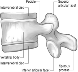
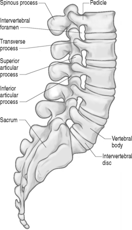
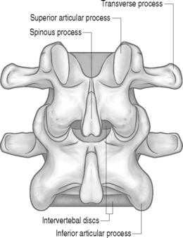
The lumbar spine is composed of five similar bony vertebral segments. They each contain a vertebral body, two laminas, two transverse processes, two pedicles, one spinous process, and two superior and inferior articular processes (Figs 80.3–80.8). The vertebral body occupies the ventral portion of the lumbar vertebrae. Its cranial appearance is that of a kidney, with the concavity facing dorsally. The lateral appearance of the vertebral body is that of a rectangular block (see Fig. 80.3). Its dorsal aspect serves as the anterior boundary of the spinal canal. Small elevations can be seen in the posterior rim of the superior surface of the body. Two small indentations can be seen on the posterior rim of the inferior surface of the vertebral body. The elevations on the superior surface are remnants of the uncinate processes that are prominently seen in the cervical spine.53 This rudimentary uncinate process allows for the commonly seen posterolateral disc protrusions in the lumbar spine. Foramens can be seen on the dorsal and anterolateral surfaces of the vertebral body. These holes serve as passageways for nutrient arteries and basivertebral veins.52 The lumbar vertebral body’s lateral diameter is typically larger than its anteroposterior diameter. The anterior height of the body is also larger than the dorsal height of the vertebral body. This configuration of the bodies is responsible for creating the normal lordotic curve that is seen in the lumbar spine.49
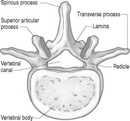
The pedicles are short cylindrical bones that project from the superolateral portion of the dorsal aspect of the vertebral body (see Figs 80.3 and 80.4). Their medial border helps form the anterolateral aspect of the spinal canal. Its caudal edge forms the roof of the segmental neural foramens.54 As the pedicle is the sole bony connection to the vertebra, it must be responsible for transmitting all the forces from the posterior bony structures to the vertebral body. In essence, the pedicle acts as a bridge between the anterior vertebral body and the posteriorly positioned facet joints and lamina. This functional importance is seen in the presence of thickened cortical walls and a trabecular bone pattern designed to support this load transference.
The lamina is a broad and thick bone that has a sheet-like appearance (see Fig. 80.4). It connects ventrally to the pedicle and projects posteriorly in a medially directed plane. The dorsal portions of the lamina merge to form the posterolateral portion of the spinal canal.54 The medial and inferior portion of the lamina has an irregular or roughened appearance due to it being the attachment site of the ligamentum flavum. The superomedial surface of the lamina is smooth with a slight curve in it.55 The ventroinferior aspect of the lamina has a bony mass which projects caudally and is called the inferior articular process. This process articulates with the corresponding superior articular facet from the vertebra below to form a facet joint.
A linear posterior projection of bone called the spinous process occurs at the dorsal junction of the lamina (see Fig. 80.4). The spinous process is the easiest of the lumbar vertebral bones to palpate, as it lies most dorsal in the spine.54 From a lateral perspective, the spinous process has a rectangular shape. Its dorsal end is roughened secondarily, and this area serving as attachment sites to numerous muscles and ligaments of the lumbar spine. Its anterior surface helps form the dorsal aspect of the spinal canal.
At the junction of the pedicle and lamina a bony process projects out laterally. This flat and rectangular bone is called the transverse process (see Fig. 80.4). It is positioned just ventral to the superior articular process and posterior to the intervertebral foramen.49 In contrast to their cervical counterparts, they do not contain a transverse foramen. The anteroposterior diameter of these bones tends to be short, making them rather thin in comparison to the lamina and pedicles. Their width narrows from a superior to inferior direction. These lateral bony processes are designed to serve as fulcrums through which muscles may exert their actions.56
A superior directed bony projection occurs at the junction of the lamina and pedicle. This bony projection is called the superior articular process (Figs 80.4–80.9). The medial surface of this articular process is lined by hyaline cartilage. This cartilage is also present on the lateral surface of each of the inferior articular processes. The cartilaginous zones of the superior and inferior articulating processes are referred to as the articular facet of each articular process.54 All lumbar superior articular processes face in a medial and dorsal direction. Their inferior articular process counterparts face in a ventrolateral direction to allow the articular processes to smoothly articulate and form a lumbar facet joint. The orientation of the superior articular processes alters as one moves from a superior to inferior direction. The upper lumbar segments tend to have a more sagittalyl oriented superior articular process, whereas the L5 and S1 superior articular processes tend to have a more coronal orientation. Physicians performing lumbar facet joint injections commonly observe this phenomenon, as they need to progressively rotate the C-arm as they target lower lumbar facet joints.55
Ligaments
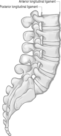
The anterior longitudinal ligament extends from the sacrum to the cervical region (Fig. 80.7). It lies on the anterolateral aspect of the lumbar vertebral bodies. In contrast to its cervical and thoracic divisions, its lumbar portion is more developed and wider in dimension.57 It consists of superficial and deep fibers. The deep fibers are short and extend a distance of no greater than one intervertebral joint. They commonly insert into the margins of the vertebral bodies. The superficial fibers can cross up to five intervertebral segments before attaching to either the superior or inferior margins of the vertebral body. In contrast to the posterior longitudinal ligament, the anterior longitudinal ligament maintains only a loose connection to the anterior portion of the intervertebral disc and ventral portion of the vertebral bodies.58 The primary function of the anterior longitudinal ligament is to resist vertical and extension forces through the lumbar spine.
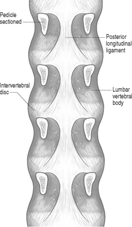
The posterior longitudinal ligament is also present from the sacrum, extending proximally to the cervical region where it becomes the tectorial ligament (see Fig. 80.8). Its fiber composition is similar to the anterior longitudinal ligament. As the name implies, it lies on the posterior aspect of the vertebral bodies and intervertebral discs. On visual inspection the ligament has a saw-toothed, or denticulated, appearance in the lumbar spine. It forms a narrow band as it crosses each lumbar vertebral body, and widens laterally as it overlies the intervertebral disc (see Fig. 80.8).59 This ligament also contains both superficial and deep fibers. The superficial fibers can span anywhere from 3 to 5 vertebral bodies in length before inserting into the margin of the vertebral body. The deep fibers typically span a distance of two intervertebral discs before inserting into either the superior or inferior vertebral body margin. Unlike the loose association of the anterior longitudinal ligament, the posterior longitudinal ligament maintains an intimate connection with the intervertebral discs’ posterior anulus fibrosus. This merging of fibers is so prevalent that gross dissection of the ligament from the posterior anulus fibrosus is extremely difficult.59 The function of the posterior longitudinal ligament is to oppose vertical distraction forces from the dorsal aspect of the vertebral bodies and to resist flexion moments to the lumbar vertebral column.56,59
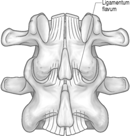
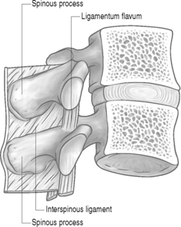
The ligamentum flavum is a paired ligament that attaches between consecutive lumbar lamina (see Figs. 80.9, 80.10).60 It is the thickest and strongest of the spinal ligaments. It inserts inferiorly into a medial and lateral attachment site of the distal lamina. The lateral portion inserts into the anterior aspect of the superior and inferior articular processes. The medial attachment occurs on the dorsal aspect of the upper lamina. It should be noted that the lateral portion of the inferior ligamentum flavum contributes to the formation of the anterior capsule of the neighboring facet joint.61 Far lateral fibers also can insert into the pedicle at the same level.49 These anterior and lateral fibers of the ligamentum flavum help to form the posterior boundary of the intervertebral foramen.62 The superior insertion site of the ligamentum flavum is into the inferior portion of the ventral aspect of the lamina and dorsoinferior pedicle one level above the inferior insertion site. The function of this ligament is to resist excessive distracting forces through the posterior aspect of the lumbar spine.63 It also aids in preventing excessive flexion moments through the lumbar spinal column. Buckling of the ligamentum into the spinal canal during extension of the spine could potentially create compression on the neurovascular structures within the canal. This buckling phenomenon is minimized by the elastic nature of this ligament. In contrast to other spinal ligaments, the ligamentum flavum is comprised of 70–80% elastic fibers.64 These fibers have the special ability to undergo deforming forces (i.e. stretch) and return to their original resting length after the stretch has concluded.65
The interspinous ligaments run between contiguous spinous processes. It is typically absent or rudimentary in the cervical spine and membranous in the thoracic spine. However, it is a well-developed ligament in the lumbar spine.67 The lumbar interspinous ligaments have been described as existing in three different parts: anterior, middle, and posterior.59,66 The anterior part of this ligament is paired (right and left) and has its superior attachment into the ventral portion of the upper spinous process. The inferior insertion of the anterior portion occurs into the ligamentum flavum at the level of the lower spinous process.59,66 The middle part of the interspinous ligament is the largest of the three portions. It inserts into the middle third of consecutive spinous processes. The posterior portion of the interspinous ligament inserts inferiorly into the dorsal half of the superior spinous process. As it courses superiorly it moves dorsally to pass posteriorly to the spinous process above the inferior insertion site. On passing the superior spinous process it merges with the supraspinous ligament.59 It functions as a weak resistor to lumbar flexion and distraction forces.67 Some authors consider these ligaments the first to undergo tearing during a hyperflexion moment through the spine.68
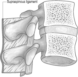
The supraspinous ligament travels just dorsally to the spinous processes in the lumbar spine where it is the strongest (Fig. 80.11). Its intersegmental presence in the lumbar spine can be variable. It can terminate at the L4 spinous process in about 73% of individuals, the L3 spinous process in about 22%, and is only seen bridging the L4–5 interspace in about 5% of individuals. It is rarely seen at the L5–S1 level.69,70 Some authors believe the fibers of the lumbar erector spinae musculature and thoracolumbar fascia replace the supraspinous ligament fibers distal to the L4 level.49 Other authors have postulated that the supraspinous ligament is not a true individual ligament, but is composed of ligamental fibers of the thoracolumbar fascia and neighboring fibers of the multifidis and longissimus thoracis muscles.49,59 It functions as a weak resistor to distraction forces through the spinous processes.67
Stay updated, free articles. Join our Telegram channel

Full access? Get Clinical Tree


