Deltoid Ligament Reconstruction
Eric M. Bluman
Richard J. de Asla
DEFINITION
Deltoid ligament deficiency is present when both the deep and superficial components of the medial collateral ligament complex of the ankle are ruptured or are insufficient.
ANATOMY
The deltoid ligament complex is a multiunit structure that provides support and restraint for the tibiotalar joint, subtalar joint, spring ligament, and talonavicular joint.
There is wide agreement that the deltoid ligament complex is made up of both deep and superficial components.
The deep portion of the complex originates from the intercollicular groove and posterior colliculus of the medial malleolus and inserts on the medial face of the talar body near the center of rotation of the tibiotalar joint. These short and stout fibers are intra-articular but extrasynovial. It is made up of anterior and posterior fascicles.
There has not been agreement over the superficial components of the complex. In one of the more detailed anatomic studies, Pankovich and Shivaram5 described the superficial layer as being made up of the tibionavicular, tibiocalcaneal, and tibiotalar ligaments. These fibers represent a triangular array originating on the distal medial malleolus and extending in a fan shape to their respective insertions. The relative contribution of these components to both ankle and foot biomechanics is still a topic of investigation.
PATHOGENESIS
The most common cause of deltoid ligament disruption is supination-external rotation (SER) ankle fractures. The most severe form of these fractures has either a medial malleolus fracture or a deltoid ligament rupture, in conjunction with a lateral malleolus fracture. The variant with an intact medial malleolus and disrupted medial collateral ligaments is termed SER IV-deltoid. This latter form is the most common form of deltoid ligament disruption.
It has been very well established that deltoid reconstruction is not indicated for disruptions that occur in conjunction with ankle fractures. Reduction and fixation of the fracture component with reestablishment of the mortise morphology leads to healing of the deltoid ligament in the vast majority of those with these combined injuries.9
A smaller proportion of patients with deltoid ligament insufficiency will have developed this as a component of stage IV AAFD.2
Deltoid ligament insufficiency without concomitant ankle fractures resulting from the acute injury has been described but will not be discussed here. This chapter concentrates on deltoid ligament insufficiency arising from degenerative causes.
NATURAL HISTORY
As the posterior tibial tendon becomes deficient, the ability to bring the hindfoot into varus actively is lost.
As the mechanical axis of the leg is shifted medially (relative to the foot) and the hindfoot deformity becomes more severe and eventually stiff, tension is progressively increased on the soft tissues of the medial ankle. The medial collateral ligament complex becomes unable to resist the loads placed on it, with eventual insufficiency and lengthening.7,8
Progression to stage IV AAFD occurs when the deltoid ligament becomes incompetent and the valgus force from the preexisting hindfoot deformity causes the talus to tilt within the mortise.
PATIENT HISTORY AND PHYSICAL FINDINGS
Aspects of the history and physical examination of stage IV AAFD will be similar to those found in the earlier stages of this AAFD.
There will be hindfoot valgus.
Because of the chronic nature of posterior tibial tendon involvement, strength will be greatly diminished and likely absent because of rupture. The patient will neither be able to resist hindfoot eversion nor actively bring the forefoot across midline.
Because of the decreased working length of the triceps surae resulting from chronic hindfoot valgus, there will be contracture of these muscles. A fixed hindfoot deformity may give a falsely optimistic impression of tibiotalar dorsiflexion. Reestablishment of ankle and hindfoot alignment without an appropriate lengthening of the heel cord will create or exacerbate an equinus deformity.
There may be significant forefoot supination.
Lateral pain may represent sinus tarsi or subfibular impingement, lateral ankle joint arthritis, or, in severe cases, distal fibular stress fracture.
Pain in the sinus tarsi is frequently unrecognized or underappreciated before palpation by the clinician.
Callosity and pain below the talar head may be present if substantial dorsolateral peritalar subluxation has caused a prominence in the medial plantar midfoot.
It is essential to determine whether the tibiotalar valgus deformity that is a hallmark of stage IV AAFD is rigid or reducible. This is further explained under Surgical Management.
Clinical determination of the presence of valgus tibiotalar deformity is greatly enhanced with radiologic examination.
The integrity of the lateral collateral ligament complex needs to be determined. A severe valgus deformity may lead to erosion and incompetence of these structures.
The surgeon must also evaluate for the presence of ipsilateral knee valgus. If this is significant, consideration should be given to correcting the proximal deformity before the foot and ankle surgery. Correction of the leg-ankle-foot axis without attention to knee deformity may not adequately relieve valgus stress through the reconstructed lower limb and result in recurrence of deformity.
Methods for examining the deltoid ligament include the following:
Palpating the area inferior to the medial malleolus. Tenderness may represent incipient or recent deltoid rupture and may only be present early in stage IV disease.
Joint line palpation. The presence of valgus tilt indicates insufficiency of the deltoid ligament.
Weight-bearing anteroposterior (AP) ankle radiographs. Valgus tilt greater than 4 degrees indicates deltoid ligament insufficiency.
IMAGING AND OTHER DIAGNOSTIC STUDIES
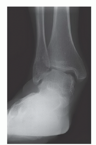
The preferred radiologic views include the three-view weightbearing series. The AP standing view will provide the most information. Patients with deltoid ligament insufficiency will demonstrate tibiotalar valgus tilting (FIG 1).
Cross-sectional imaging is required only when plans are made for performing reconstruction using native peroneus longus tendon (discussed later). In this case, magnetic resonance imaging (MRI) is used to confirm the integrity of the peroneus brevis before the longus is harvested.
Selective intra-articular blocks often help the clinician localize the exact source of pain.
DIFFERENTIAL DIAGNOSIS
Stage II or III AAFD
Medial malleolus fracture nonunion
Tibiotalar arthritis (with eccentric lateral joint erosion)
Osteonecrosis of the talus with lateral collapse
Distal tibial supramalleolar valgus malalignment (resulting from distal tibiofibular fracture or pilon fracture)
Valgus malunion of pronation-abduction-type ankle fracture with lateral plafond impaction or comminution
NONOPERATIVE MANAGEMENT
In contrast to acute deltoid deficiency presenting in conjunction with an ankle fracture, we believe that nonoperative care has a very limited place in patients with chronic deltoid ligament insufficiency resulting from degenerative causes (eg, stage IV AAFD). All but patients with medical comorbidities contraindicating surgery should undergo surgical reconstruction.
Conservative therapy may also be needed to relieve pain and temporize deformity while related orthopaedic conditions are corrected.
Should conservative therapy be chosen, custom-molded rigid orthotics that extend to the calf, such as the Arizona brace, provide the best chances of preventing progression of the disease.
Although halting the progression of the disease may be possible with conservative therapy, the deformities of stage IV cannot be corrected with bracing alone.
SURGICAL MANAGEMENT
Stay updated, free articles. Join our Telegram channel

Full access? Get Clinical Tree


