11 Deep dry needling of the leg and foot muscles
Introduction
This chapter covers deep dry needling of TrPs in the leg and foot musculature. These muscles are in a particularly challenging location as they are included in the referral patterns of multiple proximal muscles, such as the gluteus minimus, glutei medius and maximus, piriformis, tensor fasciae latae, adductors longus and brevis, vastus lateralis, sartorius, semitendinosus and semimembranosus and biceps femoris muscles (Travell et al. 1992, Dejung et al. 2001),which means that TrPs in leg muscles may be activated secondarily to TrPs in these more proximal muscles (Simons et al. 1999). Furthermore, TrPs in leg muscles can act as a link in this TrP chain, starting in proximal muscles and ending in foot muscles. The TrP chain has also been described running in the opposite direction (Lewit 2010), especially when pain and dysfunction caused by TrPs in foot and calf muscles induce gait alterations that overload muscles higher in the lower limbs and in the spine.
On the other hand, muscles in the foot and leg are the first line of defense of any anatomical or biomechanical problems occurring in the foot and, consequently, become easily overloaded by these issues leading to the development and activation of TrPs (Travell et al. 1992, Saggini et al. 1996). Joint dysfunctions and inappropriate shoes can also either cause or add to these problems. In other words, the treatment of TrPs in the leg and foot muscles is necessary, but usually insufficient to solve our patients’ problems, since proximal muscles, as well as many other different perpetuating factors, must also be addressed for a complete and long-lasting relief of the symptoms.
Clinical relevance of TrPs in leg and foot pain syndromes
Although many different possible etiologies are proposed for calf cramps, TrPs in the calf muscles, particularly in gastrocnemius muscle, seem to be important contributors (Travell et al. 1992, Ge et al. 2008, Xu et al. 2010). A small sample (n=24) clinical trial showed that xylocaine injections of TrPs in the gastrocnemius muscle induced a significantly better long-term efficacy on calf cramps than oral quinine (Prateepavanich et al. 1999).
Plantar heel pain, often diagnosed as plantar fasciitis, is commonly due to TrPs in the calf and foot musculature. Several reports have confirmed this close relationship and proven that conservative treatment of TrPs in calf muscles is useful in the treatment of plantar heel pain and plantar fasciitis (Nguyen 2010, Renan-Ordine et al. 2011). Although there is no solid evidence on the effectiveness of invasive treatments (Cotchett et al. 2010), some reports suggest that needling and injections of TrPs in the calf and foot muscles could be helpful in the management of this condition (Imamura et al. 1998, Kushner & Ferguson 2005, Sconfienza et al. 2011).
An older study attributed medial tibial stress syndrome (shin splints) to overload of the attachments of the soleus muscle (Michael & Holder 1985). One study showed that tension in the soleus, tibialis posterior, and flexor digitorum longus muscles caused a tenting effect, that exerted a force on the distal tibial fascia directed to its tibial crest insertion (Bouche & Johnson 2007). Another study demonstrated that the plantar flexors of the ankle were significantly weaker in medial tibial stress syndrome (Madeley et al. 2007). Increased tension and weakness are two cardinal features of muscles affected by TrPs. Nevertheless, the possible involvement of TrPs in these muscles in medial tibial stress syndrome has not yet been established unequivocally, and no clinical trial to date has proven that TrP treatment can be of help in the management of this condition.
Although ‘there is a strong possibility that, in muscles prone to developing a compartment syndrome, TrPs may make a significant contribution’ (Travell et al. 1992), there is no actual evidence in the literature. On the other hand, the safety of using dry needling, which could potentially cause some bleeding in the muscle, has not been established either in patients with compartment syndrome; hence, more aggressive dry needling approaches, such as Hong’s Fast-in and Fast-out technique, screwed-in/out techniques, or dry needling with thick needles may be considered possible contraindications.
Forty per cent of patients with CRPS in the lower extremities presented with active TrPs in their proximal muscles (Allen et al. 1999). In this study only the lumbar paraspinous and gluteal musculature were examined, which raises the question what percentage of active TrPs would have been found if other lower extremity muscles had been included in the examination. Early treatment of TrPs is usually recommended in patients with CRPS I to decrease their pain intensity and disability (Dommerholt 2004). Although the use of dry needling has never been reported in this indication, a recent report of two cases of upper limb CRPS I showed promising results with treatment of proximal TrPs with botulinum toxin injections (Safarpour 2010).
TrPs in several leg muscles, such as the popliteus, plantaris and gastrocnemius, can also contribute to posterior knee pain, which is often attributed to knee joint problems. TrPs in the proximal part of the gastrocnemius muscle are responsible for posterior knee pain in patients before (Mayoral et al. 2010) and after (Aceituno 2003) total knee replacement surgery and after knee arthroscopy (Rodríguez et al. 2005).
Further research is needed to elucidate the possible contribution of TrPs to the above conditions or to other structural problems such as hammer toes (Travell et al. 1992) or hallux valgus, or to different leg and foot nerves entrapments (Crotti et al. 2005, Saggini et al. 2007).
Dry needling of the leg and foot muscles
Popliteus muscle
• Anatomy: The popliteus muscle is an obtuse triangle in shape. Laterally and proximally it attaches to the lateral condyle of the femur, to the posterior capsule of the knee joint, to the lateral meniscus, and to the head of the fibula. Medially and distally it attaches to the posteromedial surface of the tibia.
• Function: Medial rotation of the tibia in open kinetic chain and lateral rotation of the femur in closed kinetic chain. It also assists marginally with knee flexion.
• Innervation: Fibers of the tibial nerve from L4–S1 spinal nerves.
• Referred pain: Typically to the back of the knee.
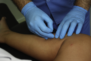
• Needling technique: The patient side lies on the involved extremity with the hip and knee flexed to 90°. The muscle is palpated right behind the proximal third of the tibia and the needle is inserted towards the TrP in a lateral direction with a slight anterior superior orientation (Figure 11.1), keeping the needle close to the posterior aspect of the tibia or even touching the bone with the tip of the needle as a reference.
• Precautions: The neurovascular bundle is in the midline of the leg, resting on the popliteus muscle, and must be avoided by keeping the needle close to the posterior aspect of the tibia. Branches of the saphenous nerve run superficial in the region where the needle is inserted. If the needle touches any of these branches, the patient will feel a superficial electrical sensation over the medial part of the leg. Should this happen during the first millimeters of needle penetration through the skin, the needle should be withdrawn and reinserted some millimeters away.
Gastrocnemius muscle
• Anatomy: The muscle is divided into lateral and medial heads. Proximally, each head anchors to the corresponding condyle of the femur and to the capsule of the knee joint. Distally, both heads insert into the Achilles tendon, which attaches to the posterior surface of the calcaneus bone.
• Function: Plantar flexion and supination of the foot. Limited contribution to knee flexion (with the knee extended) and to knee stabilization. In closed kinetic chain it contributes to knee and ankle stability.
• Innervation: Tibial nerve by fibers from S1 and S2.
• Referred pain: Most TrPs in this muscle refer pain locally. TrPs in the belly of the medial head tend to refer pain to the instep of the foot, sometimes spreading to the lower posterior thigh, the back of the knee, and the posteromedial aspect of leg and ankle.
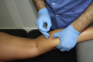
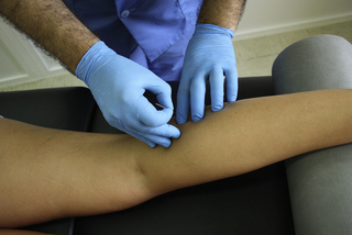
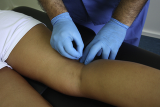
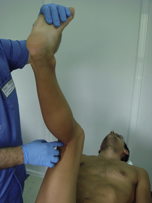
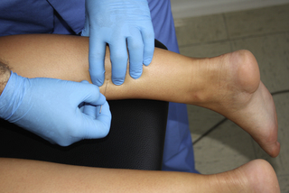
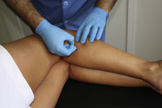
• Needling technique: The patient lies in the prone position, with the knee slightly flexed and the leg supported by a pillow. For TrPs in the central part of the medial head, a pincer palpation is used to locate and fix the taut band and the TrP and the needle is angled medially, towards the fingers located in the opposite side (Figure 11.2). For TrPs in the central part of the lateral head, a flat palpation is more commonly used to locate and fix the taut band and the TrPs. The needle is directed perpendicular to the skin aiming towards the TrP in a posteroanterior direction with a slightly lateral angulation (Figure 11.3). For TrPs in the proximal part of any of both heads, flat palpation is used to locate and fix the TrPs. The needle is directed toward the TrP in a posteroanterior orientation (Figure 11.4).
• Precautions: The sciatic nerve usually splits into the tibial and peroneal nerves in the lower third of the thigh. The tibial nerve, together with the popliteal vessels, runs along the popliteal fossa between the proximal parts of both heads of the gastrocnemius muscle. The peroneal nerve runs downward close to the biceps femoris muscle tendon. This means that the proximal part of the medial head of the gastrocnemius muscle (and therefore its TrPs) lies between the tibial nerve (along with the popliteal vessels) and the tendons of the semitendinosus and semimembranosus muscles. On the other hand, the proximal portion of the lateral head of the gastrocnemius muscle and its TrPs are between the peroneal nerve and the tibial nerve. If needling of these proximal gastrocnemius TrPs is considered necessary, the popliteal fossa must be palpated thoroughly prior to the needling procedure to locate the nerves and tendons. Anatomical variations, such as a premature split of the two divisions of the common peroneal nerve at the popliteal level, may be identified, and needling TrPs in the proximal part of the lateral head of the gastrocnemius muscle or into the plantaris muscle would not be indicated. The recommended position to palpate this region is shown in Figure 11.5. Clinicians should identify the available safe needling space between the semitendinosus muscle tendon and the tibialis nerve, and the space between the peroneal and tibialis nerves and their relationship with the biceps femoris muscle tendon. In the prone position, only the tendons are palpable (especially when the patient slightly contracts the knee flexors) and by using the tendons as landmark, the needle can be directed into the safe spaces.
When needling the proximal portions of the gastrocnemius muscle, it is conceivable that the needle may touch the back part of the capsule of the knee joint, hence it is recommended to increase the antiseptic measures to avoid creating a joint infection (see Ch. 4).
Plantaris muscle
• Anatomy: Proximally, this muscle attaches to the upper part of the lateral condyle of the femur. Distally, its long tendon anchors to the medial aspect of the calcaneus, blending with the fibers of the Achilles tendon.
• Function: Plantar flexion and inversion of the foot. In a closed kinetic chain, it assists in knee flexion.
• Innervation: Tibial nerve, through a branch containing fibers from L5–S2.
• Referred pain: To the back of the knee, sometimes reaching mid-calf level.
• Needling technique: Since it is partially covered by the upper part of the lateral head of the gastrocnemius muscle, the needling technique is similar to this (see gastrocnemius muscle above and Figure 11.4).
• Precautions: The tibial and peroneal nerves and popliteal vessels must be avoided.
Soleus muscle
• Anatomy: The muscle originates in the posterior aspect of the head and proximal third of the fibula, in the popliteal line of the tibia and in the tendinous arch between both bones. The soleus fibers attach distally to a superficial tendinous sheet, which continues directly to the Achilles tendon, which in turn attaches to the posterior part of the calcaneus.
• Function: Plantar flexion and inversion of the foot.
• Innervation: A branch of the tibial nerve containing fibers from L5–S2.
• Referred pain: Mostly to the distal part of the Achilles tendon and the posterior and plantar surfaces of the heel. Its TrPs can also refer pain to the upper half of the calf and, very rarely, to the ipsilateral sacroiliac joint. Simons et al. (1999) mentioned an exceptional referral pattern to the ipsilateral jaw area.
• Needling technique: For distal medial or lateral TrPs, the whole muscle can be held in a pincer between the thumb and two fingers, with the patient either in prone (Figure 11.6) or in a side lying position. The needle is directed towards the opposite finger or thumb. Proximal TrPs can be needled towards the fibula with the patient lying on the uninvolved side (Figure 11.7).
• Precautions: When needling the medial part of the muscle, care must be taken to avoid needling the tibial nerve.
Stay updated, free articles. Join our Telegram channel

Full access? Get Clinical Tree


