Darrach Procedure
Charles Cassidy
Leonard K. Ruby
Indications
Distal radioulnar joint (DRUJ) arthritis, secondary to
Inflammatory arthritis
Posttraumatic arthritis
Osteoarthritis
Unreconstructable distal ulna fracture
Contraindications (Relative)
Ulnar translocation
Distal radioulnar joint instability in the patient without rheumatoid arthritis
Undoubtedly, inflammatory arthritis of the DRUJ remains the most common indication for a distal ulna resection (Darrach procedure). However, the DRUJ may also be affected by osteoarthritis and trauma. Consequently, the relative indications, contraindications, surgical tactics, and outcomes are best considered with respect to the underlying cause of the DRUJ disorder.
In the rheumatoid patient, DRUJ involvement is common and, in general, these patients do well following the Darrach procedure (1). Refractory symptoms, despite 3 to 6 months of adequate therapy, including medications and splinting, are an indication for surgery. Synovectomy with ulnar head retention may be considered in selected patients without radiographic arthritis. However, associated digital extensor tendon ruptures, termed the Vaughn Jackson lesion, are best managed with concomitant distal ulna resection regardless of the radiographic appearance of the DRUJ. Pre-existing ulnar translocation is a contraindication to an isolated Darrach procedure (Fig. 32-1) (2,3). Furthermore, bone loss at the ulnar corner of the radius or accentuated radial inclination may be considered risk factors for ulnar translocation following the Darrach procedure unless the radiocarpal joint is stabilized (4,5). Options include a combined radiolunate or total wrist arthrodesis at the time of the Darrach procedure. Some authors have felt that ulnar head resection may be unwise in the rheumatoid patient even in the absence of ulnar translocation. They cite the importance of the ulnar head in maintaining the ulnar carpal ligaments, and acting as a buttress, preventing carpal migration ulnarly and palmarly. This point remains controversial. A long-term retrospective study (6) of patients with rheumatoid arthritis following distal ulna resection suggests that ulnar translocation is more likely caused by progression of the rheumatoid disease, whereas ulnar stump impingement is caused by excessive resection.
The Darrach procedure is less predictable in patients who are osteoarthritic and posttraumatic patients and tend to place more demand on their wrist. Consequently, the surgeon must consider joint-preserving surgical options in these patients. Ulnocarpal impaction can be treated by ulnar-shortening osteotomy or the Feldon wafer procedure (7). Loss of forearm rotation following appropriate treatment of a distal radius fracture can be treated by DRUJ capsular release (8) if the radioulnar joint is congruent and not arthritic. When forearm contractures are associated with a distal radius malunion, corrective osteotomy of the radius may be preferable. In such instances, DRUJ is assessed intraoperatively following radius osteotomy and a distal ulna resection is performed, if necessary.
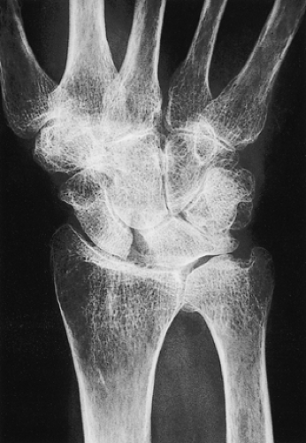 Figure 32-1 Ulnocarpal impaction in a patient with rheumatoid arthritis. Ulnar translocation is evident and may be exacerbated by distal ulna resection. In such an instance, radiolunate or total wrist arthrodesis should be performed in conjunction with the distal ulna resection. |
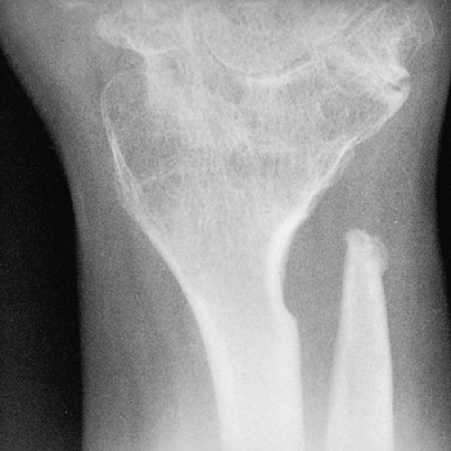 Figure 32-2 Patient with symptomatic ulnar stump impingement on radius status after ulnar head resection and radiocarpal fusion. |
Ironically, although Darrach described the distal ulna resection in the management of a DRUJ dislocation (9), DRUJ instability is considered a relative contraindication to the Darrach procedure. Invariably, stump instability will be at least as symptomatic for these patients as their original problem. Similarly, ligamentously lax patients are at risk for stump instability (10).
Adverse outcomes following the Darrach procedure are primarily related to three factors: poor patient selection, excessive ulnar resection (Fig. 32-2), and inadequate soft-tissue stabilization. We concur with Dingman (11) and others (12,13,14) that a minimal resection of the ulna is ideal: the osteotomy must be made at the metaphyseal flare. In the rheumatoid and ligamentously lax patients, soft-tissue stabilization is critical and must address not only the ulnar stump but the carpal supination and relocation of the extensor carpi ulnaris (ECU) tendon.
A number of options are available to address the soft tissue challenge. Bowers (15) has described his modification of ulnar head resection in which only a portion of the distal ulna is removed, leaving intact the radioulnar and ulnar carpal ligaments as well as the triangular fibrocartilage attachment to the ulnar styloid base in an effort to preserve stability of the remaining ulna. He also described interposing tendon, muscle, or dorsal capsule in the resected area. He labeled this procedure the hemiresection interposition arthroplasty.
Watson and Gabuzda (16) described a further modification of distal ulnar resection, for which they coined the phrase matched distal ulnar resection. In this procedure, although the ulnar styloid and most of the head of the ulna are removed, an attempt is made to preserve ulnar length to the level of the radial articular surface and to taper the ulnar shaft for a distance of 5 to 6 cm into the shape of a pencil tip. This shape presents no bony surface for impingement on the radius or the ulnar carpus and exposes maximal cancellous bone so that the ulnar carpal ligaments can reattach to the ulnar remnant. Both techniques are reasonable alternatives to the method of distal ulna resection described in this chapter.
The senior author (Ruby) reported on the results of an ECU tenodesis to stabilize the ulnar stump (17). The early results were encouraging. However, the observation that the tenodesis eventually
attenuated in some of the patients led him to develop the pronator quadratus interposition arthroplasty. This is our currently preferred technique for augmenting stability of the distal ulna (18). The rationale for the interposition of the pronator quadratus is threefold: interposition material is placed between the ulnar stump and the radius; the direction of pull of the pronator is such that it will tend to depress the ulna or elevate the radius to keep the two bones in more physiologic alignment; and the repair tends to stabilize the ECU over the distal ulna. Of interest, Kapandji, in his original paper (19), described placing the pronator quadratus between the resected ulna stump and the distal end of the ulna in an effort to prevent regrowth of the resected ulna.
attenuated in some of the patients led him to develop the pronator quadratus interposition arthroplasty. This is our currently preferred technique for augmenting stability of the distal ulna (18). The rationale for the interposition of the pronator quadratus is threefold: interposition material is placed between the ulnar stump and the radius; the direction of pull of the pronator is such that it will tend to depress the ulna or elevate the radius to keep the two bones in more physiologic alignment; and the repair tends to stabilize the ECU over the distal ulna. Of interest, Kapandji, in his original paper (19), described placing the pronator quadratus between the resected ulna stump and the distal end of the ulna in an effort to prevent regrowth of the resected ulna.
Preoperative Planning
Plain radiographs of the wrist are taken after the method of Epner et al. (20): obtain posteroanterior and lateral views with the shoulder at 90 degrees abduction, the elbow at 90 degrees flexion, and the forearm and wrist at neutral (Fig. 32-3A, B


Related posts:


Stay updated, free articles. Join our Telegram channel
Full access? Get Clinical Tree
