As the aging population grows, vertebral compression fractures are becoming an important source of pain and dysfunction. Management can be complex, because care may require multiple treatment modalities and the treatment plan must be tailored to the individual’s pain, functional limitations, and goals. Treatment options usually involve a combination of medications, bracing, and physical therapy. This article reviews current recommendations for managing vertebral compression fractures. Indications, complications, and treatment options, including vertebral augmentation, are discussed.
Epidemiology and natural history
A recent US Census Bureau report shows that in July 2004 there were 36.3 million people in the United States over 65 years of age, accounting for 12% of the total population. It is estimated that in 2050 this number will rise to 86.7 million, comprising 21% of the total population, a 147% increase during half a century . Accompanying this increasing age will be increases in the health risks already affecting the elderly population. The National Osteoporosis Foundation has reported that osteoporosis already affects 10 million people, and another 34 million are at increased risk. The silent disease of osteoporosis will predispose this aging population to fractures. Vertebral fractures will be the most commonly encountered: vertebral fractures account for 700,000 of the 1.5 million annual fractures . Data collected in the 1990s reported that the direct medical cost of vertebral compression fractures in the United States was greater than $746 million. This expense will continue to rise . The physical and emotional consequences that accompany a vertebral compression fracture can be devastating. Many vertebral compression fractures are asymptomatic. As a result, osteoporosis often remains silent, and the incident is attributed to a “back strain.”
Only 23% to 33% of fractures become clinically evident . In those that are symptomatic, the first symptom usually is back pain, which sometimes can be mistaken for a muscle strain or arthritis. After an acute fracture, the degree of vertebral body height loss can progress and, as a result, so may the spine deformity. Regular surveillance is important to treat current symptoms and prevent future complications. Compression fractures that cause intractable pain cause immobilization. In addition, acute complications, such as transient ileus, urinary retention, and, occasionally, spinal cord compression, can occur . Chronic effects include kyphosis with occasional breathing difficulties, deconditioning, insomnia, and depression. Because osteoporosis remains a silent disease process, a painful vertebral compression fracture may be the first time the diagnosis of osteoporosis is confirmed. Every such patient should be referred for evaluation of bone mineral health with appropriate medical intervention for the treatment. Physiatrists may need to function as “watch dogs” to ensure that patients have been evaluated and medical treatment options are offered. Surgical management is reserved for cases of severe spinal instability or neurologic compromise. For all other cases, conservative management should be a structured and ongoing attempt to treat symptoms, monitor for neurologic injury and further vertebral body collapse, and restore function.
Acute fracture management
Diagnostics
The history and physical examination are important in identifying the possibility of the development of a new vertebral compression fracture. Any elderly patient or a patient who has a history of osteoporosis with a new onset of low back pain or trauma associated with pain should be evaluated for a fracture. Obtaining the proper radiographs is the first step in evaluating vertebral compression fracture(s). Radiographs of the lumbar or thoracic spine should include both anteroposterior and lateral views and flexion and extension views if instability is suspected.
The initial plain film evaluation includes properly locating the vertebral body. The level of the fracture can be misidentified when anomalies such as sacralization, lumbarization, or hypoplastic ribs are present. The approximate vertebral loss of body height should be documented and expressed as a percentage. A decrease in height of 20% or more or a decrease of at least 4 mm compared with baseline height has been used to confirm a compression fracture . Definite fractures that have less marked collapse may be confirmed by MRI. Documenting the type of fracture(s) by the height loss of the anterior vertebral body, biconcave, or plana and the estimated amount of height loss is helpful in managing future problems. Reference to previous radiographic hip examinations can help distinguish between progression of a previous fracture and the onset of a new fracture. Evaluating the posterior vertebral line also is very important, because retropulsion can be a devastating complication of vertebral compression fractures.
When retropulsion is suspected, it is essential to obtain either an MRI or CT scan to assess the patency of the spinal column adequately and to evaluate the vertebral discs and nerve roots, which may concurrently be irritated or compressed.
Flexion extension views of the fracture site are important to assess for instability in the setting of a fracture complicated by retropulsion. The health care provider must keep in mind that patients who have a retropulsion associated with the fracture may present with clinical symptoms of spinal stenosis. In this setting, pain may increase with extension, and leg symptoms may be apart of the symptom complex.
It often is difficult to determine the age of a compression fracture, especially when a specific trauma or fall has not occurred. This difficulty becomes an even greater issue when multiple compression fractures are found. A bone scan or MRI will help localize more active compression fractures, but both technologies have limitations. A bone scan may not become positive for up to 10 days after fracture, therefore leading to false-negative findings if a scan is obtained too soon after an acute fracture. With MRI, the compression fracture usually shows decreased signal on T1 sequences and marrow edema on T2 fat-saturation sequences. The physical examination and MRI findings can help determine effective treatment, because not all the patient’s pain may be related to an obvious fracture. Pain related to degenerative processes, neurogenic pain, and pain related to deformity may be concomitant or isolated sources in this patient population. One possible scenario is neural foraminal narrowing at the level of the compression fracture, which may refer pain down the dermatomal distribution of the respective nerve root.
Another more recent observation is the dynamic mobility of these compression fractures. McKiernan and colleagues first reported that 35% of the fractures that group evaluated were mobile, as assessed by a supine cross-table lateral radiograph centered on the fractured vertebra. Some patients used a foam bolster to help facilitate this spinal extension, a technique that has led some investigators to try postural reduction of a fracture before performing a vertebral augmentation procedure . The results are encouraging and may offer a valuable new technique for a conservative management plan.
Medications for pain control
Adequate pain control following a vertebral compression fracture is crucial. Narcotics can relieve pain well, but short- and long-acting agents need to be regulated in timing and frequency. Side effects, especially in the elderly population, can be debilitating. Cognitive impairment, nausea, and constipation can be especially problematic. Patient and family education is essential for safe administration. Not all pain responds to narcotic medication. Disorders related to inflammation often respond inadequately to narcotics. Patients who have compression fractures may have pain related to inflammation within the periosteum and soft tissue changes as a result of the fracture that may cause an unrelenting cycle of pain and muscle spasm that is difficult to relieve. As a result, patients who have recent fractures may respond to nonsteroidal anti-inflammatory medications. Again, gastrointestinal side effects such as nausea, gastritis, and ulcers can be problematic. Cyclo-oxygenase 2 inhibitors may help avoid some of these effects. If local inflammatory mediators around the nerve root of a compression fracture are a concern, corticosteroids may be introduced to the area by an epidural injection with a transforaminal approach .
Patients who have vertebral fractures can develop radicular pain caused by compression by a retropulsed fragment combined with general inflammation occurring in the region. The exiting nerve root is susceptible to this compression bilaterally as a result of neural foraminal narrowing. On physical examination, a straight leg raise or dural tension test may reveal radicular pain, even when strength and reflex testing are found to be normal. Management of radicular pain should include anti-inflammatory medication, narcotic pain medication if needed, and a fluoroscopically guided contrast-enhanced nerve-root block or transforaminal epidural injection of steroid and an anesthetic. These injections often can minimize patients’ radicular pain quickly. If the pain becomes chronic in nature, other medications that address neurogenic pain should be used. The pharmacologic classes that are helpful in addressing neurogenic pain include antidepressants, anticonvulsants, and alpha-2-agonists. The antidepressant medications include tricyclics, selective serotonin-reuptake inhibitors, and monoamine oxidase inhibitors. The tricyclics have been studied most thoroughly for pain. Several studies have demonstrated the analgesic effect of the tricyclics separate from their antidepressant effects . Their analgesic effect often is achieved at lower doses than required for an antidepressant effect. Tricyclics reduce pain by modifying the reuptake of norepinephrine and serotonin. Common side effects include anticholinergic symptoms including drowsiness and dry mouth. Other less common adverse reactions are hypotension, cardiac dysrhythmias, and urinary retention. The tertiary amines such as amitriptyline tend to have more side effects that the secondary amines such as desipramine. Patients requiring assistance with sleep regulation might respond best to a tertiary amine, whereas those who complain of hangover symptoms may respond better to a secondary amine. Other antidepressants such as trazodone, citalopram, and the selective serotonin-reuptake inhibitors can help modify pain by their effects on serotonin , but few controlled studies have been completed to verify the many anecdotal reports. The initial dosage of a tricyclic medication should be small and increased gradually over several weeks to monitor side effects appropriately. If a selective serotonin-reuptake inhibitor is added to a regimen that already includes a tricyclic, the dose of the tricyclic may need to be halved to maintain the same blood level because of the drug–drug interaction. Discontinuing these medications requires tapering over several weeks to avoid withdrawal phenomena such as mood change or insomnia. Anticonvulsants suppress spontaneous neuronal firing rates through their actions on ion channels and/or neurotransmitters. How anticonvulsants achieve analgesic effect is not entirely clear, but the effect is thought to be related to this modification of the neurons. There are many drugs in this class that act on various sets of receptors. As a result, response to one drug does not predict a response to others in this class. Gabapentin and pregabalin have become commonly used adjunct pain medications with proven analgesic effect on neuropathic pain syndromes .
Pregabalin reduces pain and improves sleep and mood disturbances in patients who have postherpetic neuralgia and provides pain relief in patients who have painful diabetic neuropathy . Gabapentin and pregabalin have a high degree of safety and are metabolized through renal excretion. The most common side effects include sedation and cognitive clouding. Treatment with gabapentin should be initiated at a low dose (100–300 mg) at night and increased gradually until analgesic benefits are noted. Dosing can be as high as 3600 to 6000 mg/d in three equal doses. Pregabalin should follow the same course of low dosing at 75 mg twice daily increasing to 150 mg twice daily. Both medications should be titrated to the patient’s symptoms. Rapidly stopping either medication can lead to seizures, so the patient should be instructed to taper off the medication. Another neuropathic agent to consider is tizanidine, which is a central alpha-2 adrenoreceptor agonist. Tizanidine may help with pain control through a variety of mechanisms, including reducing the release of substance P in polysynaptic pathways, inhibition of the synaptic transmission of nociceptive stimuli to the spinal cord, and postsynaptic reduction in excitatory transmitter activity. Tizanidine is related structurally to clonidine, and therefore cardiovascular side effects such as hypotension and bradycardia must be monitored. Liver function tests also must be monitored regularly, because there have been reports of elevation throughout the treatment episode .
As pain lessens and the ability to perform functional activities improves, the treating physician should continue to monitor the patient’s need for pain medication and taper the medications as indicated. Removing narcotics and some anticonvulsants rapidly can cause significant morbidity. Gradual tapering adjusted to the patient’s symptoms is necessary to ensure a smooth transition.
In summary, it is important to tailor medications to patients’ needs based on the level and quality of pain. Taking a careful patient history can assist the physician in prescribing an appropriate pain medication or combination of medications safely. Understanding the indications and side-effect profiles is essential for effective management of pain related to vertebral compression fracture.
Physical therapy
Physical therapy plays a vital role in treating painful vertebral compression fractures. First, education about ways to avoid pain in activities of daily living and mobility is essential for this population of impaired patients who often are elderly. Therapeutic exercise can help reduce pain, build strength and endurance, and prevent future fractures . Aerobic exercise and weight-bearing physical activity are important in maintaining general health and bone health, but resistance training has a more potent impact on bone density . Often, the initial focus of physical therapy is improving posture and body mechanics to reduce compressive loads on the spinal column; therapy then progresses to core-strengthening exercise that facilitates truncal stability and strength .
Spinal extensor strengthening can help reduce pain by reducing compressive loads and helping maintain bone mineral density . Sinaki and colleagues reported on a prospective study in which postmenopausal women participated in a 2-year back exercise program and were followed for 8 years after completion. Benefits of this program included a reduced risk of compression fractures and improved bone density. In a randomized, controlled trial, Papaioannou and colleges found that women who had vertebral fractures and who complied with a home exercise program improved their quality of life in the domains of symptoms, emotion, and leisure and social activity over a 6-month period . Adding dynamic proprioceptive training can help reduce pain and the risk of falls in patients who have kyphosis related to osteoporotic compression fractures . The physical therapy prescription for patients who have radicular pain should be modified to treat the radicular and back pain. This modification includes avoiding activities involving positions that provoke pain. Strengthening of abdominal, gluteal, and hip muscles is important to support spinal structures with noncompressive forces and can be done by integrating the exercises into a more functional rehabilitation program. Functional exercises that use all planes of motion and simulate activities of daily living may be more beneficial for the patient . Once pain has improved, the patient should be given a home exercise program that facilitates neutral spine posture and improves strength and endurance. Adapting an aerobic conditioning program to the patient’s capabilities helps the patient reduce fear of incurring a new vertebral compression fracture or of progression of a current one.
Bracing
For appropriate patients, bracing after a vertebral compression fracture may be an important part of the treatment program. Braces generally are used to facilitate pain control, promote appropriate posture, and provide support for patients who have significant muscular deconditioning. Bracing may facilitate neuromuscular re-education and provide comfort. Although it is not always necessary, bracing for compression fractures can help reduce pain by decreasing postural flexion that causes increased loading on the painful fractured periosteum. There also can be significant relief from lumbar and thoracic paraspinal muscle spasm that develops secondary to the pain and inflammation. The brace must be tailored to fit the patient’s need for comfort and function.
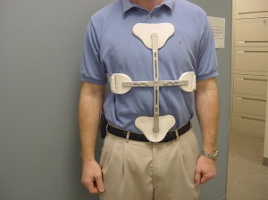
Several braces are available. In some patients, bracing can help reduce motion enough to allow the patient to tolerate natural healing and avoid further invasive intervention. After vertebral augmentation, bracing also may facilitate pain reduction and axial support for patients who have poor activity tolerance because of muscle fatigue. Bracing is very helpful for patients who have poor muscular endurance or thoracic kyphosis in which pain might be reduced by facilitating spinal extension. Problems with bracing can include a poor fit in obese patients, patient acceptance, expense, and difficulty in putting on and removing the brace. A brace that fits poorly or is too difficult to use often will end up unused. The type of brace should be directed by the patient’s level of activity and comfort. Traditionally, the Jewitt and cruciform anterior spinal extension (CASH) orthoses ( Figs. 1 and 2 ) have been used because the three-point contact facilitates thoracic and lumbar neutral positioning while decreasing flexion. For stable thoracic fractures, the authors often recommended a posture training support ( Fig. 3 ) to facilitate spinal extension and scapular retraction . This brace is easy to put on and remove, can be worn under regular clothing if desired, and is relatively affordable. Most insurance companies and Medicare will pay a portion of the cost. Small weights can be added to this backpack-like device as the patient’s strength increases and tolerance improves. A patient should start out with a single 1-pound weight and increase the weight weekly, as tolerated, but not above 3 pounds. If more limitation in motion is required because of pain, a Jewitt (see Fig. 1 ) or cruciform anterior spinal extension (CASH) brace (see Fig. 2 ) can be prescribed. When spine stability is a concern, a custom thoracic lumbar spinal orthosis can be fabricated. For lumbar compression fractures, a simple corset with a moldable plastic posterior shell can be used to facilitate appropriate spine posture and avoid chronic flexion . The purpose and importance of bracing needs to be reviewed with the patient. Not all patients require bracing, however, and not all patients have the body habitus to accommodate available orthoses.

A trial of bracing with the physical therapist working with an orthotist is helpful in determining the appropriate fit while not impeding function. As pain improves, discontinuing the brace usually is easiest when weaned in increments of decreased wearing time. Some patients find that continuing to wear the brace for specific activities after the fracture has healed may help reduce continued pain and facilitate good spine mechanics.
Injections
In addition to the treatment plan outlined previously, some patients may require further intervention to reduce pain and improve function. Patients may present with neurogenic symptoms consistent with radiculopathy. When these symptoms are experienced in combination with motor weakness, spine stability should be confirmed first. Other causes include vertebral body retropulsion as a result of the fracture causing central or lateral recess narrowing ( Fig. 4 ). Disc protrusions may be evident at, below, or above the level involved. These radicular symptoms may be managed with epidural steroid injections directed at the level of the patient’s symptoms and correlated with imaging findings. Because of the anatomic variations associated with compression fractures, the authors recommend that these injections be performed under fluoroscopic guidance with contrast enhancement to ensure accurate placement of the medication and to avoid subdural puncture ( Fig. 5 ). In patients who have intractable radicular pain, epidural steroid injections pain often may help the timeliness of the patient’s recovery.
Vertebral augmentation
Vertebral augmentation is being used more widely for nonneoplastic vertebral compression fractures . Vertebroplasty, the transpedicular percutaneous infusion of methylmethacrylate into vertebral bodies, has been used in the United States for spinal vertebral compression fractures caused by osteoporosis and cancer ( Fig. 6 ). Prospective and retrospective studies report pain improvement. Kyphoplasty, a procedure that involves inflating percutaneously placed balloons within the fractured vertebra and then infusing cement within the cavities created by the balloon, is another type of vertebral augmentation. The publicized advantage of kyphoplasty over vertebroplasty is the potential restoration of vertebral body height, which ultimately reduces deformity. With less deformity, it can be inferred that subsequent fracture rates would diminish after the procedure. Several studies have shown that this height restoration is measurable, although small in increment . Gaitanis and colleagues reported a mean increment of 4.3 mm for anterior wall restoration and a mean increment of 4.88 mm for midvertebral height restoration. This study looked exclusively at pathologic fractures. Although these increments are small, the physiologic and biomechanical advantages still need to be investigated. A recent study by McCann and colleagues found no difference between vertebroplasty and kyphoplasty in restoration of vertebral height in cadaveric vertebral compression fractures. In another study, McKiernan and colleagues treated 46 patients who had a total of 66 painful nonneoplastic vertebral compression fractures with vertebroplasty and measured height restoration after the procedure. At 6 months, there was no difference in pain or quality of life measures in patients who had vertebral height restoration and those who had not. No correlation has been found between pain reduction and the amount of cement infused or vertebral body correction . Fribourg and colleagues completed a follow-up study of 38 patients, 13 of whom had received kyphoplasty, and found a higher rate of fracture after the procedure than seen with the natural history of fracture. One study showed that the vertebral body in a cadaver model augmented with kyphoplasty was less stiff than before augmentation, suggesting one possible cause for subsequent fractures . The topic of subsequent fractures after vertebroplasty and kyphoplasty remains controversial . There are many factors involved in subsequent fractures after both procedures, including extent of osteoporosis, kyphosis and scoliosis, motion at the fracture site, posture, activities, and collaborative treatment. Future study of these contributing factors is necessary to decipher risks and prognosticate outcomes better. As with any procedure, patient selection for the procedure and patient management before and after the procedure are mastered with experience. Although known complications of vertebroplasty such as infection, discitis, radiculitis, vertebral fracture, and pulmonary embolism are uncommon, there are fairly straightforward treatment guidelines to follow should they occur . Although uncommon, rib and sternal fractures have been reported with both procedures . The overall incidence of procedural complications with vertebroplasty has been reported to be 2.4% , compared with 0.9% with kyphoplasty . Some of this variation involves the recognition of leakage, which some sources call a complication; others, however, find small amounts of leakage commonly present without symptoms . The incidence of transient increases in pain after the procedure has been reported to range from 4% to 23.4% . Transient pain increase has been attributed to increased pressure in the fractured vertebral body, inflammatory reaction to polymethylmethacrylate, and osseous ischemia .
Related posts:
 The Female Athlete Triad and Cardiovascular Dysfunction
Musculoskeletal Disorders of Pregnancy, Delivery and Postpartum
Rehabilitation in Women with Breast Cancer
Exercise for Health and Wellness at Midlife and Beyond: Balancing Benefits and Risks
The Role of Physical Activity in Bone Health: A New Hypothesis to Reduce Risk of Vertebral Fracture
Acetabular Labral Tears of the Hip in Women
The Female Athlete Triad and Cardiovascular Dysfunction
Musculoskeletal Disorders of Pregnancy, Delivery and Postpartum
Rehabilitation in Women with Breast Cancer
Exercise for Health and Wellness at Midlife and Beyond: Balancing Benefits and Risks
The Role of Physical Activity in Bone Health: A New Hypothesis to Reduce Risk of Vertebral Fracture
Acetabular Labral Tears of the Hip in Women
Stay updated, free articles. Join our Telegram channel

Full access? Get Clinical Tree


