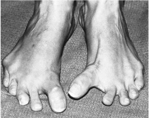
Complications of Hallux Abducto Valgus Surgery
Gerard V. Yu
Molly Schnirring-Judge
Jeffrey E. Shook
Surgical correction of hallux abducto valgus and related deformities is commonly performed. Unfortunately, complications of hallux abducto valgus surgery may develop, many of which are unpredictable. Common complications of hallux valgus surgery include recurrence of the deformity, hallux varus, and complications of bone healing, such as delayed union, nonunion, or malunion. The purpose of this chapter is to provide an overview of and insight into the diagnosis and treatment of some of the more common complications of hallux abducto valgus surgery.
RECURRENCE OF DEFORMITY
Definition, Incidence, and Etiology
Experience has shown that even when strict criteria are followed for the repair of hallux abducto valgus deformity, the condition can recur. Generally, patients with recurrent hallux abducto valgus may be divided into two groups: those patients who manifest with hallux abducto valgus early in the postoperative period, perhaps in the first few months to 1 year, and those patients who present some time later with recurrent deformity. Although one or two factors may play a dominant role in the development of recurrent deformity, others may also be involved to a lesser degree.
Early Recurrent Deformity
In general, early recurrence of the deformity may be attributed to one of several different factors: (a) an error in judgment in the selection of procedures; (b) inadequate execution of the procedure; (c) events during the postoperative care, including patient noncompliance; and (d) failure to recognize or to address concomitant deformities such as metatarsus adductus.
Most cases of hallux abducto valgus deformity are caused by a combination of dynamic soft tissue factors as well as structural factors. Although some bunion deformities have a more dynamic cause, others have a structural component as the primary etiologic factor.
The degree and extent of displacement of the sesamoidal apparatus may be strong indicators of the dynamic component of the bunion deformity. Failure to release the plantar lateral soft tissues of the first metatarsophalangeal joint can increase the incidence of recurrence after hallux valgus correction. With contracture of adductor hallucis muscle and other periarticular structures, the sesamoid apparatus displaces in a lateral direction relative to the first metatarsal head and contributes to further progression of the deformity. The failure of the surgeon to release these lateral soft tissue structures properly, a failure that inhibits relocation of the sesamoidal apparatus beneath the first metatarsal head, significantly increases the risk of recurrence. Even in cases with an adequate release of the plantar lateral structures, the surgeon may find that the sesamoid apparatus is not adequately mobilized, and consequently fibular sesamoidectomy may be necessary.
Kitaoka et al. studied 49 feet that underwent simple bunionectomy for hallux valgus deformity. The primary reason for treatment failure was recurrence, and 14% of these patients underwent revisional surgery as a result. Patients who had a lateral capsulotomy had less likelihood of experiencing recurrence of the deformity (1).
Restoring integrity to the medial joint capsule is helpful in maintaining rectus hallux alignment. As such, a deliberate reinforcement or medial joint capsulorrhaphy is sometimes performed to restrain the tendency for recurrent valgus drift of the hallux. In a cadaveric study, Kura et al. investigated the functional significance of the medial capsule and the transverse metatarsal ligament in hallux valgus deformity. A three-dimensional imaging technique was used to track the effect of sectioning these structures to assess their influence in deformity about the first metatarsophalangeal joint. No significant deformity was noted when the transverse metatarsal ligament was sectioned. Valgus deformity of the hallux increased an average of 22 degrees when the medial
capsule was sectioned, a finding that underlines the contribution of this structure to joint stabilization. However, this does not imply that medial capsulorrhaphy is the critical aspect of the soft tissue procedure responsible for maintenance of correction (2), although it is an adjunctive measure that may assist in restoring overall balance to the joint. The study does indicate the concern for addressing lateral joint contractures, because compromise of the medial joint structures is required to alleviate the bunion prominence.
capsule was sectioned, a finding that underlines the contribution of this structure to joint stabilization. However, this does not imply that medial capsulorrhaphy is the critical aspect of the soft tissue procedure responsible for maintenance of correction (2), although it is an adjunctive measure that may assist in restoring overall balance to the joint. The study does indicate the concern for addressing lateral joint contractures, because compromise of the medial joint structures is required to alleviate the bunion prominence.
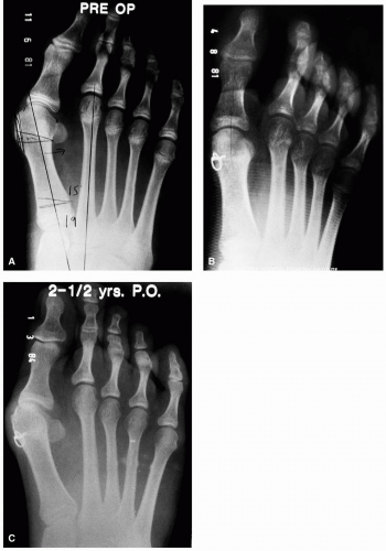
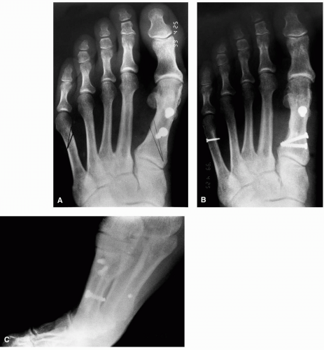
Perhaps the most common cause of recurrence is an error in judgment in the selection of the surgical procedure for the correction of a hallux valgus deformity (Fig. 1) (3,4). Typically, a capital osteotomy was employed, yet it proved inadequate to provide full correction of the intermetatarsal
angle (Fig. 2). Often, this is not so much a recurrent deformity as a residual hallux abducto valgus that was not fully corrected with the original procedure.
angle (Fig. 2). Often, this is not so much a recurrent deformity as a residual hallux abducto valgus that was not fully corrected with the original procedure.
The recurrence rate after distal metaphyseal osteotomies has been proposed to involve approximately 10% of patients undergoing these surgical procedures (3). Historically, distal metaphyseal osteotomies have been indicated for the correction of mild to moderate bunion deformities with an intermetatarsal angle of 12 to 15 degrees (4, 5, 6, 7), although surgeons commonly employ these types of procedures in patients with larger intermetatarsal angles if the deformity is flexible. Meier and Kenzora found that 94% of the patients undergoing distal metaphyseal osteotomies with a preoperative intermetatarsal angle of less than 12 degrees had a satisfactory result, compared with 74% of the patients with an intermetatarsal angle greater than 12 degrees (8).
Clearly, in some cases, a proximal osteotomy is far more effective in reducing the intermetatarsal angle than a distal metaphyseal osteotomy. Historically, procedures may have been selected based on radiographic findings alone, such as the intermetatarsal or hallux abductus angle. However, strict radiographic criteria should not serve as the sole basis for the selection of a procedure. Appreciation of the intermetatarsal angle can be better assessed intraoperatively after a complete release of the periarticular structures.
Inadequate postoperative care may also encourage the development of a recurrent bunion. Immediate postoperative bandaging should be employed to help maintain the great toe in a rectus position. When the toe is not splinted properly during first several weeks after surgery, the likelihood of recurrence of the deformity is increased. Inadequate splinting and poor maintenance of alignment allow the medial
capsular structures to undergo stretching, whereas the lateral structures attempt to recontract and shorten, with a resulting recurrence of the deformity in the earlier stages after bunion surgery (3). Obviously, the cooperation of the patient is essential during this period, to ensure the best possible result.
capsular structures to undergo stretching, whereas the lateral structures attempt to recontract and shorten, with a resulting recurrence of the deformity in the earlier stages after bunion surgery (3). Obviously, the cooperation of the patient is essential during this period, to ensure the best possible result.
Delayed Recurrence of Deformity
Failure to address concomitant deformities associated with hallux valgus deformity has also been associated with recurrence of the deformity over time (3,4,9,10). Hypermobility of the first ray may contribute to recurrence of the deformity if it is not addressed either surgically or postoperatively with orthotic control or other measures. Other deformities and conditions associated with recurrent hallux valgus may include ankle equinus, collapsing pes valgo planus deformity, metatarsus adductus, spasticity, or hyperelasticity or ligamentous laxity, such as in Ehlers-Danlos syndrome (3,4,9,10).
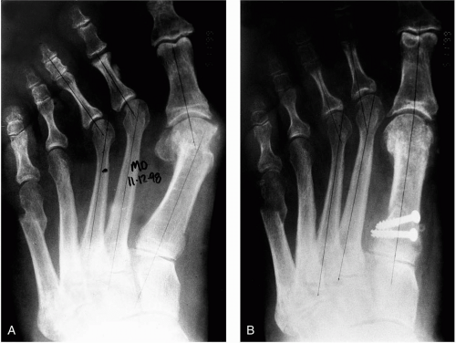
In particular, patients with concomitant hallux abducto valgus and metatarsus adductus present a challenging problem. Failure to recognize the existence of metatarsus adductus before surgical intervention is apt to result in a less than a satisfactory outcome. Recurrence and undercorrection are common and vary depending on the severity of the underlying metatarsus adductus and digital abduction (Fig. 3).
In radiographic evaluation of such feet, the intermetatarsal angle must be considered to be significantly greater than that determined by actual measurement (11, 12, 13, 14). It is not uncommon to strive to obtain a reduction of the intermetatarsal angle intraoperatively, to 0 to – 2 or – 3 degrees. In some cases, an opening wedge osteotomy of the first metatarsal or medial cuneiform may be an appropriate procedure, although we have not found this approach necessary (15).
Clinical experience shows that even when the intermetatarsal angle has been reduced to a slightly negative value, an increased separation between the first and second metatarsals may be seen later when full weight-bearing function has been restored to the foot. This ultimately results in a final intermetatarsal angle of approximately 0 to 5 degrees.
Patients who undergo surgical correction of a hallux abducto valgus deformity in the presence of a structural metatarsus deformity frequently have a residual bunion deformity, or clinical hallux abducto valgus deformity, without any radiographic evidence of such. In many cases, the surgeon may identify full correction and normal values of most radiographic parameters. The degree to which the clinical appearance of a residual bunion and hallux abducto valgus deformity occurs is proportional to the degree of metatarsus adductus deformity and the degree of compensation present (10,16, 17, 18, 19, 20, 21). The greater the metatarsus adductus deformity,
the greater is the abduction of the hallux and lesser digits on their adjacent metatarsal (10,16,22, 23).
the greater is the abduction of the hallux and lesser digits on their adjacent metatarsal (10,16,22, 23).
Clinical and Radiographic Evaluation
The clinical and radiographic findings associated with a recurrent hallux abducto valgus deformity are usually not different from those seen before the first surgical procedure, with one exception: they are usually worse. Both the type and the intensity of the pain as well as the clinical deformity are more extreme than the original presentation. Patients may also complain of a “tingling, numbness, or pins and needles” sensation suggestive of an entrapment neuropathy of the medial proper digital branch of the medial dorsal cutaneous nerve or the terminal branches of the saphenous nerve. A sharp focal area of pain may be caused by a residual osseous prominence. Pain along the plantar aspect of the joint, especially at the medial portion, may indicate an abnormal articulation between the sesamoid and the first metatarsal head.
The hallux may underride or overlap the second digit, a feature that may not have been present with the original deformity. The bunion prominence is typically larger even though aggressive resection of bone may have already been performed. Lateral bowstringing of the extensor hallucis longus tendon may be seen. Although the original deformity may be purely transverse, increased valgus rotation of the hallux is often seen in patients with a recurrent deformity.
Of significance is the degree to which the deformity is reducible and the joint range of motion is maintained while in the corrected position. A patient with a limited range of motion associated with pain and crepitation may require a joint-destructive procedure regardless of the radiographic findings. An assessment of the reducibility and flexibility of the deformity not only is helpful in identifying the most appropriate procedure, but also it may signify the propensity or likelihood that a transverse plane hallux varus deformity will develop. An inability to reduce the deformity strongly suggests tight plantar lateral structures, most notably the adductor hallucis and secondarily the lateral head of the flexor hallucis brevis muscles.
Patients should also be observed while they are in a relaxed stance position. It is not uncommon to see an exacerbation of the deformity when the foot is fully loaded. In addition, one gains a much greater appreciation of the plane of the deformity (i.e., purely transverse versus a combination of transverse and frontal). Clinical observations are then correlated with the radiographic findings.
Finally, the presence of concomitant deformities is assessed. Emphasis is placed on the presence of metatarsus adductus, as well as any pronatory changes in the foot consistent with severe collapsing pes valgo planus deformity that could require treatment to ensure correction of the recurrent hallux abducto valgus deformity, especially in the juvenile or adolescent patient.
Treatment Considerations
Most patients with symptomatic recurrent hallux abducto valgus deformity do not respond well to conservative treatment modalities. The surgical correction of a recurrent deformity is more challenging than that of the original condition. Although the goals of the revisional operation should be to establish a congruous, pain-free, functional first metatarsophalangeal joint, this cannot always be achieved. Joint-salvage procedures can only be employed when the patient has sufficient bone and stable architecture at the first metatarsophalangeal joint. Patients who have had excessive resection of the medial eminence may not have adequate bone stock to support a traditional distal metaphyseal procedure. In such cases, proximal osteotomies may be necessary despite a relatively low intermetatarsal angle. In cases of significant arthrosis, joint compromise, or instability, a resection arthroplasty, implant arthroplasty, or arthrodesis may be required. Joint-preservation procedures are preferred whenever possible.
Meticulous surgical technique is critical to achieving the desired outcome. In most cases, a dorsomedial skin incision is employed. Every attempt is made to separate the skin and subcutaneous tissues from the deep fascial layer, to prepare for anatomic closure. One must be cautious to avoid the dorsal cutaneous nerves and pertinent vascular structures. In patients with clinical evidence of nerve entrapment, the nerve is carefully explored, and any surrounding scar tissue is released; when necessary, nerve resection may be performed.
Unless the fibular sesamoid has been previously excised, attention is directed to the first interspace. Complete release of the plantar lateral structures is performed. In most cases, this operation involves release of the adductor hallucis tendon and fibular sesamoidal ligament. In more severe cases, release of the lateral head of the flexor hallucis brevis muscle may also be necessary. If adequate immobilization of the sesamoid apparatus cannot be achieved, then considerations should be given to removal of the fibular sesamoid.
After release of the intermetatarsal space, the deformity must be reassessed. When a recurrent contracture of the plantar lateral structures has occurred, or an inadequate release was performed initially, a significant improvement in the alignment of the hallux on the first metatarsal is generally seen. After plantar lateral release, one can more readily appreciate the true structural components of the deformity and decide on the need for additional procedures to achieve adequate joint congruity and position.
Various medial capsular approaches can be employed for exposure of the joint. The choice depends on the experience and preference of the surgeon. The first metatarsal head and phalangeal base are inspected. The orientation of the articular cartilage on the first metatarsal head is inspected and should be compared in position with the long axis of the metatarsal shaft.
Although some contouring of the dorsal and medial aspects
of the first metatarsal head may be necessary, aggressive resection of the metatarsal head should not be performed. Further “staking” of the metatarsal head only compromises function.
of the first metatarsal head may be necessary, aggressive resection of the metatarsal head should not be performed. Further “staking” of the metatarsal head only compromises function.
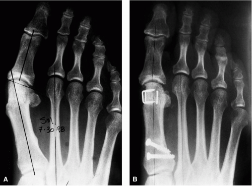
If excessive resection of bone was not performed in the initial surgical procedure, then a distal metaphyseal osteotomy may be appropriate for correction of any residual splaying between the first and second metatarsal. The type of osteotomy performed depends on several factors. In patients with significant deviation of the articular cartilage on the first metatarsal head, a Reverdin-type osteotomy may be used to reposition the articular surface properly. This form of osteotomy may also be considered to rotate the remaining cartilage into a more effective position when the first metatarsal head has been staked. Any remaining intermetatarsal splay is corrected by a more proximally oriented procedure (Fig. 4).
Structural splaying between the first and second metatarsal is usually addressed by a proximal procedure. The most common procedure is a closing base wedge osteotomy, not only to reduce the intermetatarsal angle, but also to minimize the amount of shortening. When postoperative elevatus has occurred in conjunction with recurrence of the deformity, the osteotomy may be modified to achieve both reduction of the intermetatarsal angle and simultaneous plantarflexion of the distal fragment. In some cases, a first metatarsocuneiform arthrodesis or opening wedge osteotomy of the metatarsal base or cuneiform may be appropriate, especially when restoration of length is an important consideration. Lengthening of the first ray segment may create tension at the first metatarsophalangeal joint and may possibly lead to limitation of motion, with or without symptoms.
In cases of recurrent juvenile or adolescent hallux abducto valgus deformity, careful consideration must be given to the surgical correction of concomitant deformities. In patients with a significant ankle equinus, a tendo Achillis lengthening or gastrocnemius recession may be performed. Patients with severe collapsing pes valgo planus deformity may also need surgical correction if they cannot be treated effectively with an appropriate orthotic device or shoe modifications. Finally, the influence of metatarsus adductus deformity cannot be overemphasized. Residual metatarsus adductus can have a profound influence on first ray disorders. Although it is rare to attempt full correction of the deformity in an adult, it may be important in obtaining the best correction of a juvenile or adolescent suffering with a painful hallux valgus deformity.
HALLUX VARUS
Definition, Incidence, and Etiology
Hallux varus is classified as either a congenital or an acquired deformity and has been a subject of study for many
years (1,3,4). Although acquired hallux varus may have several causes (i.e., postsurgical, trauma, rheumatoid arthritis), this discussion focuses on those cases noted after surgical correction of hallux abducto valgus or related deformities. Regardless of whether a hallux varus deformity is congenital or acquired, common denominators exist. In virtually all cases, one appreciates a muscle imbalance in which the abductor hallucis muscle gains a significant mechanical advantage over its antagonistic muscle, the adductor hallucis. In a postsurgical hallux varus, the abductor hallucis muscle usually is responsible for deformity, although in some cases the deformity may occur primarily as a result of structural alterations of the first metatarsal, proximal phalanx, or both.
years (1,3,4). Although acquired hallux varus may have several causes (i.e., postsurgical, trauma, rheumatoid arthritis), this discussion focuses on those cases noted after surgical correction of hallux abducto valgus or related deformities. Regardless of whether a hallux varus deformity is congenital or acquired, common denominators exist. In virtually all cases, one appreciates a muscle imbalance in which the abductor hallucis muscle gains a significant mechanical advantage over its antagonistic muscle, the adductor hallucis. In a postsurgical hallux varus, the abductor hallucis muscle usually is responsible for deformity, although in some cases the deformity may occur primarily as a result of structural alterations of the first metatarsal, proximal phalanx, or both.
We use the term hallux varus (or hallux adductus) to describe purely transverse plane, medial deviation of the great toe with the apex of deformity at the metatarsophalangeal joint. This condition is readily identified both radiographically and clinically. The term hallux malleus is used to describe a great toe with an extension contracture at the metatarsophalangeal joint and concomitant flexion contracture at the interphalangeal joint. When the conditions occur simultaneously, we refer to the condition as hallux varus with a concomitant hallux malleus deformity (Fig. 5). Regardless of the terminology, one must be aware of the apex of deformity and the presence of combination deformities to ensure adequate surgical selection and treatment.
The combination of the transverse and sagittal plane deformities usually represents the end point of the deformity. The rate of progression and the ultimate severity of the condition depend on several factors, including inherent flexibility, the time elapsed since the surgical procedure, the degree of musculotendinous imbalance, the amount of structural malalignment, and other underlying concomitant disorders such as neuromuscular or connective tissue diseases.
Iatrogenic hallux varus alone or in combination with a hallux malleus deformity can be more painful, disfiguring, and disabling than the original hallux valgus condition. Cosmetic complaints are rare when the varus deformity is less than 10 degrees (24). Pain associated with the deformity is often coincident with significant joint incongruity or degenerative joint disease.
McBride first described the complication of hallux varus in the follow-up study of his procedure and reported an incidence of 5.1% (25). The incidence of postoperative hallux varus reported by other authors approximates 1% (26, 27, 28). In a review of 878 first metatarsophalangeal joint procedures, Feinstein and Brown reported 10 cases of hallux varus, with an overall incidence of 1.13%. This study involved procedures for the correction of hallux abducto valgus and hallux limitus. Other studies, which reviewed procedures performed only for the correction of hallux abducto valgus, cited a similar incidence of postoperative hallux varus. Combining two separate reviews, 21 cases of hallux varus were discovered in 1,400 postoperative cases for an overall incidence of 1.5% (26,27).
In more recent literature, the reported range is from 2% to 17% (29, 30, 31). The length of the first metatarsal has been purported to predispose to the development of hallux varus (28). Janis and Donick found 18 cases of hallux varus in a review of 1,110 bunion operations. These investigators concluded that all but 2 cases were in patients with a long first metatarsal and deduced that this feature was influential in the development of a hallux varus deformity. However, considering that the normal metatarsal protrusion with respect to the first and second metatarsals is plus or minus 2 mm, with scrutiny of the results, one finds that the first metatarsal was abnormally long in only 50% (9 of 18) of these cases. In another study of 10 cases of hallux varus, the first metatarsal measured more than 2 mm longer than the second in only 2 cases. In the remaining 8 cases, the length pattern of the first metatarsal was not considered a significant cause (26). Therefore, it appears that the length of the metatarsal may not predispose a patient to hallux varus, as was once suspected.
The shape of the first metatarsal head, specifically a round first metatarsal head, has also been implicated as a factor leading to the formation of hallux varus after bunion operations (28,31). A round metatarsal head seemingly implies an absence of the sagittal groove or plantar, medial condyle. Although the morphology of the sagittal groove and plantar medial condyle may vary from patient to patient, we have yet to perform an initial bunion operation and find these structures to be absent. Furthermore, no reports exist in the literature that correlate the architecture and shape of the first metatarsal noted intraoperatively with the preoperative radiographs. The concept of a round first metatarsal head as a contributory factor to hallux varus is strictly a supposition based on retrospective radiographic analysis and clinical experience. The apparent shape of the first metatarsal head is easily altered by positional changes of the foot or by changing the tube head angle in x-ray acquisitions. Therefore, the
validity of a round metatarsal head as a contributory factor to the development of iatrogenic hallux varus deformity should be questioned.
validity of a round metatarsal head as a contributory factor to the development of iatrogenic hallux varus deformity should be questioned.
The flexibility of the first ray may certainly be a contributory factor in the development of postoperative hallux varus deformities (32). A foot that is extremely mobile and has an excessive sagittal plane range of motion may be more susceptible to postoperative deformity. It seems logical that patients with an excessive range of motion of the first metatarsophalangeal joint (90 to 100 degrees or greater) and first ray will tend to be flexible in the transverse plane as well.
Certain connective tissue disorders (i.e., Marfan’s syndrome and Ehlers-Danlos syndrome) are associated with an inherent joint laxity that predisposes the patient to recurrent deformity or hallux varus. In addition, patients with disorders such as Down’s syndrome or other neuromuscular diseases such as cerebral palsy may also be predisposed to developing a postoperative hallux varus condition.
Stay updated, free articles. Join our Telegram channel

Full access? Get Clinical Tree


