Complex Regional Pain Syndromes and Related Disorders


Complex Regional Pain Syndromes and Related Disorders


Jeffrey C. Christensen


Complex regional pain syndrome (CRPS) is a term designated to encompass the terms reflex sympathetic dystrophy (RSD) and causalgia (1,2 and 3). The hallmark of this syndrome typically is severe spontaneous pain associated with local dysautonomic features that occurs after a trauma or operation to a limb. The associated signs and symptoms are considered an exaggerated response to injury that has variable expression between individuals (4,5,6 and 7). Potentially it can become a devastating progressive disorder that leads to significant disability, emotional disturbance, and irreversible trophic changes to the extremity (8,9,10,11 and 12).

This syndrome comprises a group of heterogeneous neuropathic conditions, which are widely recognized in their chronic state but are poorly understood (10). The five major components are pain out of proportion to the injury, edema, autonomic dysfunction, movement disorder, and trophic changes (12,13,14 and 15). CRPS usually results from incomplete nerve trauma or soft tissue injury (8,16) and most commonly affects the hands and feet (15,17).

Managing a patient with CRPS in the lower extremity is a very perplexing and challenging task for the foot and ankle clinician. Early recognition seems to be critical to the outcome (6,7 and 8,11,12,15,18,19,20,21,22,23,24,25,26,27,28,29,30,31 and 32), but at times, CRPS is most difficult to discern even for the most experienced clinician. It is not uncommon for the early stages to express an incomplete clinical picture, which only faintly resembles the mature disease. Once the diagnosis is made, the clinician must now manage a recalcitrant condition with notoriously poor treatment outcomes. The condition is burdensome for the patient and physician alike and requires extensive education and counseling for patients and concerned family members. Typically, these patients are emotionally challenged with feelings of anger, confusion, frustration, or trepidation. They frequently convey urgency to resolve their dilemma, often actively searching for “the cure” for their condition. Such desperation can lead to ineffectual or misguided treatments and worsening of the condition.

While historical literature can be very useful in reviewing classical clinical observations, proper weighting of these papers is essential to begin to understand these disorders. While the classic causalgia papers have valuable descriptions of cases with a common causative mechanism, other earlier works had ambiguous study designs, less diagnostic uniformity, and a lack of controls. Thus, data and conclusions from earlier investigations may be inaccurate and misleading. Over the past two decades, significant advances have been made to enhance the understanding of CRPS and promote potentially effective treatments. This chapter focuses on integrating these various sources of information, both old and new, into a useable format for the foot and ankle clinician with the hope of facilitating a more accurate understanding of CRPS and effect improved treatments for this challenging disorder.


DEFINITIONS AND NOMENCLATURE


EVOLUTION OF PAIN NOMENCLATURE

Some of the early clinical descriptions of chronic pain syndromes are still worth reviewing to understand that some of the same issues and clinical challenges encountered decades ago are still present today. While there have been earlier accounts of painful limbs after injury, the genesis of modern pain nomenclature can be traced back to 1864 when Silas Weir Mitchell and his associates published elegantly detailed case descriptions of severe limb pain after gunshot wounds during the American Civil War (5) (Fig. 68.1). Mitchell was regarded the preeminent neurologist of his time. He ran The U.S.A. Hospital for Injuries and Diseases of the Nervous System in Philadelphia dedicated to war-related conditions. The battles provided his wards with numerous cases, representing almost every type of unusual nervous disease, especially injuries to peripheral nerves. Many of the soldiers developed chronic pain conditions from their wounds. They were admitted for weeks and months at a time, which permitted Mitchell’s physician team to make daily observations and to develop incredibly detailed case reports that will never be equaled. What is now known as CRPS, they first described as erythromelalgia, and later Mitchell coined the term causalgia (4). Cases of causalgia induced from high-velocity missiles have been documented in many wars since the civil war (8,18,33,34).

Numerous authors over the last 145 years have observed posttraumatic pain disorders that varied from Mitchell’s descriptions of causalgia. In 1900, Paul Sudeck used early radiographic techniques to discover associated bone atrophy with causalgia (Sudeck atrophy); however, he regarded osteoporosis as one of
several late consequences of this disorder and felt that the condition was an exaggerated inflammatory response rather than sympathetic overactivity (35,36 and 37). DeTakats in 1937 described a condition called reflex dystrophy involving pain with vasomotor disturbances, trophic changes, and relief of pain with sympathetic blocks (38). Evans in 1947 reviewed a large series of cases and described a similar series as RSD (39). Unfortunately, while this entity has become the most widely described posttraumatic pain syndrome to date, its clinical description has lacked refinement since its inception.


Figure 68.1 Photograph of Silas Weir Mitchell. (Courtesy of the National Library of Medicine.)

The evolution in our conceptual understanding of these disorders has lead to nearly continuous controversy and disagreements since Mitchell’s early descriptions. The ongoing debates regarding nomenclature have been the most heated. This is most exemplified by the numerous terms authors have used to describe causalgia-like disorders (Table 68.1). Doupe et al in 1944 stated:


The confusion caused by using the term causalgia for different conditions and by describing cases of causalgia under different names indicates the need for the classification and segregation of different post-traumatic states. However, a classification, no matter how sorely needed, cannot be satisfactory so long as it is based on symptoms” (40).

Unfortunately, their advice was never heeded. More recent classifications have been further complicated by indiscriminate use of terms and inadvertent erroneous labeling of patients with improper diagnoses. This resulted in researchers and clinicians becoming completely dissatisfied with pain nomenclature as terminology became increasingly ambiguous and clinically misleading. The diagnosis of RSD has been abused and has become a “catch-all” term to describe virtually any regionalized chronic pain syndrome with almost any combination of clinical criteria (41,42,43,44 and 45).


TABLE 68.1 Historical Synonyms for CRPSs


Acute atrophy of bone


Posttraumatic painful osteoporosis


Algodystrophies mineures


Posttraumatic spreading neuralgia


Algodystrophies reflexes


Posttraumatic sympathetic dysfunction


Algodystrophy


Posttraumatic sympathetic dystrophy


Algoneuodystrophy


Posttraumatic vasomotor disorders


Ascending neuritis


Pseudoarthritis


Causalgia


Pseudorheumatism


Causalgia-like states


Reflex arterial spasm


Causalgic state(s)


Reflex aldodystrophy


Chronic segmental arterial spasm


Reflex dystrophy


Chronic traumatic edema


Reflex neurovascular dystrophy


Erythromelalgia


RSD


Etat Physiopathique of Vulpian


Reflex sympathetic imbalance


Homans minor causalgia


Reflex trophoneurosis


Major traumatic dystrophy


Shoulder-hand syndrome


Major causalgia


Sudeck-Leriche syndrome


Mitchell causalgia


Sudeck atrophy


Mimo-causalgia


Sudeck dystrophy


Minor causalgia


Sympathalgia


Minor traumatic dystrophy


Sympathetic neurovascular dystrophy


Minor Sudeck syndrome


SMP


Neurodystrophy


Sympathetic overdrive syndrome


Pain-dysfunction syndrome


Synesthesialgia


Painful osteoporosis


Thermalgia


Peripheral acute trophoneurosis


Traumatic angiospasm


Postinfarctional sclerodactyly


Traumatic edema


Posttraumatic dystrophy


Traumatic neuralgia


Posttraumatic osteoporosis


Traumatic osteoporosis


Posttraumatic pain syndrome


Traumatic vasospasm


Posttraumatic painful state


Trophic edema


Because causalgia, RSD, and Sudeck atrophy have been abused as descriptive and diagnostic terms, the limits of their definitions become less clear. Therefore, some patients are correctly diagnosed, while others are not. Tragically, the effects of this definition erosion adversely affect outcomes and treatment effectiveness.

Since the late 1980s, there was a relative explosion of new research, which yielded important knowledge and insight into posttraumatic pain mechanisms. Exponential growth of papers (using the keywords of RSD, sympathetically maintained pain (SMP), sympathetic nervous system, and pain) was seen to rise by a factor of six to sevenfold between 1966 to 1977 and 1990 to 1995 (46). This growth in knowledge fueled the desire to revise the classification of these related disorders. In 1986, in an effort to lessen the clinical ambiguity, the International Association for the Study of Pain (IASP) redefined RSD as “a state of continuous pain in a portion of an extremity after soft tissue or osseous trauma without major nerve injury, associated with sympathetic hyperactivity” (47). Roberts, in the same year, suggested the term sympathetically maintained pain as a more appropriate diagnostic expression since the sympathetic nervous system is directly involved (48). In cases in which sympathetic overactivity did not contribute to pain, it was termed sympathetically independent pain (SIP). Yet controversy lingered with these terms and required further refinement 7 years later. The IASP again revised the RSD/causalgia/SMP taxonomy. It was clarified that neuropathic pain is a clinical symptom associated with a presumed underlying nerve injury. Finally, it was determined that terms causalgia and RSD were antiquated, misleading, and still improperly utilized. Thus, a new taxonomy using CRPS was introduced in 1993 via a consensus workshop of the IASP (2,3).

From this consensus workshop, CRPS was divided into two groups. CRPS type I was designated to correspond to RSD and CRPS type II corresponded to causalgia. Specific criteria for each were agreed upon (Table 68.2) and provisions made to allow for future flexibility in the taxonomy. Future modifications can include variants within each type and possibly other types as well. It should be noted that RSD and causalgia are now transitional terms, which may still be used but not as primary descriptors. As a further clarification, SMP is now looked upon to include CRPS and other related disorders (Fig. 68.2).


TABLE 68.2 CRPS-Diagnostic Criteria


TYPE I


(2-4 must be satisfied)


1.


The presence of an initiating noxious event or a cause of immobilization


2.


Continuing pain, allodynia, or hyperalgesia with which the pain is disproportionate to any inciting event


3.


Evidence at some time of edema, changes in skin blood flow, or abnormal sudomotor activity in the region of the pain


4.


This diagnosis is excluded by the existence of conditions that would otherwise account for the degree of pain and dysfunction.


TYPE II


(All three criteria must be satisfied)


1.


The presence of continuing pain, allodynia, or hyperalgesia after a nerve injury, not necessarily limited to the distribution of the injured nerve


2.


Evidence at some time of edema, changes in skin blood flow, or abnormal sudomotor activity in the region of the pain


3.


This diagnosis is excluded by the existence of conditions that would otherwise account for the degree of pain and dysfunction.


Stanton-Hicks M, Janig W, Hassenbusch S, et al. Reflex sympathetic dystrophy: changing concepts and taxonomy. Pain 1995;63:127-133.


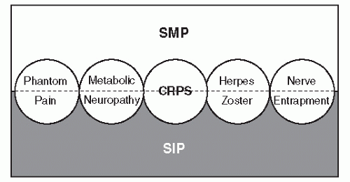
Figure 68.2 Schematic diagram of SMP as it functions as an umbrella term for a group of related disorders including CRPSs.


NEUROANATOMY AND PHYSIOLOGY


ANATOMY OF PAIN TRANSMISSION

Nociceptors Specialized unencapsulated nerve endings (Fig. 68.3) that initiate pain transmission when specific thresholds of thermal, mechanical, or chemical stimuli are received. Stimulation of peripheral nociceptors indicates potential or actual tissue damage (49,50). The pain perceived from this mechanism is referred to as nociceptive pain.


Figure 68.3 Drawing depicting appearance of various nociceptors and mechanicoreceptors found in cutaneous and subcutaneous tissues.


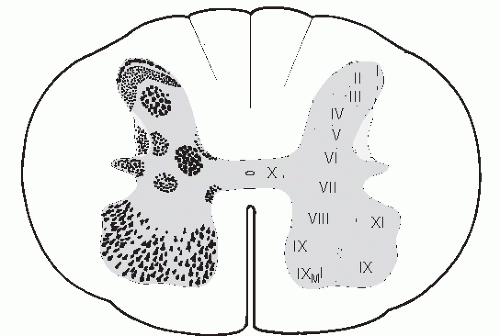
Figure 68.4 Diagram representing cross-sectional neuroanatomy of the spinal cord and the systematic location of laminae within the gray matter as described by Rexed. Laminae I, II, and V are highlighted to illustrate location of nerves involved with pain transmission and modulation within the spinal cord. Most of the WDR neurons are found in Lamina V.


Nerve Fibers Morphology

There are two basic groups of nerve fibers: myelinated and unmyelinated. These groups of fibers can be further broken down into three types of fibers based on diameter and conduction velocity. The thickest fibers are large myelinated (Aδ) fibers that conduct the fastest, medium-thickness fibers are thin myelinated (Aδ), and the thinnest are unmyelinated (C) slow-conducting fibers (49,51). C-fibers are found in the sympathetic and sensory nerves. The sensory C-fibers carry dull pain signals, whereas medium-sized Aδ fibers carry fast pain signals. Each fiber type has a unique specialized function and, due to their sizes, are differentially susceptible to various pathologic processes.


Nociceptive Transmission in the Spinal Cord

Most nociceptive signals follow afferent fibers that enter the spinal cord in the lateral dorsal root. The fibers bifurcate and travel both anteriorly and posteriorly. The majority of fibers terminate in Rexed laminae I, II, and V (Fig. 68.4). Nociceptive signals are then processed or modified by interneurons and transmitted to tract or projection cells (50).


Dorsal Horn Cells

There are two kinds of cells found in the dorsal horn, nociceptive specific (NS) found in lamina I and wide-dynamic-range (WDR) in lamina V. As implied, the NS cells only respond to noxious stimuli, whereas the WDR cells have the capacity to respond to noxious and innocuous stimuli. The WDR cells are involved with the pathogenesis of CRPS (see Pathophysiology).


Autonomic Nervous System (Sympathetic Division)

The thoracolumbar segment of the autonomic nervous system is part of the sympathetic division. This segment provides autonomic function to the lower extremities. It consists of groups of motor nerves whose cell bodies are located in ganglionated chains on each side of the spinal column (Fig. 68.5). The chain consists of 11 thoracic and 4 to 6 lumbar ganglia. From these chains, the nerve fibers travel along the spinal nerves and innervate blood vessels, sweat glands, and pilomotor structures. The sympathetic fibers in the periphery exert their influence via the release of norepinephrine. The organization of the preganglionic cells is complex and may involve the neuron traversing several levels of paravertebral ganglions along the chain before synapsing with postganglionic neurons. There are distinct functional classes of sympathetic innervation, which may explain why neuropathologic conditions associated with the sympathetic nervous system are clinically diverse (52,53).


Figure 68.5 Diagram of lumbar sympathetic chain positioned along the anterior lateral aspect of the vertebral segments.


Ascending/Descending Systems

There are multiple tracts that conduct nociceptive information in the spinal cord (49). The most important is the spinothalamic tract, which projects into the posterior lateral thalamus. A descending antinociceptive system functions to modulate nociceptive information at the level of the dorsal horn.


PERIPHERAL AND CENTRAL INTERACTIONS

It is widely believed that the pathophysiologic mechanisms associated with SMP can be ultimately traced to various neurotransmitters that “turn on” the disorder. In a partial nerve injury, the nerve segment can produce vasoactive substances, excitatory amino acids (L-glutamate, L-homocysteate, and L-aspartate), neurokinins, (substance P, neurokinin A and B, and other peptide neurotransmitters such as vasoactive intestinal peptide and calcitonin gene-related peptide). These substances and others are likely involved with connecting the events taking place at the peripheral injury site with various spinal cord structures.

Roberts set forth a hypothesis for SMP that took into account interactions between peripheral and central systems after peripheral nerve injury (48). He described a mechanism for which neurons in the spinal cord are activated and tie central processes with peripheral nerve dysfunctions. These neurons are referred to as WDR neurons located in the dorsal horn of the spinal cord. Cutaneous afferents including myelinated Aδ fibers and unmyelinated C-fibers both project into nociceptor specific and WDR neurons in the spinal cord. The WDR neurons ascend up the spinal cord and are capable of projecting pain signals. Continuous depolarization of C-fibers can induce sensitization of the WDR via transport of neurotransmitters; this in turn enlarges the cutaneous receptive field and leads to a greater responsiveness within that field to innocuous stimuli. This phenomenon involving a maladaptive change at the spinal cord level has been referred to central sensitization (14,100,101,102,103 and 104). An activated WDR in turn can activate the diffuse noxious inhibitory controls (DNICs) in the spinal cord, which functions as a filter and amplifier. This enhances the production of pain signal transference centrally by inhibiting background signals and extracting random innocuous signals entering the dorsal horn. With central sensitization, innocuous stimuli to an injured extremity can be improperly coded by the WDR and are projected as ascending pain signals in the spinal cord. The DNIC and WDR also are effected by inputs from emotional stimuli (76).


CLINICAL MANIFESTATIONS

Clinical signs and symptoms are remarkably variable among patients (12,61,105,106) and follow an individual timeline. The development of constant severe pain is usually expressed regionally on an extremity with a distal predominance. Pain severity often appears disproportionate to the described injury or event. Five major components seen clinically are pain, edema, autonomic dysfunction, movement disorder, and trophic changes (12,13,14 and 15). Typically, the pain has a burning quality and can be elicited from innocuous mechanical or thermal stimuli (allodynia) (107). Motor impairment can comprise loss of function, tremor, or dystonia (36,62,98,108,109,110,111,112,113,114 and 115). Additionally, early in the clinical course of CRPS, the pain may be sympathetically maintained and responsive to sympathetic blockade or sympathectomy.


CLINICAL PAIN CHARACTERISTICS

Pain is the most prevalent characteristic for the majority of patients. Most notable is pain at rest. The pain usually is continuous and spontaneous and can be provoked by a variety of stimuli. Patients will frequently relate common everyday intolerances due to their increased sensitivity. Increased extremity pain can be experienced with the most trivial stimulus like a sudden gust of wind. Typical nonpainful sensations such as pulsations from shower water, pressure from bed sheets, loud music, air conditioning, or automobile road vibrations can become painful experiences to the patient with CRPS. Mitchell in his early descriptions of causalgia was most impressed with this unusual hypersensitivity (4). The excessive hypersensitivity coupled with minor mishaps and negative reinforcement results in patients becoming strikingly overprotective of their injured part. In many patients, a classic protective pose or extreme avoidance measures are observed.

Upon clinical examination, the patient may often describe a pain distribution that follows no anatomical or dermatomal pattern (14). In the foot and ankle, patterns typically are more severe dorsally and anteriorly and less severe plantarly
and posteriorly (14,116). The pain described typically is more severe than anticipated from the injury. Over time, the pain will tend to spread if untreated and include a component of mechanical and thermal hypersensitivity.


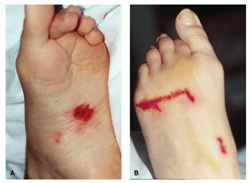
Figure 68.6 Photographs of isolated hemorrhagic bullae that ruptured on both the plantar and dorsal foot. Lesions developed within hours after severe flare of condition from overzealous physical therapy. Residual ulcers healed quickly over the following week.


DERMATOLOGIC CHANGES

There can be a wide spectrum of dermatologic changes observed in CRPS such as altered cutaneous temperature, nail and hair growth, skin color, and subcutaneous integrity (12,14,117,118 and 119).

In patients with active CRPS, vesicular eruptions, bullae, and ulcers are occasionally observed (120,121) (Fig. 68.6). Bullous eruptions seem to be somewhat associated with glossy skin and a reduction of burning pain when they are present (4). Schwartzman also described a small percentage of cases displaying Gardner-Diamond syndrome (autoerythrophagocytosis), which appeared explosively in the involved area as large tender purpuric areas that appeared like bruises (122). Additionally, occasional cases can also develop spontaneous hematomas and relapsing infections (105). The mechanisms for these spontaneous changes have not been elucidated.

The skin may have a sclerodermal-like appearance, which occurs earlier in the clinical course and is more severe in cases in which extremities are cold dominant (105). Glossy skin is often observed in CRPS, and in these instances, the skin is tightly drawn and often hairless without wrinkles. Mitchell was fascinated with this finding and described the skin as “…shining as though it had been skillfully varnished” (4).

Nail changes are common after nerve injury. After section of a nerve, nails are likely to club and slow down in growth, while with partial nerve injuries, nails are particularly prone to increased incurvation, which can lead to paronychia (4). In CRPS, nails also tend to become brittle, ridged, and dull (8,122). Incurvated nails have been reported in both unaffected and affected limbs in 8% of the patients with CRPS (105).

Edema is more prevalent in acute stages. Oftentimes, edematous changes can obscure underlying muscle atrophy (105). While hair loss or increased hair growth is frequently observed, piloerection in CRPS is often ignored by clinicians (123). Dependent rubor can be very impressive, and evaluation in a dependent position can often be helpful in the initial workup (Fig. 68.7).


VASCULAR CHANGES

In the more acute setting, there are often signs of vasomotor disturbances, which may manifest themselves in several ways (20). The red, warm, and edematous skin with vasodilation in the affected extremity is caused by a local inflammatory reaction. However, in more chronic situations, the skin may also be cold and mottled due to vasoconstriction with livedo reticularis, cold intolerance, and induration. Skin temperature asymmetry has often been cited as a very important clinical finding; however, in some investigations, the temperature was highly variable on each individual and followed no significant pattern (124,125 and 126). Furthermore, based on experimental study, skin temperature was not necessarily correlated with sympathetic activity and it would be “clinically dangerous” to conclude on the basis of a cold extremity that there is increased sympathetic tone (127).

Skin temperature measurements are an easy measure to assess cutaneous sympathetic vasoconstrictor activity (128). The clinical challenge is the spectrum of variation seen in the syndrome and the daily variation experienced by individuals. Recent studies have demonstrated that dynamic sympathetic response in CRPS patients is impaired compared to contralateral limbs regardless of temperature dominance of the limb (69,119,128).


MOTOR IMPAIRMENT

Motor abnormalities in CRPS are common and can be one of the earliest signs observed (4,12,98,105,113,129,130). The clinical expression seen is variable and may include muscle weakness, spasms, intention tremor, difficulty initiating movements (pseudoparalysis), and dystonia (97,98,108,109,113,115,131,132).
Veldman et al, in a large prospective series of 829 patients suffering from CRPS, found tremor and muscle incoordination in approximately 50% of the patients (105). A perceptible lag time can be seen between asking a patient to move the injured part and the first observed movement (98). Often a quivering in the digits with paradoxical contracture of opposing muscle groups is observed (i.e., extensors firing when digits are being flexed). With careful questioning, patients with CRPS will often relate problems with movement. They may say something like “my brain is telling it to move, but it won’t”; however, with milder expressions, movement disorders may be less apparent. Carefully observed clinical motor testing will reveal these subtle movement deficit (see “Physical Examination” section). Bilateral motor impairment (“mirror image” distribution) has also been reported (98).


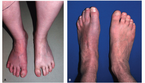
Figure 68.7 A: Photograph of an acute CRPS type I 1 month post fifth digit arthroplasty. Note dependant rubor of the right foot. Dotted line depicts medial boundary of allodynia. Hyperalgesia was present up to mid tibia. Early diagnosis was critical in this case, which quickly went into remission with pharmacologic treatment and physical therapy. B: Photograph of foot with deep dependant rubor in chronic case of CRPS type II, 2 years after surgical repair of Lisfranc fracture dislocation. Note subtle avoidance of full plantargrade weight-bearing and diminished hair growth.

Tremors are often seen especially with the initiation of movement (129). These tremors are usually not present during sleep (98,115,129). These flexion-extension tremors have an observed mean frequency of 7.2 ± 0.4, which is not statistically different from normal controls; however, the amplitude of the tremors are nearly 10 times greater (129). All of the tremors can be abolished by a successful sympathetic block and return when the effects of the block wear off (98,129). Deuschel et al, quantitatively analyzed tremors with CRPS I patients and regarded the tremor being an enhanced physiologic tremor similar to a thyrotoxemia and not of central origin (129).

Dystonia occurs when an injured extremity suffers from sustained spasms, which in turn cause a fixed dystonic posture. Dystonia is less common and usually has a later presentation with a worse prognosis (98,109,112,113,130). Veldman et al observed dystonia in 25% of patients with CRPS of long duration (105). This compares favorably with Scwartzman’s and Kerrigan’s findings of 21% rate of involvement (98). Patients with dystonia are extremely resistant to therapy and their dystonia can be so powerful that it can break casts and dislocate joints (14,76). The patterns of dystonia are variable (Fig. 68.8); not only can it be localized (i.e., involving one digit), but it can dominate an extremity or spread to other extremities. Bhatia et al investigated 18 patients with dystonia (16 were females). The authors reported patterns of spread that followed either a hemiplegic (5 cases), transverse (2 cases), or triplegic (2 cases) distribution (109).


PSYCHOLOGICAL RESPONSES TO CRPS

Like any other chronic illness, CRPS significantly impacts the life of patients and their families. They not only exhibit physiologic disturbances but also display significant motivational and emotional behaviors (133). The disorder can be emotionally taxing and produce patterns of anxiety and depression, erratic sleep patterns, and episodes of increased pain. For many, even the slightest emotional stressor will exacerbate pain (8,59). Therefore, some will become withdrawn and somatically preoccupied, while in others feelings of guilt, fear, or anger pervade, and they are often more behaviorally erratic in the search for “the cure.” It should be remembered that the mind plays a role in all types of suffering; the mind either copes and adapts or fails to varying degrees (134). The seemingly strange behaviors coupled with the often minor precipitating event has led some physicians to consider the condition psychogenically induced and the behavior malingering. Yet most of these patients, prior

to their injury, never had any documented psychological issues (109). Like any chronic pain, CRPS can shape behavior that can lead to pathologic emotions. Just the anticipation of pain without experiencing the stimulus can trigger the behavior. This learned pattern of operant reaction to painful experiences will reinforce a decay of activity and advance of pain avoidance (133). All CRPS patients suffer from this phenomenon in varying degrees; however, patients with magnified psychological responses will be inherently more difficult to treat. While some patients have psychological problems that can complicate their care, in over 90% of the cases, these are the consequence of CRPS and not the cause of their disability (6).


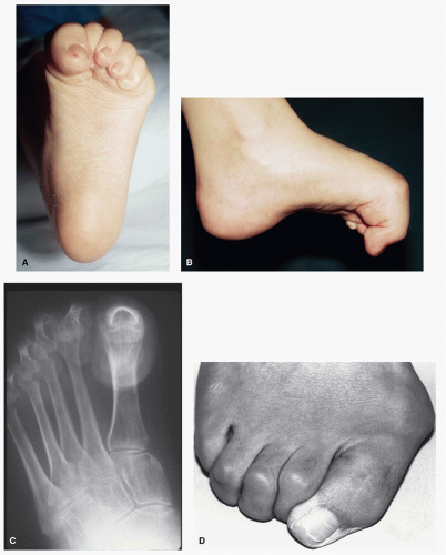
Figure 68.8 Photographs depicting examples of various dystonia patterns. A-C: CPRS type II, rapidly progressive digital dystonia with dislocated metatarsophalangeal joints. Etiology was an ankle sprain. D: CRPS type I with intrinsic foot muscle dystonia with progressive hallux valgus after foot contusion.


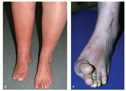
Figure 68.8 (Continued) E: CRPS type II with anterior tibial tendon dystonia after surgery at level of tarsal tunnel. F: CRPS type II and dystonia of extensor hallucis longus and anterior tibial muscles. Etiology was digital entrapment subsequent to hallux valgus surgery.


SYNDROME PROGRESSION

Late in its course, CRPS frequently becomes sympathetically independent (14,76). If untreated or nonresponsive to treatment during the acute stage, the condition may become refractory to almost any form of treatment. Trophic changes are usually a late finding and may include all structures from skin to bone including tendons, aponeuroses, muscles, and joint capsules (12). In end-stage disease, obvious sympathetic abnormalities are not clinically present unless stimulated so that a delayed appearance of skin changes (sudden edema, abrupt cyanosis, livedo reticularis, etc.) 5 to 15 minutes after provocation is observed (135). In severe cases, multiple extremities can be involved (4,5,55,105) (Fig. 68.9). In multiextremity involvement, there are mirror image patterns as well as upper and lower extremity involvement, but this is rare. Veldman et al reported 39 of 829 cases of multiple limb involvement in which five cases had three or four limbs involved (105).

The spreading of CRPS from a lower extremity to an upper extremity or vice versa has been reported (122,136,137). This indicates that extension of this process occurs beyond adjacent spinal cord segments. Reports of development of rapid onset of CRPS at secondary sites or with elective surgery outside the injury zone seem more likely to occur once the primary disease is established (76). Patterns of spread in CRPS have been reported and are variable. Maleki et al in a retrospective study looked at patterns of spread in 27 patients with CRPS type I. Contiguous spread was noted in all patients (gradual enlargement of involved area), whereas independent spread was documented
in 70% (19 patients) to a distant location from the initial site. Mirror image spread was noted in four patients (15%) (137).


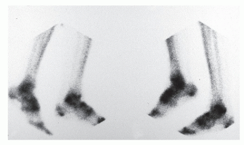
Figure 68.9 Photograph of a bone scan image of a 45-year-old female with a history of a unilateral ankle sprain and symmetrical CRPS I of both ankles. Note mirror image involvement of her delayed image of the ankles with diffuse uptake of the tarsals. This is a typical pattern of CRPS that results after ankle sprains. She ultimately had a successful placement of a spinal nerve stimulator.


TABLE 68.3 Known Precipitating Factors of CRPS in Extremities


Ankle sprain (60,98,116,132,140,141,164)


Cast immobilization (12)


Contusion (4)


Crushing trauma (60,142)


Dislocations (4,60,116)


Elective surgery (60,98)


ˆ


Ankle arthroscopy (143,144,145 and 146)


ˆ


Ankle stabilization (116)


ˆ


Bunionectomy (147,148)


ˆ


Laser wart excision (116)


ˆ


Neuroma excision (98,116)


ˆ


Os trigonum excision (149)


ˆ


Tarsal tunnel release (116,150)


ˆ


Tumor excisions (151)


Electric injury (152,153)


Exuberant bone callus (4)


Fractures (60,154)


Frostbite (60)


Infection (60)


Laceration (4,60)


Munchausen syndrome (155)


Overuse injury (116)


Overzealous physical therapy (156)


Phlebitis (60)


Projectiles (4,18,33,34)


Puncture wounds (4)


Spinal anesthesia (60)


Stress fractures (140)


Tumors (4,151,157)


Venipuncture (98,158,159 and 160)


CAUSATIVE FACTORS AND EXACERBATIONS


PRECIPITATING EVENTS

CRPS type II can be triggered by various evoking events; the most common is nerve trauma from fractures and sprains (12,29,39,55,60,138,139) (Table 68.3). Patients with CRPS type I will usually describe an injury, noxious event, or immobilization period prior to the expression of the disorder (14). Often the reported trauma can be trivial (42,60,161,162). While traumatic incidents are normally elicited, there are 10% to 25% of cases in which no known cause can be determined (6,60,163). The possibility of repetitive injury has also been reported to explain some of these cases (70,138,151).

Soft tissue injuries such as ankle sprains are a common inciting mechanism of CRPS (29,60,164). Schwartzman considers that the ankle sprain is the most common precipitating event (˜50% in his series) for CRPS disorders; however, he could not explain the mechanism of this occurrence (29,60,164). Yet in the sports medicine literature, there are reports of traction injury with confirmed conduction deficits to the peroneal nerve branches that result from stage II or III ankle sprain mechanisms (165,166). This indicates that CRPS caused from this mechanism is likely type II CRPS rather than type I. To further support this concept, Christensen reported on 90 cases of CRPS and described a distinct subset of cases that were induced from an ankle sprain and had a pattern of anterior ankle allodynia with the focus of pain over the intermediate dorsocutaneous nerve (116).

While immobilization is often described as an inciting or causal event in CRPS (12), there is no available evidence to confirm this relationship. It is more likely that a preexisting trauma with the potential for developing CRPS is worsened by the immobilization process. While it has been reported that overzealous physical therapy can cause CRPS (156), therapy is usually very beneficial and is considered a cornerstone of treatment strategies (see “Functional Restoration in Treatments” section).


PREDISPOSING FACTORS

It has not been fully elucidated if genetic factors can predispose the development of CRPS. However, there have been several associations between the clinical expression of CRPS and major histocompatibility complex (MHC) markers. HLA-DR13, HLA-DR2, and HLA-DQ1 have been reported to have an association with CRPS (167,168 and 169). The complete genome of the MHC region has been mapped, revealing over 100 microsatellite loci, which can permit fine-mapping of genetic loci with genetic predisposition (170). Fine-mapping of patients with CRPS and dystonia has provided additional support for HLA-DR13 as an important marker in CRPS and suggests other microsatellite alleles that are either predisposing or protective in the clinical expression of CRPS (171).

There is evidence of increased seroprevalence of parvovirus B19 IgG (172). Further research is needed to determine the clinical significance of this finding in 39 patients. It would be interesting to do a larger series and combine genetic testing of patients as well.

In CRPS type II, a common reported etiologic mechanism is a high-velocity projectile (4,8,18). This usually occurs above the knee or elbow and involves a partial nerve injury. It has been shown that bullets wounding tissues adjacent to nerve tracts can still cause nerve traction injury via cavitation (18,173). The shock wave from the projectile can cause significant nerve displacement, which injures the nerve without loss of continuity.

There have been many widely varied opinions about the possibility or probability that CRPS has a psychopathologic etiology (174,175,176,177,178 and 179). The most commonly advanced theory pertains to the psychological uniqueness of CRPS. However, there is very little support for such theories. Critical review of CRPS literature prior to 1991 revealed that studies were not prospective and of poor quality; therefore, there was no available data to support a psychopathologic etiology (180,181). More recently, there have been prospective investigations that revealed no evidence of abnormal psychiatric illnesses (110,182). Additionally, CRPS patients have been compared with other non-CRPS groups, and these investigations concluded that there was no evidence to suggest that patients with CRPS were psychologically unique (183,184,185,186 and 187).

Ochoa and Verdugo proposed that many cases of CRPS were inappropriately diagnosed and many cases were likely psychologically based. They suggested that the concept of RSD was a “naïve pseudoscientific concept.” They described the entity of “psychogenic pseudoneuropathy” that was common but frequently undiagnosed by clinicians (176). They characterized this entity as nonanatomic motor or sensory involvement and
most common with mononeuropathy. Unfortunately, he cited their own group’s work to build their case and did not offer any psychiatric evidence to support their findings.


CRPS IN CHILDREN

CRPS in children is an accepted diagnostic entity that has been widely held to differ intrinsically from the adult condition (188). However, clinically, there are relatively few disparities (189). The average age of onset is 12.5 years with a range from age 3 to 18 years (190). It has been reported to occur less often than in adults (108,191). Indeed, the first CRPS cases in children were reported in the 1970s with greater recognition of the disorder occurring in the 1980s (190,192). In children under 18 years of age, CRPS occurs in females more often than in males (4:1) (190) and affects the lower extremity significantly more frequently than the upper (189,190) (5.3:1).

While there is little evidence of any major psychopathology in childhood CRPS, there is a significant incidence of family dysfunction and secondary gain at home with these patients (192,193). In many cases, there can be unusual stresses pertaining to organized sports, academics, marital conflict, or sibling rivalry. These stresses and the family’s reaction to an injury seem to amplify the symptoms. Thus, it becomes necessary at times to enter the patient into a formal multidisciplinary program to address all facets of the disorder (189,190,191,192,193 and 194).

Effective treatment for the child with CRPS usually includes individual and family counseling combined with an intense physical therapy program (192,193 and 194). Even after initial effective treatment, a portion of patients can expect recurrences in the first 6 months after treatment (194). Generally, invasive treatments are not necessary (194); however, there have been reports of pediatric CRPS cases treated with standard pharmacologic treatments and lumbar sympathetic blocks (195). However, in more intractable cases, an indwelling catheter can be placed epidurally or along the sympathetic chain (190), to permit twice-a-day physical therapy sessions. Successful outpatient treatment has been reported with use of transcutaneous nerve stimulation in pediatric patients with CRPS (192,196,197).


EXAMINATION, TESTS, AND STAGING


HISTORY OF ILLNESS AND SYSTEMS REVIEW

CRPS can almost always be associated with some prior injury event or surgery. It is important to document temporal events and the progression of pain associated with the prior inciting event. The onset of neurologic symptoms and signs usually follows within hours to several days after the injury, although longer delays are not uncommon (8). Pain is always in the area of the injury or event, and as a rule, the pain involves areas larger than the original injury site.

The most pronounced CRPS symptom is constant ongoing pain. This pain is described as aching, burning, or viselike, although other vivid descriptions are frequent. In addition to unremitting pain, there may be pain paroxysms, or spontaneous exacerbations, described as sharp, shooting, or electric-like in quality. Encourage the patient to describe all the components of pain in his or her words. Special attention should be paid to pain paroxysms, evoked pain, and rest pain. It is very important to distinguish between spontaneous pains (neuropathic etiology) and hyperalgesia (nociceptive amplification) to assist in determining if therapies targeted to nociceptive endings are going to be potentially effective (198). Pain rating scales can be helpful to measure clinical progress; however, the clinician should use a scale in a consistent reproducible manner. Furthermore, the issues surrounding pain should be probed. This should include quality of pain, duration, impact, severity (rating scale), aggravating factors, and response to daily activities. The McGill pain questionnaire can be helpful in gathering this information in a consistent manner (199). This is especially true in patients who are poor historians or have difficulty verbalizing their pain experience.

Most patients complain of hypersensitivity with exacerbation of the ongoing pain or provocation of new pain upon such stimulation as touching or movement or upon exposure to temperature changes, either cold or warm. Most often, it is best to question situations they have learned to avoid. These experiences have already shaped their avoidance behaviors and are more accurate in recalling the quality and intensity of the pain. Inquiring about sensitivity from bed sheets, shower water, application of ice, weather changes (both temperature and barometric change), and vibration (i.e., riding in a car, etc.) can help the examiner correlate subjective with objective findings. Response to these factors can help profile the type of pain involved and give clues to effective treatments.

There are other subjective complaints in addition to pain. These may include weakness, fatigue, incoordination, clumsiness, shaking, and abnormal postures. Frequently in CPRS, patients develop stiffness of the affected limb or body part, as well as stiffness in the muscle and joint pains in the surrounding area. Patients with paroxysms of pain frequently will confuse them with muscle spasms and cramps.

Jul 26, 2016 | Posted by in MUSCULOSKELETAL MEDICINE | Comments Off on Complex Regional Pain Syndromes and Related Disorders

Full access? Get Clinical Tree

Get Clinical Tree app for offline access