Clinical examination of the lumbar spine

36


Clinical examination of the lumbar spine


image


History


Introduction


Assessment of backache and/or sciatica is never easy. Despite the increasing accessibility of highly sophisticated technical aids, diagnosis of lumbar problems still relies on the principles of history and clinical examination.


Taking the history is by far the most significant tool in the diagnostic procedure. Whereas examination techniques – both clinical and paraclinical – give current information only, the history also puts the evolution of the disease in the picture. History not only is the record of past and present suffering but also constitutes the basis of future treatment, prevention and prognosis. Furthermore, it also gives information about the degree of disablement the problem has produced and will produce.


For these reasons, a careful history, detailed and in chronological sequence, cannot be taken in a hurry. The examiner must make time to listen and have the patience to unravel complicated stories. Most patients have difficulty organizing their story or remembering every detail. Sometimes they are even unable to give precise responses to simple questions, and mix up past and present symptoms, pain and disablement, and physical, emotional and social disturbances. For a satisfactory diagnosis, however, it is essential to obtain a description of the past and present pain in meticulous detail. Therefore the examiner should develop a specific technique of questioning which is chronological and precise. The answers (both positive and negative) form a pattern that is related to knowledge of applied anatomy, biomechanics and pathogenesis. Taking a history thus translates the patients’ subjective complaints into an anatomical and biomechanical context which correlates with one of the well-known syndromes.


The importance of the chronological order in which the symptoms present is illustrated by the following (simple) case report: a patient states that he developed a sudden backache, which was followed a few days later by severe pain in the left calf. Two days later he noticed numbness of the outer two toes and difficulty standing on tiptoe. By that time the backache had ceased. This is the story of an S1 root compression with neurological deficit, almost certainly caused by a large disc protrusion at the level of L5–S1. This chronological description pinpoints not only a diagnosis but also the therapy: manipulative treatment will be of no help and the patient should either be treated by epidural local anaesthesia or surgery, or await spontaneous recovery.


In practice, the patient’s symptoms are not always as obvious as in this idealized case but it does give an idea of the value of a good history. After the history has been taken, the examiner should have a fair idea of the diagnosis or at least be able to distinguish activity-related backache from non-activity-related, referred or functional pain.


Pathogenesis


In lumbar spine problems, the mechanism of causation is usually reflected in the behaviour of the pain. Localization of the symptoms, their evolution and the relation to activity and posture differ according to the tissue involved. Pain in the lumbar and pelvic–gluteal area is usually of local origin but may also be referred from intra-abdominal or pelvic lesions. Sometimes lumbar pain is devoid of any organic basis and is then labelled as non-organic or ‘functional’. Local organic disorders may or may not be related to activity. The former are called activity-related spinal disorders, the latter non-activity-related spinal disorders (Box 36.1).


Activity-related spinal disorders (Ch. 38)

These are caused by a mechanical dysfunction: discodural and discoradicular lesions, capsular or ligamentous lesions, and spinal stenosis.


Discodural interactions may cause all three major syndromes: lumbago, backache and sciatica. Both articular and dural symptoms are present and have a strong tendency to evolve over time: because the discal displacement seldom remains stable, localization and intensity of the symptoms vary with localization and intensity of the dural impingement.


Lesions of the facet joints are characterized by localized pain. There is absolutely no change in localization. Dural and root symptoms are also absent.


Ligamentous pain typically occurs in relatively young people. It is created by prolonged or increased postural stress and abolished by correction of posture. In long-standing cases, movements also become painful at full range. In disorders of the lumbar ligaments, pain is always intermittent and vague and never referred below the upper buttocks. Lesions of the iliolumbar or sacroiliac ligaments, however, may give rise to slight reference of pain in the groin and the back of the upper thigh, respectively. Again, neither dural nor root symptoms are present.


In central spinal stenosis, chronic and vague lumbo-sciatica is brought on by walking or standing and relieved by stooping or sitting. Pain is often associated with feelings of numbness and weakness in both legs. These patients are never under 30 years old and more often are over 60.


In stenosis of the lateral spinal recess, a middle-aged or elderly patient complains of unilateral sciatica coming on during standing and walking. Sitting or bending forwards alleviates the pain immediately. As in central spinal stenosis, dural symptoms and signs are absent, as are root signs.


Non-activity-related spinal disorders (Ch. 39)

These include:


In ankylosing spondylitis, pain may vary in an unexpected way. One day the patient awakes without any discomfort and is able to do any kind of heavy work. The next week, the patient may wake up early with a painful back and disablement persists for the rest of the day. This differs markedly from the patient with discodural backaches, in whom pain starts on getting up or when an attempt is made to put on socks or tights. The back thus aches during certain movements or positions, whereas others ease the pain.


In rheumatoid conditions, the pain is typically experienced most severely in the morning and improves throughout the day.


In malignant disease, pain is unremitting and worse at night. Lumbar pain increases steadily even after root pain has set in; its distribution is not confined to a single dermatome.


Pain referred to the back

Pain in the back that is completely unrelated to movement or posture and displays hardly any temporal pattern suggests a referred source from intra-abdominal or pelvic lesions, such as those of the aorta, and the genitourinary and gastrointestinal tracts. In the initial stages of these diseases, the history may signal the disease via warning signs. Some of these are briefly discussed here.


Problem solving


While taking the history, the examiner endeavours to find an answer to the following questions:


Because low back pain is most often caused by a soft tissue lesion and so is frequently attributed to disc disorders, the history serves in the first place to verify whether this is the case. ‘All discs are alike, all other lesions are different’ is Cyriax’s statement, which has been proved true in orthopaedic practice. Therefore, in disc displacements of all types, confirmation of the facts detailed in Chapter 33 is expected.


The history also provides an opportunity to interpret the credibility of the patient’s story, again by looking for ‘unlikelihoods’ – facts that do not correlate with one of the well-known syndromes. If, during the history, the slightest doubt arises, the clinician should be on the alert. More ‘unlikelihoods’ should then be sought in the patient’s story or during the following clinical examination. In contrast, when patients state exactly what is expected and normal for a comparable case, there is certainly no reason to doubt their reliability; nor will a search for possible psychoneurotic components be necessary. Patients devoid of a physical cause will rarely escape detection if the history is properly taken. In these patients, none of the well-known physical patterns emerges – the rules and facts of referred pain do not fit. They do not so much describe their symptoms as the degree of suffering. They fail to supply relevant answers and, if the examiner insists, questions are often resented.


During the history the interviewer should obtain specific data on the following:


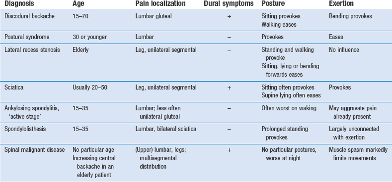
Age and activities of daily living


Disc lesions causing backache and sciatica are most common between the ages of 20 and 50 years. Over 60, the frequency decreases. Under the age of 20, discodural interactions are rare, although not impossible.


Sciatica caused by a posterolateral disc protrusion can be expected from adolescence to old age.


In elderly patients, lateral recess stenosis is to be more frequently expected as the cause of root pain (Table 36.1). Also, degenerative spinal stenosis is a disease that occurs predominantly in the elderly.


Spondylolisthesis can provoke posterior ligamentous pain in the young. Postural ligamentous pain is also more frequent in young patients with a standing job.


Ankylosing spondylitis typically provokes alternating sciatica between 15 and 35 years of age. It is 4–9 times more frequent in men.1


The activities undertaken as part of the patient’s profession, hobbies and sports will give additional information that is extremely important in judging the actual functional incapacity and in designing a treatment strategy. Most patients do not suffer from pain, rather from the disability the pain provokes. It is obvious that discodural backache will produce more disability in a truck driver who has to sit for the whole day than in a patient who does light and varying work. For some, normal activities are unrestricted but their favourite sport is impossible, and this is a major concern.


Profession and activities are also important in relation to treatment, recurrences and prophylaxis. If a bricklayer gets lumbago every second year, his back can be judged to be reasonably stable and manipulation will help him sufficiently each time there is a new attack. If, by contrast, an office worker has attacks of lumbago five times or more a year, these repeated events indicate that the back is very unstable. Although manipulation may solve the problem for a short time, it is obvious that stronger prophylactic measures will be necessary; successful manipulation should be followed by sclerosing injections, back school and/or a good lumbar support.


Routine of history taking


In disorders of the lower back, symptoms can diversify. The clinician must try to obtain a clear impression not only of present discomfort but also of former events (see Box 36.4). Pain is the most common and important symptom and is usually what forces the patient to seek medical help. Other symptoms are not always mentioned spontaneously but should be asked about: the presence of paraesthesia, numbness, a cold foot or incontinence.


Symptoms are usually presented by the patient in a very disorganized way. The interviewer then tries to create ‘order in the dis-order’. The best approach is chronological, the patient being asked about events leading up to the onset of the symptoms and then recounting chronologically what has happened since.


Pain


All the different aspects of pain should be investigated: localization, onset, evolution and duration of the perceived ‘current’ pain; influence of movement and posture; and the presence of dural symptoms. It is also very useful to obtain information on the same factors in previous attacks (Box 36.2).


In disorders of the lower back, pain may be experienced as backache, as gluteal pain with or without reference to one or both legs, or as typical root pain.


Current pain


Side and level

Patients are first asked if they feel any pain at the present time and to point to its location. The method chosen may give information on emotional status. A stable patient generally places the palm of the hand at the site of maximal pain and moves it across the body to demonstrate the route of radiation. A psychologically unstable patient never touches the painful area but only points it out vaguely with the thumb.2


Back pain may be felt centrally, unilaterally or bilaterally. Central pain can never be referred from a unilateral structure – for example, a facet joint or a sacroiliac joint. Bilateral pain also hardly ever has a central origin. Unilateral pain in one buttock is typical of a discodural problem. Sometimes the sacroiliac joint is responsible but a strained muscle is a rarity.


Bilateral, vague leg pain is usually dural pain. Segmental pain in both legs can sometimes be caused by two protrusions. However, bilateral root pain is more often the result of spondylolisthesis, spinal stenosis, lateral recess stenosis or metastases. Bilateral osteoarthritis of the hip joints and intermittent claudication due to thrombosis in the iliac arteries may also create pain in both legs.


The level of the pain is also important. In backache with dural reference, pain is usually situated in the lower lumbo-sacral region, and may radiate downwards to one or both buttocks.


If the patient points to the upper lumbar area, the investigator should immediately be on the alert. Malignant diseases in the lower back have a great preference for this area (see p. 536).


In sacral, coccygeal or perineal pain and numbness, compression of the S4 root is a real risk and constitutes an absolute contraindication to manipulation.


In sciatica, unilateral pain in the relevant dermatome results from pressure on the dural sleeve of one of the lower lumbar nerve roots. It is important, though not always easy, to distinguish radicular pain from dural pain. The latter is extrasegmentally referred and therefore experienced over a larger area, not restricted to one dermatome. It may even spread upwards to the chest or down both thighs, sometimes reaching the ankles. By contrast, the segmental pain of root compression is easily recognized when a patient presents with a severe ache in the leg clearly situated within the borders of the respective dermatome. The difference between radicular and dural pain is extremely important in both diagnosis and treatment. Every effort should therefore be made to obtain a precise description of the localization and characteristics of the pain. To a patient, a leg is a leg, and most are not precise about whether the pain is in the front or back of the thigh, whether it spreads beyond the knee and whether it is localized or generalized.


Pain in one lower buttock only is rarely dural; more commonly, it is a segmental reference from S2.


Onset of pain

Low back disorders may be acute, chronic or recurrent. The patient should identify the first time that the symptoms occurred. ‘When did your back problem start?’ is thus an important question.


A long history of, say, 20 years of ‘suffering’ from backache requires further questioning on whether the problem is continuous or intermittent. It may be that there was just one attack of acute lumbago 20 years ago with a second attack 2 days ago; or a constant and daily ache may have been present over the whole period of 20 years; or the problem may be recurrent, incapacitating backache six times a year. Although in all three instances the history extends over a period of 20 years, it is obvious that the diagnostic and therapeutic approach will differ, as will the prophylactic measures to be taken.


Information about the duration of symptoms is also extremely important in cases of sciatica. There is no limit to the duration of radicular pain resulting from lateral recess stenosis but in discoradicular interactions a course terminating in spontaneous recovery is the rule. Once the protrusion has shifted to one side, symptoms tend to abate; the protrusion has settled itself outside the intervertebral joint and there it lacks nutrition and shrivels away. As a rule, the patient recovers within 12 months of the onset of radicular pain. However, this only applies in patients under 60 years of age.


The next question concerns the speed of onset: ‘How did it start: was the onset sudden or gradual?’


Backache coming on over some hours, or even the morning after doing heavy work involving much stooping and lifting, suggests a soft disc lesion slowly increasing in size, i.e. a soft nuclear displacement. Cyriax used to say: ‘Pulp oozes, cartilage subluxates in an instant.’ In this type of discal displacement, traction is usually the treatment of choice, except in very acute cases where it is strongly contraindicated. An epidural injection is then the alternative.


Acute lumbago starting suddenly, at the moment the patient bent forwards or lifted a weight, is typical of a hard, annular disc displacement. In a case that is not too acute, manipulation is almost always successful in one or two sessions.


Course of pain

In most discoradicular interactions, pain typically starts in the back and sooner or later shifts into one leg. ‘Shifting pain’ of this nature strongly indicates a disc lesion. If backache gradually increases and after some time extends into one leg and finally involves the back and both legs, a progressive lesion such as a tumour is very likely and the examiner should be on the alert.


Pain starting in the leg, slowly getting worse over a period of months and finally spreading upwards to the posterior aspect of the thigh, is very suggestive of a primary posterolateral protrusion compressing the S1 root. The lesion occurs in young adults and is always nuclear.


Chronic sciatic pain in the elderly, extending over months to years, is typical of radicular pain from a narrowed lateral recess.


‘Alternating pain’ in the legs suggests bilateral sacroiliac arthritis, which is usually a manifestation of early ankylosing spondylitis. Less often, it indicates a disc lesion in a very unstable lumbar intervertebral joint.


What factors influence the symptoms?

In activity-related spinal disorders, it is obvious that there is a relationship between the symptoms and posture or exertion.


Posture and exertion


In a discodural interaction, the ache is increased by stooping, lifting, sitting or coming upright after sitting and is relieved during walking and in recumbency. However, there are some characteristic histories that are slightly different from the usual findings for disc lesions. For example, a patient between 20 and 40 years of age may awake without any pain and remain asymptomatic over the next few hours, even on exertion. During the day backache comes on, slowly getting worse. On going to bed, the pain ceases after an hour or so. This is the typical history of a ‘self-reducing’ disc lesion.


A patient may complain of pain in the lower back which comes on with prolonged standing. The ache gets worse and is finally followed by bilateral root pain, eventually with paraesthesia in both feet. Sitting or lying down results in cessation within a minute. In young patients this pattern suggests a spondylolisthesis. If the patient is elderly, stenosis of the spinal canal should be suspected.


In ‘ligamentous postural’ syndromes, pain is particularly increased by maintenance of a particular posture, whereas altering the position relieves the pain. Moreover, the longer the position is maintained, the more intense the pain becomes. Barbor3 described the discomfort of ligamentous pain as ‘the theatre, cocktail party syndrome’: it is impossible to sit at the theatre or stand at the cocktail party without low backache occurring. In contrast, the symptoms are relieved by activity. This syndrome is typically found in the young.


Coughing and sneezing


Another factor that may influence symptoms is raised intra-abdominal pressure during coughing and sneezing (Box 36.3). Pain in these circumstances may be a dural sign produced by sudden increased intradural pressure, which in turn causes sudden expansion of the dura pressed against the protrusion. Although it is very often related to a disc protrusion, it is clear that any space-occupying lesion in the lumbar spinal canal compressing the dura mater (e.g. a neuroma or malignant tumour) may evoke the same response. Often the patient will not mention it spontaneously, so the investigator must enquire about coughing and sneezing.


In disc lesions, coughing and sneezing normally increase the lumbar or gluteal pain. However, when they also increase the pain in the leg, manipulative reduction will almost certainly fail. Epidural injections should then be tried. A neuroma may evoke the same sign but the pain is usually felt more in the leg than in the back.


In active sacroiliitis, pain on coughing is felt in the buttock and sometimes radiates into the thigh. It results from the painful distraction of the joint caused by the momentarily increased intra-abdominal pressure.


‘Early morning’ pain


This wakes the patient and eases on getting up, after which it is possible to do fairly heavy work; the pattern is suggestive of ankylosing spondylitis. The pain is felt centrally in the whole lumbar region and varies from day to day.


Sometimes early morning pain is caused by a discodural interaction. The increased hydration of the disc during recumbency possibly exacerbates an existing small posterior bulge, which then slowly compresses the dura mater, so waking the patient before dawn. This type of disc lesion is best treated by epidural local anaesthesia.


It should be emphasized that these histories are quite different from the usual story for a patient with a small disc lesion who has a painful stiff back on getting up in the morning. Turning in bed is also mentioned as causing a twinge and is quite different from increasing pain in a recumbent position.


Duration of pain

Lumbago usually recovers spontaneously within a week because of the strong counterpressure exerted by the posterior longitudinal ligament, which gradually reduces the large posterocentral displacement.


In backache, there is no predictable time limit: the protrusion is small and remains more or less in contact with the rest of the intervertebral content of disc, end-plates and ligaments, thus receiving sufficient nutrient supply to maintain it. Because of a decrease in intervertebral height, the counterpressure exerted by the posterior longitudinal ligament becomes less effective. As a result, the discal tissue may remain displaced for years, with or without remission. Spontaneous recovery probably never occurs, whereas reposition by manipulation or traction is simple and is indicated, regardless of how long the patient has had the problem.


In sciatica, the protrusion has slipped posterolaterally and becomes extra-articular. Consequently, the bulge is cut off from its nutrient supply. Slow but continuous shrinking sets in from the moment the backache ceases, and results in spontaneous remission of the sciatic pain within 8–12 months. Therefore treatments such as manipulation and traction are worth trying but only during the first 6 months. Once this time limit has passed, they are not relevant because the process of spontaneous recovery becomes more and more likely to succeed. Later recurrences at the same level are no longer likely. In major or long-standing sciatica, epidural local anaesthesia may relieve the pain during the period of spontaneous recovery. If this fails and pain remains unbearable, surgery is indicated.


Root pain that does not get better within a year or even gets worse after, say, 8 months is suggestive of conditions other than disc lesions, such as neuroma or lateral recess stenosis.


Previous attacks

Previous attacks should be related to age, localization, origin, evolution, influence of movement and posture. Also important are the frequency of attacks, as well as the type of treatment and its result.


Localization of symptoms

In disc lesions, the localization of symptoms is determined by the site of the pressure on the dura mater or nerve root. In a new episode, the pain may have similar localization. However, as a disc may easily shift and compress sensitive structures at other places, repeated attacks of backache caused by the same disc may provoke pain on different sides. Reports of former attacks, not located on the same side, point to a disc protrusion as the cause of the problem. A shifting pain means a shifting lesion, and only the disc is free to move from one side to another.


Pain changing from one buttock to the other is also seen in early ankylosing spondylitis with involvement of the sacroiliac joints.


In capsular and ligamentous disorders or in spinal stenosis, localization is fixed, and remains unchanged over a long period.


Discodural interactions

The history may also serve to obtain an idea of the degree of discodural interactions.


Marked articular symptoms (twinges) and postural deviation

These are characteristic of intense discodural contact. The deformity is noticed by the patient or by others. The typical case is acute lumbago in which the patient is painfully locked in flexion by a large central protrusion at the posterior aspect of a lumbar intervertebral joint. Any attempt to extend the lumbar spine squeezes the protrusion further backwards and increases the already painful pressure on the dura mater. Adoption of the flexed position decreases the pressure exerted by the subluxated part of cartilage on the dura mater.


A large posterolateral protrusion is accompanied by some deviation of the lumbar spine in lateral flexion, so projecting one hip sideways. The patient is not able to move in the opposite direction. Such lateral deviation suggests a lesion at the fourth or perhaps the third lumbar level.


When the lumbar spine is fixed in flexion or in lateral flexion because of root pain, all conservative treatment is likely to fail and surgical management is indicated.


If a patient with acute lumbago states that twinges are felt on even the slightest movements, therapists should be on their guard. Although manipulation can be tried safely, it will not always be tolerated. Traction, however, should definitely be abandoned, because it makes matters worse, usually at the moment it is released. The safest and most effective treatment is epidural local anaesthesia. It almost always affords immediate relief, although the large displacement remains present, continuing the marked deviation and limiting joint movements. Manipulation, carried out from the next day on, is much better tolerated and usually gives good results.


Numbness and/or weakness

The patient states that the foot flops during walking or that standing on tiptoe is impossible: this suggests a large posterolateral protrusion not reducible by manipulation or traction.


Box 36.4 summarizes the routine of taking a pain history. Table 36.2 outlines some typical histories.


Paraesthesia


When the patient states that there are ‘pins and needles’, this is pathognomonic of pressure on or inflammation of the peripheral nervous system. In practice, the cause is pressure. These symptoms are extremely important but are often not mentioned by the patient, so the examiner must enquire about them.


Two different syndromes causing paraesthesia must be considered: nerve root compression and pressure on the spinal cord.


Pressure on a nerve root results in a typical set of symptoms: pain and paraesthesia, strictly related to the segment involved. Pressure on the dural sleeve of a nerve root causes severe segmental pain. Pins and needles indicate that the nerve fibres are irritated and they are always felt in the distal extremity of the dermatome. For this reason, it is vital to determine their exact areas; an accurate dermatomal distribution of paraesthesia is always a better pointer to the affected nerve root than is the pain itself.


In external compression of the nerve root, the sheath is compressed before the fibres and pain will therefore appear before paraesthesia. In discoradicular interactions, the sequence of segmental pain first, followed later by pins and needles and numbness, is therefore an ‘inherent likelihood’. If the paraesthesia appears before the pain begins, other lesions such as a neuroma or tumour should be suspected. In lateral recess stenosis, pain and paraesthesia usually appear simultaneously. Also, the symptoms do not tend to change over months or years.


If the paraesthesia is painless, a lumbar disc protrusion is an unlikely cause. Multiple sclerosis, diabetes, pernicious anaemia or cord compression is more likely. In these circumstances, pins and needles are also more diffuse in both feet or in all four limbs. The symptoms extend beyond the borders of innervation of any root or peripheral nerve. In cord compression, neck flexion may also bring on pins and needles.


Danger to S4 nerve roots


These roots are situated in the midline of the spinal canal, well protected by the posterior longitudinal ligament. In a large posterocentral protrusion, this ligament is placed under increased pressure. Finally, the ligament may rupture and damage the S4 roots (cauda equina syndrome).


Because mobility tests for the fourth sacral roots do not exist, it is almost impossible to evaluate their function. The diagnosis of cauda equina syndrome should therefore be made entirely on the history. Patients typically present with a classic triad of (1) saddle anaesthesia, (2) bowel and/or bladder dysfunction, and (3) lower extremity weakness.4 Some patients are timid and do not mention these symptoms, so it is important to ask about them in the three types of case in which a large posterocentral protrusion is to be suspected: acute lumbago, acute perineal pain and bilateral sciatica. It should be re-emphasized that manipulation is absolutely contraindicated; even traction is not at all safe if the slightest suspicion of compression of the fourth sacral roots arises. Prompt surgery is required and any delay results in substantial morbidity.5


The patient’s reaction to the symptoms


History taking should also determine how far the patient is disabled by the symptoms. Some patients are stoical, while others react in a hypersensitive way. Before active therapy such as manipulation is instituted, the presence of pronounced psychological factors must be established. For this reason, patients should be encouraged to relate how far their daily activities are disturbed. Later on, during the clinical examination, it will be established objectively how bad the handicap really is. If disablement is severe for a minimal lesion, it is likely that psychological problems are responsible for the symptoms and should be treated first. Furthermore, it is unwise to manipulate or inject a patient who seems to demonstrate psychoneurotic behaviour or is involved in a compensation claim.


Inspection


One important feature of this part of the examination is gaining an idea of the degree of disability. The clinician should observe the patient from the moment he or she enters the consulting room. In particular the following are noted:


Next, the patient undresses so that posture can be observed, especially the lower back, pelvis and lower extremities. This is best done in good and uniform light; light falling from a unilateral source will give unilateral shadows, which may give a false idea of shape and posture.


The shape of the normal trunk


The patient should be observed posteriorly and laterally. From the posterior aspect, the shoulders and pelvis should be level and equal, and the soft tissue structures on both sides should be symmetrical (Fig. 36.1a). The thoracic and lumbar vertebrae should be vertically aligned. The angles of the scapulae should be level with the seventh thoracic spinous process; the iliac crests should line up with the fourth lumbar vertebra. The lower extremities should share the body load and be in good alignment: the hip joints not adducted or abducted, knees not bowed or knock-kneed, feet parallel or toeing out slightly, and the calcaneal bones neither pronated nor supinated.


From the side (Fig. 36.1b), the thoracic kyphosis and lumbar lordosis are observed and should have a normal curve. The pelvis should be in the neutral position, i.e. the anterior superior iliac spines lie in the same vertical plane as the symphysis pubis. Hip, knee and ankle joints should be neither flexed nor hyperextended.


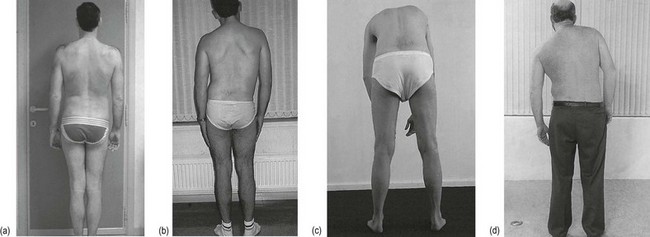
The pathological trunk
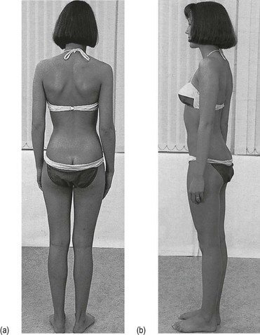


Posterior view

Many lumbar spinal disorders present with asymmetrical posture. This asymmetry may be in the vertical plane – the spinous processes do not align, or in a horizontal plane – the iliac crests, the anterior and posterior superior spines and the greater trochanters are not level in relation to each other. A pelvic tilt may be caused by anatomical changes above or below the greater trochanter, such as changes of the femoral head and neck or anatomical leg length discrepancy from growth disturbance. A lateral shift or list may have several causes.


Static scoliosis (Fig. 36.2a)

The origin of the list is a pelvic tilt due to a leg length difference; placing boards of various thicknesses under the foot of the shorter limb levels the pelvis, making the list disappear.


There is no clear evidence as to the significance of differences in leg length in the generation of spinal symptoms. If a platform under the shorter limb eases or even abolishes the pain while standing or on lumbar flexion or extension, a raised heel is advised. Some physicians recommend correction of any kind of leg length inequality. However, most investigators agree that mild leg length inequality of up to 15 mm is not a factor that contributes to low back pain.6,7 Correction is therefore only of importance in recurrent attacks of lumbago and in the presence of a difference of more than 15 mm.


Sciatic scoliosis (Fig. 36.2b)

The lateral shift caused by mechanical dysfunction and muscle spasm in the lower lumbar spine is called sciatic scoliosis. It usually results from painful impingement of dura mater or nerve root. Most often, a shifted disc is responsible but it is good to remember that any space-occupying lesion in the vertebral canal can cause such an impingement.


In disc lesions, gross lateral deviation usually results from displacements at the L4 or L3 levels. Disc lesions at L5–S1 seldom result in marked lateral deviation because of the stabilizing action of the iliolumbar ligaments on the joint, although some pelvic tilt remains possible.


In lumbar disc displacements, six possible types of deviation (sciatic scoliosis) exist:


• Towards the painful side. This shows that the displacement is situated medially, i.e. at the axilla of the nerve root.


• Away from the painful side. In this case, the protrusion lies lateral to the nerve root, which is drawn away by the deviation of the trunk.


• Alternating deviation. This demonstrates that the dura mater slips from one side to the other of a small midline protrusion. It is also diagnostic of a protrusion at the fourth lumbar level.


• Deviation on standing, which disappears during flexion.


• No deviation when standing erect but marked deviation on attempted trunk flexion. This is often seen in root pain.


• A momentary deviation when the trunk is flexed halfway. The patient is seen to deviate suddenly at a particular moment during flexion, returning to a symmetrical posture as this point is passed. Usually pain is felt at the moment of deviation but occasionally it is not. This sign indicates that a fragment of disc alters its position at the back of the intervertebral joint and temporarily touches the dura mater.


Jun 5, 2016 | Posted by in ORTHOPEDIC | Comments Off on Clinical examination of the lumbar spine

Full access? Get Clinical Tree

Get Clinical Tree app for offline access