Clinical Evaluation of Shoulder Problems

CHAPTER 4 Clinical Evaluation of Shoulder Problems


From our first days in medical school, we are taught that establishing a correct diagnosis depends on obtaining a meaningful and detailed medical history from the patient. This requires the physician to ask specific questions while at the same time actively listening to the responses from the patient. Often physicians formulate their next question without listening to and interpreting the answer to the previous inquiry. Obtaining a good patient history is, in itself, an art that requires experience and patience. I vividly recall one of my mentors stating that all patients come to your office and tell you exactly what is wrong with them when they answer but four or five questions. Our task is to decipher their answers to those few questions.


Time is perhaps the most valuable—and least available—commodity in our medical lives in the 21st century. We employ physician extenders to help our efficiency and we ask patients to fill out reams of paperwork with numerous questions while we are seeing another patient. We thus lose the advantage of directly listening to our patient, observing their expressions and interpreting their body language. Each of these facets can offer valuable information about the diagnosis of their shoulder problem. We must recognize that often the answer to one question leads to the formation of the next question. This valuable opportunity is lost in the hustle of managing medical care in this era, and it is indeed a lost opportunity. Our duty to our patients is to inquire, listen, examine, test, and then formulate a diagnosis. When performed in this logical fashion, the diagnosis is nearly always straightforward and the treatment then easily rendered.


In evaluating the patient we also must bear in mind that we really are assessing the patient not just interpreting radiographic studies or laboratory values. In this increasingly technological world, it is often easy to lose sight and begin treating magnetic resonance imaging (MRI) scans without treating the patient. For example, nearly every MRI of the shoulder we have seen in a patient older than 30 years suggests acromioclavicular joint pathology. Perhaps it is then not surprising that the most overdiagnosed and overtreated condition about the shoulder relates to the acromioclavicular joint.


As clinicians we must evaluate the patient’s history and perform a thorough physical examination to establish a strong correlation in the features of each pathologic process. Our confidence rises when the patient’s history of the complaint is consistent with the majority of the physical findings. This confidence rises even more when radiographic and laboratory studies are also consistent with the initial diagnosis. When each of these features of the patient evaluation point to the same diagnosis, our certainty of the correct diagnosis becomes assured. It is obviously much more disconcerting when a patient’s history suggests impingement syndrome, the physical examination is more consistent with instability, the radiographs document osteoarthritis and the laboratory values suggest gout.


We hope the methods described in this chapter for taking a history and performing a physical examination allow any clinician to determine which pathology is primarily responsible for the patient’s complaints.


PATIENT HISTORY


Taking a history from a patient is an art. We must ask specific questions, actively listen to the response, and only then formulate the next question. The answer to each successive question should ultimately lead the physician to a correct diagnosis. It is important not to get tunnel vision and lead the patient toward the diagnosis that you think is present.


Recall that many widely varying diagnoses manifest with similar symptoms and only after a complete history and examination can differentiation of diagnoses be made. For example, if a patient presents complaining of an inability to elevate or externally rotate the arm, the physician might immediately diagnose a frozen shoulder. Sending that patient, who really has advanced osteoarthritis, to physical therapy to increase the range of motion (ROM) ensures a therapeutic failure. A diagnosis is only established after each phase of the evaluation is complete. Anything else in the name of expedience and efficiency does a disservice to our patients.


Age


Most, if not all, disease processes occur in specific patient age ranges. Although malignancies and traumas can occur at any chronologic age, even these processes tend to stratify by age. Surgical neck fractures of the humerus are typical of a postmenopausal woman with osteoporosis rather than an 18-year-old male football player. Osteosarcomas of the proximal humerus are more common in a 20-year-old than in a geriatric patient. The younger, athletic person in the second or third decade of life more likely has instability, whereas the 60-year-old golfer with a painful shoulder more likely has rotator cuff disease.


Though not fully studied or accurately analyzed, the perception is that osteoarthritis of the shoulder is occurring at ever-younger ages. Not only is it in the domain of the 70- and 80-year-old patient; often patients present in their 50s and younger with osteoarthritis. Avascular necrosis, infections, and rheumatoid arthritis can occur at any age, and thus age is a poor discriminator. Spontaneous hematogenous septic arthritis may be slightly more common in youth, but its clinical presentation is usually so specific that age of the patient need not be considered.


Shoulder instability and its subsets of pathology are much more common in the youthful years. Labral tears, superior labrum anterior and posterior (SLAP) tears, and biceps tendinitis are commonly seen in patients younger than 30 years. However, some activities that span the entire age range, such as downhill skiing, offer much opportunity for acute shoulder dislocations. Nevertheless, the implications of a traumatic shoulder dislocation are age specific. In the younger patient who sustains a glenohumeral dislocation, the more likely associated injury involves the labrum or biceps anchor. Conversely, in the older patient, the acute glenohumeral dislocation is more commonly associated with a rotator cuff tear. Similarly, trauma to the shoulder can afflict the acromioclavicular joint. In the younger patient, disruption of the joint is more common, whereas in the older person, clavicular fracture may be more common.


Less-common conditions afflicting the shoulder still display a predilection for certain age groups. Gout and symptomatic calcific tendinitis usually occur in middle age. Adhesive capsulitis appears in midlife (more commonly in women), and diabetic neuropathic disease is more common in the older person. Cuff tear arthropathy clearly favors women in their mid-70s.


Sex


Most pathologic processes that afflict the shoulder know no gender boundaries. Trauma can occur to anyone; arthritis, infection, cuff tears, avascular necrosis, calcific tendinitis, and gout can likewise occur with equal frequency in male and female patients. However, three conditions have a significantly higher prevalence in female patients. Although none of these maladies is exclusive to women, their prevalence strongly favors them.


Multidirectional shoulder instability is seen many times more often in young female patients between the ages of 15 and 25 years than in male patients of the same age. Why this occurs remains unclear. Male patients might present with clinical evidence of multidirectional laxity, but perhaps because of stronger and better-conditioned muscles they are better able to compensate for their ligamentous laxity in ways female patients cannot or do not. In this condition, it remains doubtful that there is a difference between sexes in the pathophysiology of the condition, but the positive biological response to the process seems to favor the male patients. A teenage female athlete who presents with shoulder complaints likely has some type of instability pathology. However, the clinician must remain open to other diagnoses and never forget to distinguish patient symptoms from clinical signs. The young person might present with symptoms of cuff tendinitis that are caused by underlying shoulder instability.


Female patients also tend to present in far greater numbers than males with adhesive capsulitis1 This is in contrast to the idiopathic stiff and painful frozen shoulder, which is equally prevalent among male and female patients and describes restricted shoulder ROM associated with pain. A frozen shoulder can result from any number of pathologic processes such as post-traumatic stiffness, immobilization, and tendinitis. Adhesive capsulitis is a specific diagnosis most prevalent in women 40 to 60 years of age. It is associated with an idiopathic inflammatory process involving the glenohumeral joint capsule and synovium that results in capsular contraction and adhesion formation.


Although massive rotator cuff tears probably occur in greater numbers in men, it is the women, classically older than 70 years, who develop the sequelae of these massive cuff tears. The diagnosis of cuff tear arthropathy as defined by painful collapse of the humeral head with superior migration (not iatrogenically provoked by prior release of the coracoacromial ligament) favors geriatric women much more than men for unclear reasons.


Presenting Complaint


When the physician inquires about a patient’s chief complaint during the initial visit, the response is most commonly one of pain. Subsequent questioning is directed toward better understanding the characteristics of that pain; a presumptive diagnosis will follow from this. Most presenting complaints related to the shoulder are defined by patients as pain, stiffness, loss of smooth motion, instability, neurologic symptoms, or combinations of these.


With respect to shoulder pathology, another chief complaint may be one of joint instability. In this case, the patient might have no pain and is only concerned by the sense that the shoulder joint is loose, sloppy, or recurrently dislocating. The patient might initially complain of numbness or tingling down the limb, which may be caused by neurologic pathology unrelated to the shoulder. Dissection of this symptom may be more challenging because pathology in the neck might have to be distinguished from shoulder pathology.


Weakness is rarely a singular presenting complaint. Painless weakness nearly always defines a significant neurologic event or pathologic process. If stiffness of the shoulder is a presenting complaint, it is nearly always accompanied by some element of pain. A patient might present with a complaint of crepitus or popping in and about the shoulder associated with activity or a specific arm motion. An isolated awareness of crepitus without pain is very rare.


Pain


The discussion of pain is challenging because it is, by definition, a completely subjective complaint. In our vast armamentarium of technology and laboratory analyses, we cannot objectify pain. Pain is a perception of data presenting to our brains. We have all experienced the reality of injuring ourselves with minor scrapes and scratches in our daily lives but been fully unaware of any event until hours later. Have we all not jumped into a pool of water only to feel cold initially? Within minutes, the initial discomfort fades as we rapidly become conditioned to the water temperature. The water temperature obviously does not change; it is our perception of the same data input to our brain that changes.


So too it is with other painful stimuli. Psychologists (and perhaps our own experience) tell us that mood can have a dramatic effect on pain perceptions. People who are depressed or sullen by nature tend to experience more discomfort and be more disabled for a given amount of noxious stimuli, and the opposite is likewise true. Energetic, optimistic, and happy patients tend to discount even significant amounts of otherwise painful stimuli.


Other societal issues are also known to affect a patient’s perception and response to pain. Specifically, issues that relate to secondary gain can have significant influence on patients’ responses to treatment of their pain. Active litigation where contested remuneration is involved can lead to perpetuation of symptoms. In much the same way, patients with workers’ compensation claims might have little incentive to report improvement in their symptoms. Yet we have no pain meter to substantiate or dispute a person’s claims.


Despite these limitations, obtaining a history related to pain is critically important and valuable. Such features as its character, onset, radiating patterns, aggravating factors, and alleviating features nearly always assist the clinician in discerning a diagnosis.


Character of the Pain

Despite our inability to measure pain, patients use similar adjectives to describe their pain. These descriptions can offer much insight into its cause. Pain associated with an acute fracture understandably causes a severe and disabling pain, often remaining for days minimally responsive to narcotic analgesics. By contrast, the pain of impingement and rotator cuff pathology is commonly described as dull, boring, and toothache-like in quality. The pain of a frozen shoulder is typified as all or none. When it is present at the endpoint of available motion, the pain is truly disabling, whereas when the arm is functioning within its available arcs of motion, pain does not exist. Patients with painful osteoarthritis describe pain that frequently alternates between a sharp stabbing pain under high compressive joint loads and a chronic lower level of pain with less-demanding activities. Patients with severely destructive rheumatoid disease are often so conditioned by the chronicity of their disease that description of their pain appears inconsistent with the degree of joint destruction. These patients tend to be more disabled by their functional loss than by their perceived pain.


Acute calcium deposition in the cuff tendons provides a characteristic type of pain. The pain is so acute and so severe that calcium deposit in the shoulder has been likened to a kidney stone of the shoulder. The pain associated with a kidney stone seems so well understood by the population at large that the pain in the shoulder associated with acute calcific deposit is easily understood as well. Patients seek a dark, quiet room with minimal competing stimulation. The pain can be nauseating and disabling enough that many patients find their way to an emergency department (ED). The clinical picture is so evident and the radiographs so predictable that the diagnosis is rarely in doubt.


Location of Pain Perception

Pain is poorly localized around the shoulder girdle. The specific location where the patient perceives the pain is rarely the site of origin of the pain. The most common location for the perception of rotator cuff disease and the associated bursitis is down the arm toward the deltoid muscle insertion. The pain and inflammation associated with bicipital tendinitis is typically down the anterior arm, although the site of pathology is proximal to the intertubercular groove.


The pain pattern of most intrinsic shoulder pathology is one that radiates down the arm to the level of the elbow. It is distinctly rare for intrinsic shoulder maladies to result in pain perceived to extend below the elbow joint. Conversely, pain of cervical origin usually radiates from the base of the ipsilateral ear toward the posterior shoulder and into the scapular region. A true cervical radiculopathy, which most commonly involves the fifth and sixth cervical nerve roots, provokes symptoms that are perceived to radiate into the forearm and hand in a dermatomal pattern. In contrast to pain derived from cervical radiculopathy, pain from adhesive capsulitis does not follow a dermatomal pattern. The pain often radiates along the trapezius muscle and periscapular muscles because these muscles become strained and fatigued by the excessive scapular rotation that must compensate for the decreased glenohumeral motion.


Pain associated with an acromioclavicular injury usually radiates medially and results in perceived pain along the mid and medial clavicle. Intra-articular processes such as osteoarthritis, avascular necrosis, and rheumatoid disease rarely result in perceived radiation of pain. Patients report that their pain is poorly localized and remains centered around the shoulder without associated arm pain.


The pain of an intra-articular infection is not unlike that associated with any joint. The pain is severe, exquisite, and maximally disabling. The clinical picture is so specific that the clinical suspicion is exceedingly high until a definitive laboratory diagnosis is confirmed.


Aggravating Factors of Pain

As a part of the history of pain, the clinician needs to elicit circumstances that seem to make the pain worse. Often the pain is influenced by arm position, which can provide insight into its cause. Patients might state that the pain is worse or aggravated when the arm is positioned above shoulder level, such as occurs when washing or combing their hair. Activities that result in a long lever arm with the elbow extended, such as reaching across the car seat or reaching out the window to use an automatic teller machine, increase the pain of a weak or torn rotator cuff. Increasing pain in the shoulder that occurs while pulling bed covers up at night is strongly associated with impingement and cuff disease. The occurrence of pain at night needs to be elicited.


There appear to be two distinct types of night pain, each associated with a different shoulder condition. The more severe and disabling type of night pain strongly suggests a rotator cuff tear. The pain is described as gnawing, incessant, and unremitting, and it not only awakens patients from sleep but it often precludes any meaningful sleep at all. Patients often relate that the only way to obtain sleep is to rest semirecumbent in a chair. In a different circumstance, patients might acknowledge night pain that is positional. They can typically fall asleep but they are awakened if they roll onto or away from the affected shoulder.


Patients with positional night pain rarely convey the degree of frustration with sleep interruption that occurs with a cuff tear. Although patients with positional night pain may be annoyed by the sleep interruption, they generally can fall back to sleep easily and don’t develop that deep sense of misery associated with persistent sleep deprivation. Positional night pain is most often associated with loss of shoulder internal rotation through muscle stiffness or loss of capsular compliance. Painful arthritis of the acromioclavicular joint can also result in positional night pain and is caused by the compressive loads borne by that joint when lying on the affected side. The pain of these conditions might also be aggravated by lying on the unaffected shoulder. While lying on the unaffected shoulder, the weight of the arm falling across the chest in adduction also results in acromioclavicular joint compression and posterior capsular stretch.


Patients with adhesive capsulitis describe pain that is characterized by its sudden severity aggravated by clearly reproducible arm positions. They have no pain until they reach the endpoint of their available motion, when their pain becomes immediate and severe. As their condition progresses they note an increasing inability to perform their activities of daily living, including reaching overhead or reaching behind their back for dressing or personal hygiene.


With an intra-articular process such as glenohumeral arthritis, patients usually note that aggravation of symptoms comes with activities associated with repetition of a similar motion. Painting, sweeping, polishing, vacuuming, ironing, and washing a car are activities that predictably aggravate the pain of arthritis and impingement. Loading of the joint while at the same time performing a repetitious act is particularly aggravating to joint maladies that result from incongruent joint surfaces such as avascular necrosis, osteoarthritis, and rheumatoid arthritis.


Although inquiry about and analysis of aggravating factors in the assessment of shoulder pain is rarely in itself fully diagnostic, it remains a very important consideration as the history taking progresses.


Factors That Alleviate Pain

In the same way that analysis of aggravating factors provides insight into the etiology of the shoulder problem, so too does inquiry into those features and factors that alleviate or improve the symptoms. Many times the alleviating factor provides the best information in arriving at the correct diagnosis. Whereas there is much overlap in diagnoses with respect to aggravating factors, it would be unusual to find one factor that solves several different problems. For example, if a patient finds that an over-the-counter antiinflammatory truly improves the symptoms, it would logically follow that the patient has an inflammatory condition. Certainly an antiinflammatory does not solve the apprehension of a shoulder instability problem, nor would it likely manage the pain of an acute fracture. Patients with a frozen shoulder characteristically state that there is absolutely no improvement in their pain with nonsteroidal antiinflammatories.


Patients with rotator cuff tears and impingement often note that in placing the affected arm over their head, they find significant improvement in their pain. Often this arm position is the only way they can find meaningful sleep. This is called the Saha position (Fig. 4-1), named for the Indian orthopaedic surgeon who recognized this phenomenon. He postulated that with the arm resting overhead, there is a balance of tension of the cuff muscles in their least tense state. When the arm is passively elevated overhead in the supine patient, the supraspinatus is subject to its least tension, and pain diminishes in many patients.


Alleviating factors can include activity modification, medications, narcotics, antiinflammatories, injections, and physical therapy. Physical therapy for stretching over long time spans usually improves symptoms and needs to be assessed during history taking.


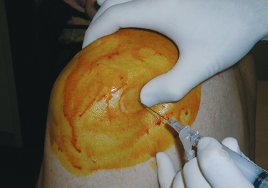
The response to local anesthetic injections when placed in specific anatomic locations around the shoulder can be very instructive and diagnostic. In a patient with chronic subacromial impingement, 5 mL of 1% lidocaine placed in the subacromial space provides immediate and dramatic relief of pain (Fig. 4-2). This response becomes diagnostic of a subacromial process, and it becomes especially valuable when trying to discern whether the patient’s perceived pain is originating in the shoulder or whether it is referred pain from the neck. A similar local injection test is useful in evaluating the acromioclavicular joint as a source of the patient’s pain. Alleviation of pain with arm adduction following an injection directly into the acromioclavicular joint strongly suggests pathology at this joint. Intra-articular injections can provide similar supporting information regarding the source of a patient’s symptoms.


These specific injection tests are valuable in defining the pathologic process, and in the case of subacromial impingement, the response to the local anesthetic can predict response to surgical treatment. Moreover, a negative response to a subacromial local anesthetic can predict a negative response to subacromial surgical treatment.


Response of Symptoms to Self-Prescribed Treatment

With the advent and ubiquity of the Internet, patients have now become more involved in their health care decisions. There are countless websites dedicated to patient information, and these help them self-diagnose, although not always with great clarity or accuracy. There are likely even more websites from which patients can receive a wide variety of treatment recommendations for their self-diagnosed shoulder malady. Searching for “physical therapy” brings up millions of hits, and searching for shoulder-specific physical therapy brings up well more than 1 million websites. No doubt then that it is the rare patient who arrives at your office without some knowledge, opinion, and effort at self-management of shoulder pain.


It is important to take time to explore what methods, medications, and modalities the patient might have tried before coming to the physician. Explore the realm of nutraceuticals and ask specifically about the common ones, including glucosamine, chondroitin, shark carti-lage, and methylsulfonylmethane (MSM), because many patients do not consider these to be medicines and do not include them in their medication lists. Patients consume seemingly countless vitamins and vitamin combinations in their effort to improve their physical well-being. With the exception of glucosamine and chondroitin, which themselves have not been subjected to the rigors of the scientific method to prove their efficacy, there is little published objective information to make recommendations to patients. Nevertheless, we have all seen patients who are certain that some combination of these herbs, vitamins, and supplements have affected their medical condition in some way or another. It is important to query and document these treatments in the overall evaluation of the patient with a shoulder problem.


Box 4-1 lists the facets of pain that need to be explored during a patient history.


Instability


In this discussion, it is imperative that the concept of instability is understood to mean the patient has symptoms of some shoulder problem. Many asymptomatic shoulders exhibit increased joint translation and are clearly loose during a physical examination. Such asymptomatic shoulders are defined as lax, not unstable. To have shoulder instability, by definition, means the shoulder is symptomatic for the patient.


In the younger and active age groups, the symptom of shoulder instability may be the patient’s presenting complaint. Although there is often a history of acute traumatic event resulting in the initial well-defined onset, in many cases no such traumatic event occurred. Indeed, it has only been since the 1980s that genetic factors in ligamentous laxity have been recognized as significant factors in patient perceptions of shoulder instability.


The diagnosis of shoulder instability can be very easy when the patient presents with an appropriate history of trauma. Nearly always there has been a trip to the ED and radiographs to document the events. However, with the increasing availability of sports trainers at most of the high school, college, and professional competitions, reduction of a dislocation by those personnel results in a history only; there are no ED records or radiographs. Although the history in these situations is still strong, an examination and radiographs even a few days following the event makes this a less-than-challenging diagnosis.


The more challenging problem occurs in the patient with a sense of slipping and looseness in their shoulder without a history of macrotrauma. More often than not, this more subtle instability pattern is associated with a nondescript level of discomfort and diffuse pain around the shoulder girdle. The discomfort is poorly localized and may be more scapular in location. The association of such symptoms with paresthesias down the arm is nearly always related to shoulder instability. A history of repetitive microtrauma is elicited. Such activities might include frequent swimming, gymnastics, and ballet. Although these activities would not appear to be highly stressful to the joint, they do demand muscle function defined by high endurance. Conventional thought suggests that when the ligament quality and integrity do not contribute to joint stability, the surrounding muscle activity and appropriate proprioceptive activity become more important to maintaining a functioning joint.


The sense of instability might occur with the arm only in certain positions or it may be present regardless of arm placement or position. True symptomatic multidirectional instability is typically symptomatic in midrange positions before ligament tension reaches the end of the range. The physician must carefully inquire about which activities and arm positions provoke the symptoms. Patients with this type of instability might have symptoms that are incapacitating enough that they tend to avoid extremes of glenohumeral motion. Pain is the more common symptom with a shoulder instability based on ligamentous laxity (AMBRI), and apprehension is more common with unidirectional traumatic instability (TUBS). (TUBS stands for traumatic etiology, unidirectional instability, Bankart ligamentous detachment, and surgical repair. AMBRI stands for atraumatic etiology, multidirectional instability, bilateral shoulders, rehabilitation with rotational strengthening, and inferior capsular tightening [surgery performed when conservative therapy fails].)


The classic patient with traumatic instability is a male athlete who sustained an identifiable traumatic event during the course of a violent activity. Football tackling, a high-speed fall or collision while downhill skiing, and a hyperextension force on an extended arm (basketball blocking shot) are very common scenarios that result in an acute traumatic shoulder dislocation. Conversely, the classic patient with multidirectional shoulder instability is the young asthenic female ballet dancer, swimmer, or volleyball player with nondescript shoulder pain that also involves the scapula and provokes paresthesias down the arm occurring in the absence of a defined traumatic event.


Isolated symptomatic posterior shoulder instability is most often associated with a very specific event or process. Although falling on the outstretched arm is a common scenario, because the arm is most often placed in the scapular plane to brace the fall and protect the head, a posterior force is only placed on the hand. As the body continues to fall to the ground, the arm is extended at the shoulder, placing an anterior force on the shoulder. This results in the much more common anterior dislocation under such circumstances; posterior shoulder dislocations are rarely associated with traumatic events that include falls.


Posterior shoulder instability is seen most often in the scenario of electric shocks and epilepsy. It appears that electrical stimulation to the muscles around the shoulder, when provided in a pathologic setting, can result in posterior shoulder dislocations. Severe electrical discharges, whether from within (major grand mal seizure) or extrinsically provided (such as an electric shock), appear to result in the posterior shoulder musculature actually pulling the shoulder out of joint. Historically, there is an associated increase in posterior dislocations of the shoulder associated with excessive use of ethanol and the social activities that can follow. Falling asleep on a park bench with the arms over the back of the bench while inebriated has been associated with posterior shoulder dislocations.


Box 4-2 lists the queries that should accompany a history that suggests instability.


Crepitus


A patient’s perception of crepitus around the shoulder is rarely seen without other associated symptoms. Chronic rotator cuff tendinitis and chronic inflammation of the subacromial bursa can result in a crunching sensation and cause the patient to report a noise coming from the shoulder. Because these are inflammatory conditions, they are nearly always associated with some perception and complaint of pain as well. Scapulothoracic bursitis and snapping scapula syndrome can cause a painful crunching sensation in the patient’s upper chest posteriorly when the patient elevates the arm. This usually is also associated with some pain.


Following surgery for rotator cuff repair, patients often become aware of painless crepitus in the subacromial space. Although the exact etiology remains unclear, it is likely related to the regeneration of the bursa that had been excised as part of the initial surgical procedure. It seems to become most apparent during physical therapy rehabilitation at about the sixth week and can linger for several months. Although patients predictably hear the crepitus and perceive the vibrato, only rarely is there an accompanying complaint of pain.


Other intra-articular processes can cause noise to be perceived in the shoulder. Minor subluxations may be perceived as a thunk; labral tears similarly can cause a low-frequency noise that a patient either hears or feels. Chasing down noises and their specific causes can be frustrating and elusive. Fortunately, many other history and physical examination features offer substantive clues as to a correct diagnosis.


PHYSICAL EXAMINATION


Cervical Spine (Neck)


The physical examination of the shoulder begins at the neck. Pathology within the cervical spine can manifest with arm pain and nerve symptoms that radiate down the arm. The patient might believe the source of the problem is somewhere other than the neck. The examiner begins by standing behind the patient and observing the neck and shoulder girdle for symmetry, muscle mass, scars, and deformity. The examiner assesses the ROM including extension, flexion, rotation, and bending. This is best done while standing behind the patient. Because it is difficult to use a goniometer to make measurements, surface relationships are commonly substituted.


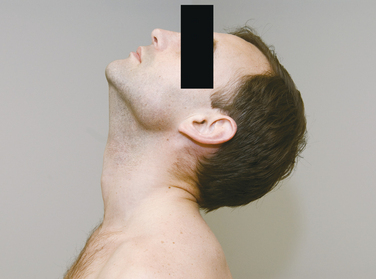
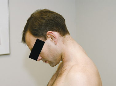
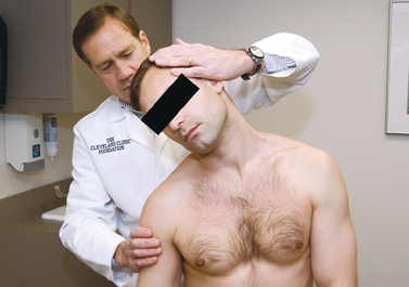
Neck extension (Fig. 4-3) is recorded by noting that the imaginary line from the occiput to the mentum of the chin extends beyond the horizontal. Flexion is recorded by noting how many fingerbreadths the chin is from the chest when the patient flexes the neck as much as possible (Fig. 4-4). The patient leans the head to the side while looking forward (Fig. 4-5), and the distance from the shoulder to the ear is recorded for lateral flexion. Lastly, the patient turns the head from side to side and the examiner notes the degree of rotation. These cervical spine motions are made actively (by the patient) rather than passively (by the examiner).


The Spurling test (Fig. 4-6) is performed by placing the cervical spine in extension and rotating the head toward the affected shoulder. An axial load is then placed on the spine. Reproduction of the patient’s shoulder or arm pain is considered a positive response.


Although a detailed neurologic examination is beyond the purview of most shoulder examinations, clinical judgment determines the degree of peripheral nerve assessment necessary to establish a correct and complete diagnosis. Examining the strength of the trapezius, deltoid, spinati, and biceps and triceps muscles suffices for most general shoulder examinations. However, in some situations a more thorough examination needs to be completed, which includes assessment of motor and sensory distributions of each peripheral nerve of the upper extremity or extremities.


Shoulder


Inspection


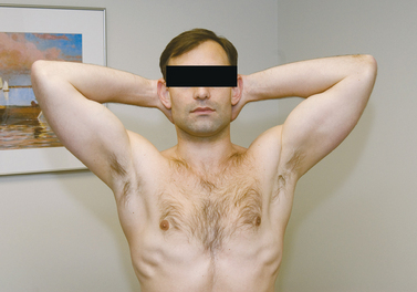
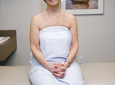
Inspection of both shoulders can reveal pathology that would otherwise go unnoticed if the examiner relied solely on the patient history or physical examination. Both shoulders need to be exposed (Fig. 4-7). First, observe the clavicles for deformity at both the sternoclavicular joint and acromioclavicular joint. A prominent sternoclavicular joint can be due to an anterior dislocation, inflammation of the synovium, osteoarthritis, infection, or condensing osteitis. A loss of sternoclavicular joint contour is consistent with a posterior dislocation of the medial clavicle, which is worked up urgently to confirm the diagnosis. The acromioclavicular joint is often prominent secondary to osteoarthritis and needs to be compared to the opposite side for symmetry.


The relative height of each shoulder is noted as the patient sits with arms by the sides. Small differences in shoulder height are often found in normal patients and can be confirmed by asking whether their shirt sleeves seem longer on one side than the other. Pathologic causes of a difference in shoulder height can be explained by problems with the articulation of the scapula and thorax or glenohumeral joint. Drooping of the scapula can be caused by trapezius paralysis, scapular winging, scoliosis, pain that results in splinting of the scapula, fractures of the scapula, or disruption of the scapula–clavicular suspensory complex. Deltoid dysfunction can cause the humerus to hang lower than on the normal side.


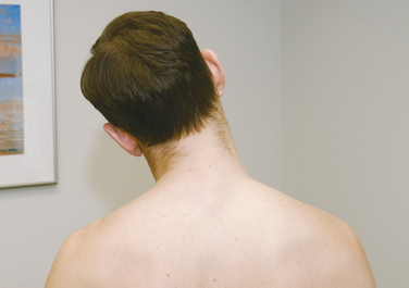
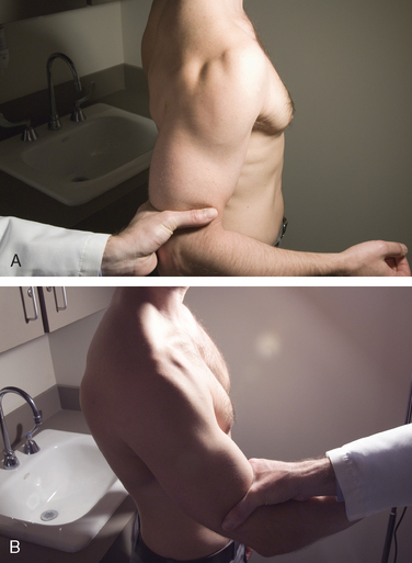
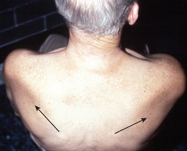
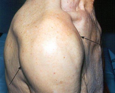
Muscle inspection begins with the three portions of the deltoid muscle. Marked atrophy is easy to identify, but deficiencies in the posterior or middle deltoid are more difficult to appreciate until active shoulder motion is initiated (Fig. 4-8). In patients with a large amount of subcutaneous tissue, palpation of the muscle belly may be the only way to distinguish a pathologic muscle contraction from the normal side. Inspection from the back reveals the muscle bulk of the supraspinatus and infraspinatus muscles, as well as the trapezius muscle (Fig. 4-9).


Once the muscle bulk has been assessed, the static position of the scapulae must be noted. If the soft tissue obscures the view of the medial border or the scapular spine, palpation of these landmarks can help visualize the attitude of the scapula at rest. Excessive lateral rotation of the scapula or an increased distance between the medial border of the scapula and the spine could be caused by trapezius palsy. This can also be accompanied by a prominent inferior tip of the scapula. A laterally prominent inferior scapula tip can be caused by serratus anterior muscle weakness related to a long thoracic nerve injury, but this might only be recognized during active shoulder motion.


The most common skin manifestations of shoulder pathology are ecchymosis, which occurs after fractures, dislocations, or traumatic tendon ruptures, and erythema, which occurs with infection and systemic inflammatory conditions. Less commonly, the skin around the anterior shoulder is swollen and enlarged due to a subacromial effusion and a chronic rotator cuff tear (Fig. 4-10). The examiner notes the presence of scars and their location and character. A widened scar can indicate a collagenopathy often seen in association with shoulder instability.


Stay updated, free articles. Join our Telegram channel

Tags:
Sep 8, 2016 | Posted by in PHYSICAL MEDICINE & REHABILITATION | Comments Off on Clinical Evaluation of Shoulder Problems

Full access? Get Clinical Tree

Get Clinical Tree app for offline access