Fig. 47.1
Male patient complaining about stiffness after primary guided motion cruciate substituting TKR (left). After single-stage cemented revision TKR (right, Peter Brehm BPKS RH)
Cementing the stems in revision TKR has many advantages. In cases of revision TKR undertaken due to infection, loading of the cement with antibiotics many be beneficial [2]. Additionally, cemented stems usually provide instant stability and fixation. Cementation of the stem may also facilitate the positioning of components in a central position avoiding overhanging components. On the other hand, shorter stems are at a higher risk for malalignment as they are less guided by the intramedullary canal [8]. A downside of full cementation is the difficulty of cement removal, in the event of future revisions [8]. Bone cement may also play a role in the development of pulmonary and cerebral microembolism during joint replacement [9].
Cementation often allows the use of shorter stems, which may accommodate the implantation in cases of deformities. These stems are usually smooth and have rounded edges like a cemented hip prosthesis. Cementless stems need to be longer and have splines and flutes to engage the diaphysis to attain the similar stabilization as achieved by a shorter-stem cemented into the metaphysis. This was shown by in vitro experiments [10]. In a recent finite element study, shorter cemented stems induced lower stresses in the tibial bone-stem interface and lowered micromotions between the implant and bone compared to longer cementless stems [11].
Full cemented stems exhibited a 10-year survivorship of 94 % as described by Whaley et al. [7]. Fehring et al. compared survival at midterm follow-up and found significantly better survival for cemented stem fixation compared to non-cemented stems [2].
47.2 Cementless Fixation
Cementless stem fixation in revision TKR also has many benefits (Fig. 47.2). A well-centered long press-fit stem facilitates component alignment by strictly directing the articular component [8]. Consequently, a revision system that does not allow for correction of the articular component position with offset stems will often produce cortical overhang or undersizing of articular components. In clinical practice most surgeons apply a hybrid fixation technique cementing the articular components and use a non-cemented, long press-fit stem [12, 13]. The long stems confer excellent stability to the implant and are easily removed if necessary [14]. Porous-coated stems also achieve excellent fixation but their removal is very challenging and as a result their use is limited in clinical practice [15].
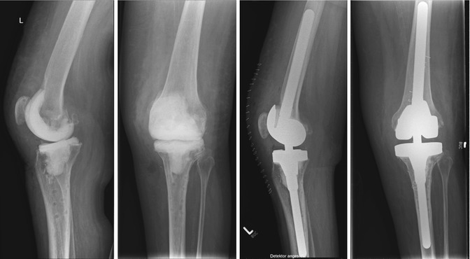
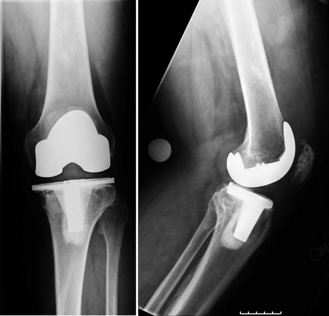
Fig. 47.2
Anterior-posterior and lateral radiographs of a female patient after first-stage revision (removal of TKR) in infected TKR following infected ORIF of fracture of lateral aspect of tibial plateau (left). Radiographs after uncemented second stage (RT Plus Modular, Smith & Nephew)
A well-centered long press-fit stem facilitates component alignment by strictly directing the articular component. Consequently, a revision system that does not allow for correction of the articular component position with offset stems will often produce cortical overhang or undersizing of articular components.
A good, full fit in the canal of the diaphysis has been suggested for long-term survival of uncemented stems [12]. The intramedullary canal should be reamed until adequate cortical contact is made to allow for press-fit fixation of the cementless stem [13]. However, there is no clinical data available to verify this.
“End of the stem pain” has been described to be a more prevalent and severe problem with uncemented stem fixation, than with cemented stems [16]. The press-fit insertion of a cementless stem may increase the risk of penetration of the cortical bone during reaming and in the event of fractures during the insertion of trials or implants.
47.3 Fixation with Metaphyseal Cones and Sleeves
In revision TKR the femoral condylar bone is often of poor quality. Bone may be osteopenic, poorly vascularized, and significantly reduced in stock due to prior surgery, infection, or osteolysis. With insufficient condylar bone stock, rigid fixation in the metaphyseal-diaphyseal junction must be obtained. To augment fixation in revision TKR, femoral and tibial metaphyseal sleeves have recently been introduced. Highly porous tantalum metaphyseal-filling cones were designed to accommodate metaphyseal bone loss [22–25]. Early clinical results with these devices are promising [26]. At short-term follow-up, structural support for the implants of a revision TKR were possible in some series [27]. Metal sleeves, which are usually used without cement, were introduced for this purpose [28]. The metaphyseal-diaphyseal junction is prepared with a broach to allow the sleeves to achieve initial axial, rotational, and coronal stability which permits immediate weight-bearing [28].
To augment fixation in revision TKR, femoral and tibial metaphyseal sleeves have recently been introduced. Highly porous tantalum metaphyseal-filling cones were designed to accommodate metaphyseal bone loss.
A short-term study was conducted in our institution using these sleeves without cement. Twenty patients were followed, who underwent revision TKR with non-cemented, porous-coated proximal tibial sleeves (Figs. 47.3 and 47.4). The use of tibial sleeves was deemed appropriate based on bone stock, age, and activity level. To measure performance, postoperative ranges of motion (ROM), Knee Society Scores (KSS), and Knee Society Functional Scores (KSFS) were taken at a minimum of 6 months follow-up. Radiographs were also evaluated for component positioning, subsidence, or loosening. Survivorship at a mean of 1.2 years follow-up (0.5–1.8 years) was 100 % without subsidence, change in component positioning, loosening, or radiographic demarcation. Despite the short follow-up period, patients who underwent revision TKR with the porous-coated proximal tibial sleeves demonstrated excellent efficacy and recovery, in terms of both pain and function. Osseointegration begins to become apparent about 6 months postoperatively. These results may justify increased use of non-cemented proximal tibial sleeves. However, long-term follow-up is necessary to show if bone adheres to and integrates fully into the prosthesis.
 Laboratory Analysis in the Assessment of Painful Total Knee Replacement
 Periprosthetic Fractures Following Total Knee Replacement
 Joint Line Restoration in Revision Total Knee Replacement
 Discussion to Chap. 34: Persistent/Recurrent Pain After TKR Not Always TKR Related
 Use of Stems in Revision Total Knee Replacement
 Periprosthetic Fracture Treatment in Total Knee Replacement


Related posts:
Laboratory Analysis in the Assessment of Painful Total Knee Replacement
Periprosthetic Fractures Following Total Knee Replacement
Joint Line Restoration in Revision Total Knee Replacement
Discussion to Chap. 34: Persistent/Recurrent Pain After TKR Not Always TKR Related
Use of Stems in Revision Total Knee Replacement
Periprosthetic Fracture Treatment in Total Knee Replacement
Stay updated, free articles. Join our Telegram channel
Full access? Get Clinical Tree
