CHAPTER 28 Carpal, Metacarpal, and Phalangeal Fractures
Carpal, metacarpal, and phalangeal fractures are among the most common injuries treated by hand surgeons. Historical data suggest that 10% of all fractures occur in the metacarpals and phalanges and that 80% of all hand fractures involve the tubular bones.1–3 Until the early 20th century, most carpal and hand fractures were treated nonoperatively. Even today, fractures that are nondisplaced or minimally displaced may be viewed as stable and treated conservatively or with closed reduced and cast immobilization. Unstable fractures require surgical fixation to maintain length, alignment, and rotation. Other options for fixation include percutaneous pinning, external fixation, traditional open reduction and internal fixation, and arthroscopically assisted reduction and internal fixation (AARIF).
HISTORICAL BACKGROUND
Arthroscopic surgery of the wrist and hand is a rapidly evolving discipline. Since Chen first reported on diagnostic arthroscopy of the wrist and finger joints in 1979 with the Watanabe no. 24 arthroscope, techniques for small joint arthroscopy have developed at a rapid pace.4 Current indications include the diagnosis and treatment of numerous wrist disorders, including fractures, soft tissue pathology, and arthritis. Arthroscopy of small joints, including the metacarpophalangeal (MCP) and interphalangeal (IP) joints, has lagged compared with the phenomenal interest generated by wrist arthroscopy. Badia theorized that minimal reporting in the literature and lack of direct teaching of this technique have contributed to the limited use of this potentially useful technology.5
In 1995, more than 15 years after Chen’s classic article, Ryu and Fagan described their experience treating the ulnar collateral ligament Stener lesion in eight thumbs with success.6 Arthroscopic reduction was achieved by reducing the lesion until the avulsed ligament sat juxtaposed to its insertion site on the proximal phalanx. After the reduction was performed, the ligament ends were débrided, and the joint was pinned. The results were excellent, with range of motion and strength comparable to those of the unaffected contralateral thumb.
In 1999, Rozmaryn and Wei presented a paper detailing the technical aspects of MCP arthroscopy, with general references to the potential use of this technique in treating juxta-articular and intra-articular fractures.7 In the same year, Slade and Gutow published a review article on arthroscopy of the MCP joint.8 They offered detailed technical explanations and emphasized that small joint arthroscopy requires specialized instruments and a thorough knowledge of the anatomy within these specialized joints. In 2006, Badia reported encouraging results treating bony gamekeeper’s thumb with AARIF.9
ANATOMY
The MCP joint is composed of several structures, including the osseous metacarpal head, the proximal phalangeal base, and soft tissue restraints, which include the stout volar plate, collateral ligaments, and a relatively thin and flimsy dorsal capsule. Strong extensor tendons run dorsal to the dorsal capsule and are held in check by capsular fibers and the sagittal bands. On either side of the joint are the insertions of the intrinsic muscles on the extensor mechanism. The digital neurovascular bundles rest volar to the joint, along with the flexor tendons; the terminal branches of the dorsal sensory nerves are less predictably centered over the dorsum of the joint.
PATIENT EVALUATION
History and Physical Examination
Particular attention should be paid to patients with frail or “rice paper” skin, such as those with chronic disease and those receiving steroids.10 If small joint arthroscopy is performed on these patients, special care should be taken to limit traction forces across the joint to prevent significant disruption of the skin surface.
Diagnostic Imaging
Plain radiographs provide most of the information necessary to diagnose lesions of the MCP joints. Special views, including oblique and Brewerton views to evaluate the metacarpal head, may be useful adjuncts to traditional posteroanterior and lateral radiographs. Computed tomographic (CT) scans aligned in the sagittal plane can help to define the step-off of intra-articular fractures, and bone scans can help to identify increased activity in the metacarpal head, which suggests possible osteochondral defects.8
TREATMENT
Indications and Contraindications
AARIF is an excellent choice for juxta-articular fractures of the carpometacarpal (CMC) and MCP joints of the thumb and digits, because these small joint injuries require anatomic reduction to prevent post-traumatic arthritis. As Slade and Gutow pointed out in their 1999 article describing arthroscopic techniques for MCP joint fixation, minimal surgical manipulation of the soft tissue envelope and preservation of the blood supply during operative exposure are indispensable for obtaining an optimal result.8 AARIF preserves vascular blood supply to bony fragments by minimizing dissection of the soft tissue envelope surrounding the joints of the hand. Open surgery of tissues surrounding these small joints may contribute to collateral ligament and flexor tendon scarring and shortening, which ultimately limit tendon excursion and lead to stiffness and suboptimal results.11
A example of a fracture pattern amenable to AARIF is the simple two-part fracture with displacement greater than 1 mm involving the metacarpal head or base of the proximal phalanx.10 Other excellent indications for AARIF are for reduction of an avulsion fracture with a rotated fragment from the collateral ligament insertion and for treatment of die-punch articular fractures of the proximal phalangeal base. AARIF enables the surgeon to use the arthroscope to clearly visualize articular reduction with a probe or Kirschner wire and to visualize subsequent percutaneous fixation.5 In pediatric cases, Salter-Harris type III physeal fractures may be reduced and secured without additional injury to the growth plate with the use of AARIF.
Slade and colleagues presented their results using AARIF in 1998 at the Annual Meeting of the Arthroscopy Association of North America (AANA). This study documented fewer complications and improved final range of motion when fractures were treated with AARIF compared with the results of standard open reduction.12
Contraindications to the use of AARIF for MCP and proximal IP fractures are poor soft tissue coverage, open fractures, and active cellulitis.10 Fractures with three or more fragments, comminution, or associated diaphyseal extension that cannot be easily reduced through percutaneous means are also disqualified for AARIF, because these fractures are better treated with standard open approaches.
Conservative Management
Most acute traumatic injuries involving the MCP and proximal IP joints can be managed conservatively with a trial of splint or cast immobilization. Fractures that result in rotational malalignment, an inability to fully extend the digits, or intra-articular displacement are best treated by some form of operative intervention, because nonoperative care may lead to long-term disability, such as pain, stiffness, or post-traumatic arthritis. As the surgeon becomes more adept at small joint arthroscopy, acute indications may evolve due to more accurate assessment of the extent of injury and more precise treatment.5
Arthroscopic Technique
Instruments
Inflow may be provided through a gravity system using a pinch pump and small joint tubing or the camera itself and a standard arthroscopy pump set to maintain a pressure between 50 and 80 mm Hg. The small radius of the inflow cannulas requires higher pressures to ensure adequate irrigation.8
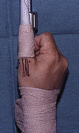
Adequate traction is vital for all joints, particularly those as small as the CMC, MCP, and proximal IP joints. Several commercial traction devices are available to adequately distract the joint and provide traction with the assistance of sterile, disposable plastic finger traps (Fig. 28-1). A mini-fluoroscope is essential to help localize the joint space, assess fracture reduction, and visualize fixation. Standard small hand instruments are also necessary, including a wire driver, mini-curettes, mini-fragment screws, and a power drill for final implant seeding.