Fig. 59.1
Brown tumor. Well-demarcated lytic lesion involving the proximal tibia
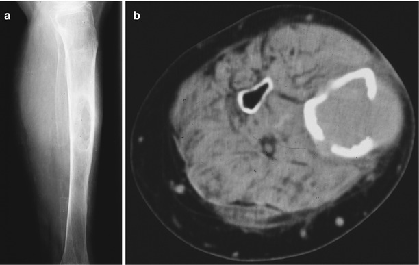
Fig. 59.2
(a) Radiolucent lesion seen in the diaphysis of the tibia. (b) CT scan: the lesion erodes the cortex and involves soft tissue
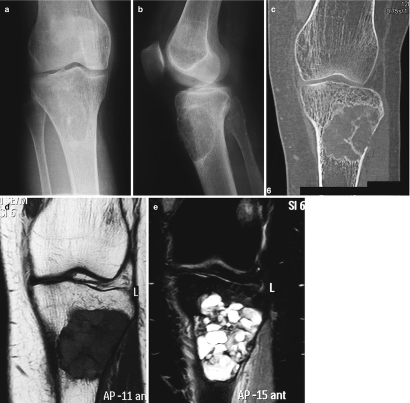
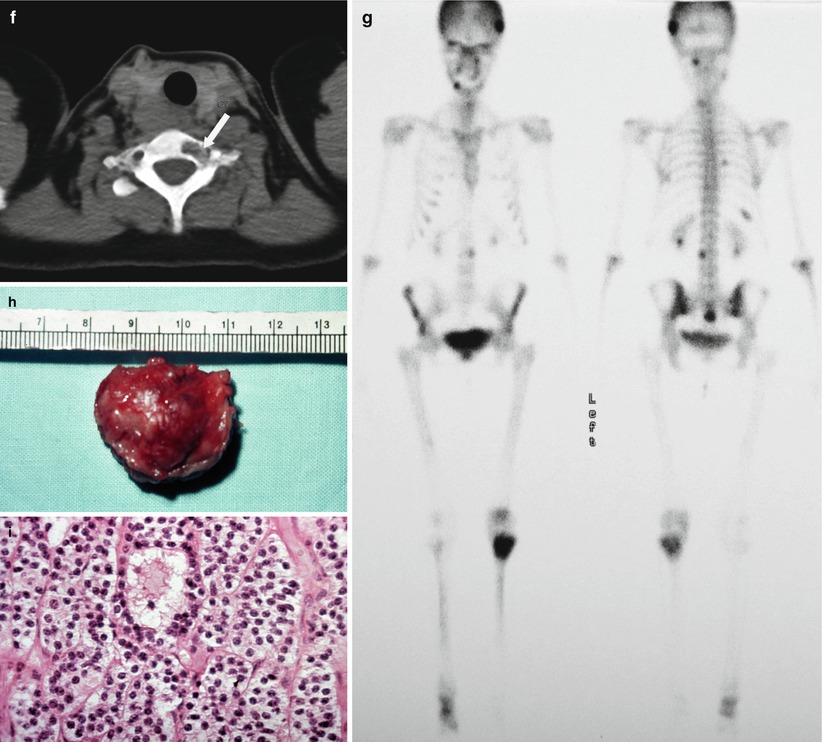
Fig. 59.3
Hyperparathyroidism. (a–e) Radiographs, CT scan, and MRI of the proximal tibia with a lytic lesion. (f) Core needle biopsy of the seventh cervical body was done (arrow). (g) Scintigram shows multiple hot spots. (h, i) Parathyroid adenoma. Gross specimen and microscopic features
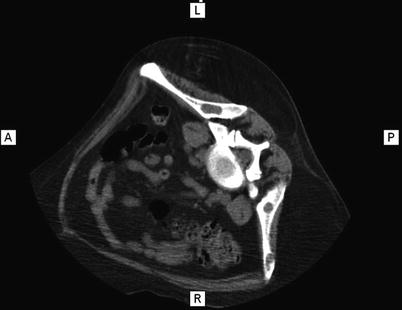
Fig. 59.4
Multiple lytic lesions in the pelvis in a patient with hyperparathyroidism
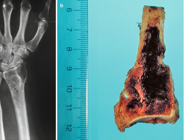
Fig. 59.5


(a) Brown tumor of the lower end of the radius. (b) Gross specimen
Related posts:


Stay updated, free articles. Join our Telegram channel
Full access? Get Clinical Tree
