Brachial Plexus Birth Palsy: Shoulder Reconstruction


Brachial Plexus Birth Palsy: Shoulder Reconstruction


CASE PRESENTATION

An 18-month-old child with known brachial plexus birth palsy (BPBP) is referred for limitations of arm function. He has been enrolled in an early intervention physical therapy program with daily home exercises since early infancy. The parents’ and therapist’s notes indicate a return of elbow flexion at 4 months of age. They state he has never been able to reach his hand above shoulder level, and he reaches his hand to his mouth only with his elbow high in the air. He did have a clavicle fracture at birth, and the parents wonder if this is part of the problem.1 On exam, he has limited passive external rotation (ER) to 10 degrees in adduction and 60 degrees in abduction. There is a bony prominence palpable in the posterior soft spot of his shoulder with internal rotation.


THE FUNDAMENTALS

Muscle imbalance in a growing child can lead to bone and joint deformity. The glenohumeral joint is no different.2 In children with incomplete recovery from BPBP, the adductor and internal rotator muscles are often out of balance with the abductor and external rotator muscle forces (Figure 21-1). The pectoralis major is dually innervated and will recover earlier than the opposing muscles in an upper plexus injury. The subscapularis, innervated by the upper and lower subscapular nerves off the posterior cord, also acts as a deforming force, through either unopposed muscle activity and tightness or, less likely, fibrosis. The latissimus dorsi (thoracodorsal nerve supply off the posterior cord) and teres major (lower subscapular nerve supply off the posterior cord) often recover in advance of the deltoid (axillary nerve off the posterior cord), supraspinatus, and infraspinatus muscles (suprascapular nerve off C5 or upper trunk). In addition, periarticular tightness and myostatic contracture can develop. This combination of forces leads to an adduction and internal rotation contracture in infancy or early childhood (Figure 21-2). Eventually, if unrestrained, this can cause progressive glenohumeral deformity and dislocation (Figure 21-3). Experiments using animal models have replicated this sequence from nerve impairment through muscle imbalance to joint deformity.3


Etiology and Epidemiology

Between 10% and 40% of infants with BPBP will have an incomplete recovery. Some will have minimal deficits; others will have profound, permanent disability. The C5-C6 (-C7) children with an incomplete recovery are more at risk for glenohumeral dysplasia and dislocation4 than either the total plexus or the minimally affected patients. Publications on shoulder dislocations and deformity date back to Stimson and Fairbank and include many modern publications.5, 6 and 7 Cumulative knowledge is now extensive. Glenohumeral dysplasia and dislocation are clearly high risk for children with continued imbalance and contracture.8 Close monitoring, evaluation, and treatment
are required. Through the radiographic advances of MRI scans, confirmed by ultrasound and arthrograms, it is clear that deformity starts very early in life and is progressive if untreated.


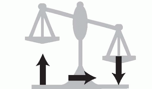
FIGURE 21-1 The return of internal rotator strength before ER muscle activity leads to an imbalance about the glenohumeral joint. This can progress to deformity and dislocation.


Clinical Evaluation

Assessing individual muscle strength in an infant can be challenging. The Medical Research Council (MRC) muscle strength grading system (0 to 5) is too exacting, as infants and toddlers cannot follow verbal instructions for resistive testing. Recognizing that muscle strength has to be inferred from observed or stimulated movement in infants and children, Gilbert and Tassin modified that MRC system to a four-point gradation: (0) no muscle contracture, (1) muscle contracture without movement, (2) movement with gravity eliminated, and (3) complete movement against weight of extremity (Table 21.1). Other classification systems include the modified Mallet scale (Figure 21-4, Table 21.2) and the Hospital For Sick Children Active Movement Scale (Figure 21-5).9, 10 and 11 These later systems have been shown to have inter- and intraobserver reliability.


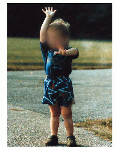
FIGURE 21-2 The affected left arm of this child with incomplete recovery reveals restrictions in ER and above horizontal shoulder function that is typical for these patients. He has to reach his mouth with his shoulder maximally abducted. He cannot reach the top of head and can only reach the left side by tilting his neck forward and to the side. His humeral head is dislocated.


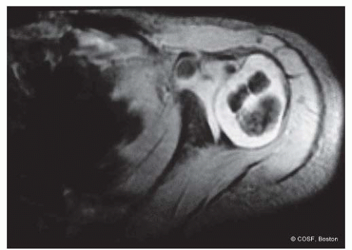
FIGURE 21-3 MRI of the affected glenohumeral joint reveals posterior subluxation of the humeral head and glenoid deformity with an early pseudoglenoid or biconcave joint. The humeral head is already beginning to change shape, and the position of the biceps insertion reveals the presence of an internal rotation contracture.

Assessing passive range of motion is an essential part of comprehensive shoulder evaluation in these patients.

Maintaining passive ER with scapular stabilization is integral to their therapy program. Conversely, those infants and children losing ER in adduction12 and/or abduction2 are most at risk for glenohumeral deformity (Figure 21-6). Before a fixed deformity develops, the shoulder instability can be palpated in the posterior soft spot with alternating internal (subluxation) and external (reduction) rotation. Later, the dislocated humeral head is palpable posteriorly with clear asymmetry to the opposite side anatomic situation (Figure 21-7). Scapular winging is present in all these children with muscle imbalance and contracture (Figure 21-8).


Table 21.1 Gilbert and Tassin’s modified MRC classification for motor strength


Muscle grade


Strength


0


No contracture


1


Muscle contracture, no movement


2


Some movement, gravity eliminated


3


Complete movement against gravity


From Gilbert A, Tassin JL. Surgical repair of the brachial plexus in obstetric paralysis. Chirurgie. 1984;110(1):70-5.


Table 21.2 Modified Mallet classification


Functional Parameter


Class I


Class II


Class III


Class IV


Class V


Global abduction


None


<30 degrees


30-90 degrees


>90 degrees


Normal


ER


None


<0 degrees


0-20 degrees


>20 degrees


Normal


Hand-to-neck ability


None


Not possible


Difficult


Easy


Normal


Hand-to-mouth ability


None


Marked trumpet signa


Partial trumpet signa


<40 degrees of abduction


Normal


Internal rotation


None


Not possible


To S1


To T12


Normal


a A trumpet sign consists of abduction of the shoulder with simultaneous flexion of the elbow.


From Mallet J. Paralysie obstetricale du pexus brachial. Traitment des sequelles.


Rev Chir Orthop. 1972;55(suppl):8-166.


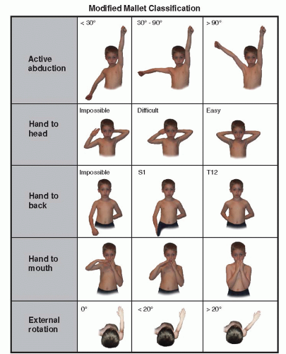
FIGURE 21-4 Modified Mallet classification for global abduction, ER, hand to neck, hand to spine, and hand to mouth. Each category is graded I to V with I, no function; V, normal function, and grades II, III, and IV depicted by illustration. Some children are not testable due to age and lack of cooperation at that particular visit. An aggregate Mallet score is a combination of the scores for all five categories, ranging 0 to 25.


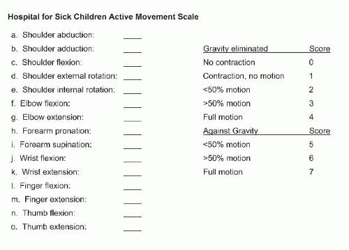
FIGURE 21-5 The Hospital For Sick Children Active Movement Scale grades each muscle movement from the shoulder to the hand by a score of 0 (no function) to 7 (normal function). A child must have full gravityassisted function (score of 4) before antigravity scoring can be obtained.


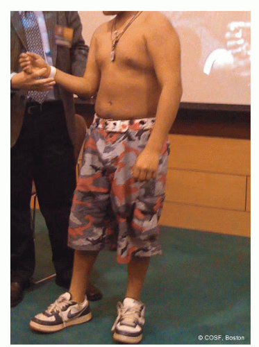
FIGURE 21-6 With the scapula stabilized, this patient has limited ER in adduction. The glenohumeral joint needs investigation for deformity.

Advanced radiographic evaluation is now a significant part of treatment planning for these patients. Plain radiographs give limited information due to the major unossified areas about the shoulder at this age. Ultrasound in skilled hands provides definitive information on glenoid dysplasia and joint alignment similar to hip ultrasounds
in developmental dysplasia. MRI scans are diagnostic (Figure 21-9).13 CT scans are faster, cheaper, and give comparable information as MRI scans but have high radiation dosing and do not show the unossified glenoid apophysis14,15 (Figures 21-10 and 21-11). Arthrograms also give comparable information16, 17, 18 and 19 (Figures 21-12 and 21-13). The clinical continuum of dysplasia at the glenohumeral joint progresses from anatomic alignment and symmetric development, increased glenoid retroversion, humeral head posterior subluxation, and biconcave or pseudoglenoid (Figure 21-14) to a frank dislocation with a flat humeral head and glenoid (Figures 21-15 and 21-16). The degree of deformity can guide the surgeon on timing and the type of operative intervention. MRI scans are more capable than physical examinations of revealing the presence and degree of glenohumeral dysplasia.


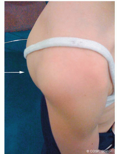
FIGURE 21-7 View of the affected right shoulder with obvious posterior dislocation of humeral head.


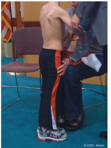
FIGURE 21-8 Marked scapular winging in this patient with dysplastic glenohumeral joint and shoulder contractures.


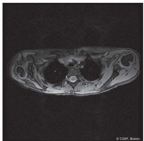
FIGURE 21-9 Obvious asymmetry between affected and unaffected glenohumeral joints by MRI scan. The affected side has marked glenoid deformity, posterior subluxation of the humeral head, and a pseudoglenoid. The unaffected side has anatomic alignment.


Surgical Indications

Indications for surgery about the shoulder in these children include (1) infantile dislocation, (2) persistent internal rotation contracture despite maximal nonoperative treatment, (3) limitation of active ER and above shoulderlevel function with plateauing of neurologic recovery, (4) progressive or marked glenohumeral deformity, and (5) a combination of any or all of these (Figure 21-17). The age at intervention is dependent on the severity of the problem and when the patient presents for care. The spectrum can be from the first 6 to 12 months of life through adolescence. We advocate early intervention when indicated to prevent progressive deformity, allow for joint remodeling, and maximize function.

Almost all publications over the past 100 plus years indicate the problem is a combination of muscle imbalance, soft tissue contracture, and bone and joint deformity. The interventions recommended in those papers, public presentations, and patient consultations are various ways of addressing those issues. Surgical guidelines that emerge are (1) in the young child (6 months to 2 years) with resistant contracture but potential ongoing ER and abduction recovery, intervention for contracture release alone (botulinum toxin A [Botox, Allergan Pharmaceuticals, Inc., Irvine, CA] injection, subscapularis slide, arthroscopic joint release, coracoacromial ligament release) is indicated20, 21,

Only gold members can continue reading. Log In or Register to continue

Stay updated, free articles. Join our Telegram channel

May 24, 2016 | Posted by in ORTHOPEDIC | Comments Off on Brachial Plexus Birth Palsy: Shoulder Reconstruction

Full access? Get Clinical Tree

Get Clinical Tree app for offline access