Bones
INTRODUCTION AND OVERVIEW
Bones are complex organs with many important functions, the most obvious being structural. They provide support for the body and the means by which muscles can insert into fixed structures in order to allow movement. They are also important in hearing, through the transduction of sound via the ear’s ossicles, and they protect other soft organs that are easily damaged, such as the brain, eyes, kidneys, lungs and spleen. Bone marrow, which is largely within the medulla of the long bones, is the centre for production of blood cells (haematopoesis) and an important site for storage of fatty acids. Bones also have important metabolic functions, including:
This chapter describes the following conditions affecting bones:
OSTEOPOROSIS
DEFINITION
In 1991, osteoporosis was defined as a metabolic bone disease characterised by ‘low bone mass, microarchitectural deterioration of bone tissue leading to enhanced bone fragility and a consequent increase in fracture risk’.1 However, the current conception of osteoporosis is as a disease of compromised bone strength that predisposes an individual to an increased risk of fracture.2 In this new definition, osteoporosis is a dynamic, rather than an anatomical, condition. The disease is thus low bone mass and deteriorated bone quality, and fracture is the clinical consequence of the disease.
Osteoporosis is highly prevalent in the general population. Using the operational definition of osteoporosis (based on measurement of bone mineral density (BMD)), the prevalence of osteoporosis in Australian men and women aged 60 years and above was estimated to be 11% and 27% respectively.3 As expected, the prevalence of osteoporosis increases with advancing age, from approximately 5% (men) and 13% (women) at 60 years of age to over 28% (men) and 63% (women) at age 80 or above.4 With a rapidly ageing population, it is expected that osteoporosis will be an even greater problem in the next 20 years or so.
The outcome of osteoporosis is fragility fracture. Fractures are a major public health problem because of their high incidence rate in the population, and because individuals with fracture are at increased risk of mortality and morbidity and reduced quality of life, and incur significant healthcare costs. In several population-based epidemiological studies, all major fractures were associated with increased mortality, especially in men.5 Even in healthy older women, clinical vertebral fractures, commonly manifested as asymptomatic fracture, and hip fractures, are substantially associated with increased mortality.6,7 A more recent study demonstrated that asymptomatic vertebral deformity is a major risk factor for subsequent fracture and mortality.8
The increase in fracture cases will result in a significant increase in healthcare costs. The annual cost of treatment of fractures and associated sequelae has been estimated at $10–20 billion in the United States and £3 billion in England and Wales. Within the next 50 years the cost of hip fracture alone in the United States may exceed $240 billion.9 In Canada, the annual cost of hip fracture is estimated at $650 million and is expected to rise to $2.4 billion by 2041.10 In Australia, the direct and indirect costs of fracture have been estimated at $7.5 billion.11
EPIDEMIOLOGY OF FRACTURES
Theoretically, any fracture related to low bone density may be considered an osteoporotic fracture. Fractures of the spine (vertebrae), hip and wrist (distal forearm) have long been regarded as typical osteoporotic fractures.12–16 Most types of fracture occur more often in patients with low BMD,17,18 and therefore most types of age-related fractures are osteoporotic in nature. According to this definition, the following fracture types are considered osteoporotic:
In both women and men, fractures occurring at the skull and face, hands and fingers, feet and toes, ankle and patella are classified as not due to osteoporosis.19
Because fragility or osteoporotic fracture is defined as fracture-related with minimal trauma (i.e. a fall from standing height or less),20,21 in the research setting, fractures clearly due to major trauma (such as motor vehicle accidents) or due to underlying diseases (such as cancer or bone-related diseases) were excluded from analysis.5,16,18,22–26
Overall, the incidence of any fracture in women and men increases with advancing age, and is greater in women than in men. The incidence of fracture also varies according to geographic variations between and within countries. Fracture rates in women aged 60 or older are six times higher than those in women aged 35–59 years; in men aged 60 or older the rate is 1.4-fold greater than in men less than 60 years of age.27
Overall fracture rates are greater in urban than in rural areas.28,29 The difference is more pronounced in those aged 60 years or over, suggesting that environmental factors have an impact on bone health.
The magnitude of osteoporosis-related fracture can be quantified by the lifetime risk of fracture. In developed countries it is estimated that the residual lifetime risk of any fracture for men and women from age 60 is approximately a quarter and just under a half respectively.16 For individuals with osteoporosis, the mortality-adjusted lifetime risk of any fracture is probably over 40% for men and 65% for women.3 The three most common sites are hip, vertebral and Colles’ fracture.
TYPES OF FRACTURE
Hip fracture
Hip fractures include femoral neck, intertrochanteric or subtrochanteric fractures. Hip fracture is the most serious consequence of osteoporosis because it incurs many subsequent complications, including pain and disability.30
It is estimated that, from the age of 60 years, 11 out of 100 women will sustain a hip fracture during their remaining lifetime. This risk is higher than the risk of being diagnosed with breast cancer (around 10%). Approximately 17% of women and 30% of men who have sustained a fracture will die within 12 months after the event.31 However, the exact causes of death among these individuals are not clear. Among women who survive the fracture, over 20% require long-term care and over a third are unable to return to their prior work,32 and have a significantly reduced quality of life.33
Vertebral fracture
Osteoporosis has sometimes been referred to as a ‘silent disease’ because individuals often do not have apparent symptoms and pain until a fracture occurs. Asymptomatic vertebral fracture can be considered a silent disease, because patients do not realise that they have a fracture and do not seek medical attention. Currently, there is no ‘gold standard’ for the identification of vertebral fracture34,35; estimates of its overall prevalence and incidence rate in elderly women and men depends in part on the definition used.36
Asymptomatic vertebral fracture can be detected by conventional radiology, but it is not an attractive means of large-scale screening, because of cost and radiation exposure. In a large-scale study,37 vertebral fracture was present in 12% of women and men. As expected, the prevalence of vertebral fracture increases with age, more sharply in women.38 The use of corticosteroid therapy doubles the risk.39 Studies have found that about 33% of vertebral fractures or deformities are symptomatic40 and that only 23% of vertebral deformities in women were diagnosed clinically.41 In other words, there are many ‘silent’ vertebral fractures that produce no obvious symptoms. Vertebral deformities, whether clinically recognised or not, are related to an increase in chronic back pain and disability,42,43 and to low health-related quality of life44 and an increase in mortality.8,45
Distal forearm fracture
A fracture of the distal forearm is one that occurs through the distal third of the radius and/or ulna. It is frequently and typically seen in women.46,47 Although its consequence is less serious than that of a hip fracture, it is associated with significant pain and may be associated with severe and long-term complications.48,49 The incidence increases rapidly with advancing age in women (but less so with men), for up to 10 years following menopause, and tends to slow thereafter.50
CLINICAL ASSESSMENT OF OSTEOPOROSIS
The clinical assessment of osteoporosis and fracture risk is based on assessment of:
Risk factors
Risk factors can be broadly classified into modifiable and non-modifiable risk factors (listed in Box 23.1). Of these, the four key risk factors are:
A prior fragility fracture substantially elevates an individual’s risk of future fracture.5,8,51,52 The elevated risk is 1.5- to 9.5-fold depending on age at assessment, number of prior fractures and the site of the incident fracture. A pre-existing asymptomatic vertebral fracture increases the risk of a second vertebral fracture and non-vertebral fracture at least four-fold.8 A study of a placebo group showed that 20% of those who experienced a vertebral fracture during the period of observation had a second vertebral fracture within 1 year. Patients with a hip fracture are at increased risk of a second hip fracture. The risk of subsequent fracture among those with a prior fracture at any site is 2.2 times that of people without a prior fragility fracture.52 A family history of osteoporotic fracture is also a major risk factor for fracture.
Bone strength
At present there is no direct method for reliably measuring bone strength. However, BMD, measured by dual-energy X-ray absorptiometry (DXA), provides a benchmark for bone strength. BMD measurement could account for up to 70–75% of variance in bone strength.53 BMD is often standardised by expressing it as a T-score, i.e. the number of standard deviations (SD) from the young normal mean, taken as aged between 20 and 30 years.
There is a strong, continuous and consistent relationship between BMD and fracture risk, such that each SD lowering in BMD is associated with a 1.6-fold increase in fracture risk in both men and women.54 In Chinese women, the relative risk of fracture for each SD lowering in femoral neck BMD was two-fold.55 There is evidence that the magnitude of association between BMD and hip fracture risk (with relative risk being 2.256 to 3.657) is equivalent to or even stronger than the association between serum cholesterol and cardiovascular disease. Measurement of BMD is therefore considered the gold standard for diagnosis of osteoporosis in elderly men and postmenopausal women.
An operational definition of osteoporosis, by which a postmenopausal woman is considered to have osteoporosis, is if her femoral neck BMD (which is more reliable than lumbar spine measurements) is decreased by at least 2.5 SD compared with the mean value in young adults.58 The WHO classification also includes definitions of osteopenia and normal BMD (Table 23.1). The operational criteria of osteoporosis for women has also been adopted for men.59 These classifications do not apply to children.
TABLE 23.1 WHO diagnostic categories for BMD in postmenopausal women
| Category | Projected proportion of population* |
|---|---|
| Normal | BMD not more than 1 SD below peak BMD in young adult mean (T-score > −1) |
| Osteopenia | BMD 1–2.5 SD below young adult mean (T-score −1 to −2.5) |
| Osteoporosis | BMD 2.5 SD or more below young adult mean (T-score at or below −2.5) |
| Established osteoporosis | BMD 2.5 SD or more below young adult mean, and the presence of one or more fragility fractures |
* Urban, 2020 (%). SD: standard deviation. BMD: bone mineral density.
Source: Nolla et al. 200260
Who should have a BMD measurement?
Mass screening using DXA scanning is not recommended or feasible without some selection of the target population. One important and difficult question is how to decide which women should undergo BMD measurement and further evaluation based on the results of BMD scan. It is suggested that a case-finding strategy be adopted, to identify individuals ideally eligible for a BMD scan61—that is:
In the absence of BMD measurement, a number of clinical prediction rules, including the Osteoporosis Self-assessment Tool for Asians (OSTA), have been developed,62,63 to identify ‘candidates’ for a BMD scan. Most of these scores, based only on age and body weight, are a simple prediction rule that can potentially be useful in identifying women at high risk of osteoporosis. These scoring systems64,65 can be useful in ruling out osteoporosis.
Bone turnover markers
Bone turnover rate in postmenopausal women correlates negatively with BMD, and markers of bone resorption are associated with fracture risk.66 A reduction in biochemical markers appears to be correlated with a decrease in vertebral fracture incidence67 in some studies, but is not necessarily always predictive of response to therapies. Nevertheless, the predictive value of biomarkers in assessing an individual patient can be limited by their high variability within individuals.68
Assessment of absolute fracture risk
At any given level of BMD, fracture risk varies widely in relation to the burden of other risk factors56 (some modifiable), such as:
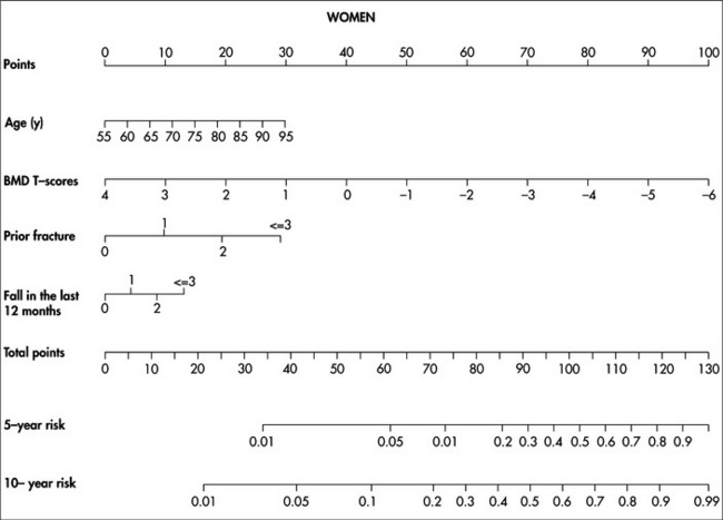
Thus, for any one individual, the likelihood of fracture depends onavv a combination of risk factors. This means that two individuals, both with osteoporosis, can have different risks of fracture because they have different non-BMD risk profiles. Likewise, an osteoporotic individual can have the same risk of fracture as a non-osteoporotic individual. A logical and appropriate assessment of fracture risk for an individual should therefore take into account the individual’s risk profile, including BMD and a history of fracture. A multivariable-based nomogram such as the one developed by Nguyen and colleagues69,70 and the WHO’s FRAX model71 can be used to estimate an individual’s absolute risk of fracture, and help select patients suitable for intervention.
The critical question of who should be treated can only be answered by a complete evaluation of an individual’s risk profile. Treatment is cost-effective if an individual’s 10-year risk of hip fracture is between 1.2% and 9.0%, depending on age.72 To facilitate the estimation of this threshold, a nomogram (Figure 23.1) is used to estimate the probability of fracture based on age, BMD and history of fractures and falls.

FIGURE 23.1 Nomogram for predicting 5-year and 10-year probability of hip fracture.69 To use, mark the age of an individual on the ‘Age’ axis and take a vertical line up to the ‘Points’ axis. Repeat this process for each additional risk factor, then sum the points. Locate the final sum on the ‘Total points’ axis and take a vertical line down to the 5-year and 10-year risk lines to determine the individual’s probability of sustaining a hip fracture within these timeframes
TREATMENT
Lifestyle advice
Nutrition and supplements
Nutrition
Higher dietary protein intake is associated with a lower rate of age-related bone loss,73 and fruit and vegetable intake is positively associated with bone density in men and women.74
Calcium and vitamin D
A combination of calcium and vitamin D reduces the incidence of hip and non-vertebral fractures.75–77 Elderly women (aged 69+ years) taking calcium (1200 mg/day) and vitamin D (800 IU) for 18 months had their risk of hip fracture reduced by 43% and non-vertebral fracture by 32%, although in a recent meta-analysis it was shown that the use of calcium or calcium plus vitamin D reduced fracture risk by 12%.55 In summary, calcium and vitamin D appear to be efficacious in reducing the risk of hip fracture and non-vertebral fractures. There is some evidence, however, that calcium supplements can lead to a moderately increased risk of myocardial infarction. Regular moderate sun exposure should also be remembered as a valuable way of maintaining adequate vitamin D levels. If vitamin D levels are very low (< 50 nmol/L2), a large loading dose of vitamin D of up to 50,000 IU per month until the level is at least > 75 nmol/L2 and then a maintenance dose of 2000 IU daily with 3-monthly monitoring.78 Some advocate a larger loading dose of 100,000 IU as being helpful in bringing up the levels more quickly, followed by a supplement of 1000–5000 IU daily, monitored at 3-monthly intervals until normal range is achieved. Then maintenance of 1000 IU daily if sun exposure has not increased.
Pharmacological
Hormone therapy
Hormone therapy (HT) has been used in treating osteoporosis for some time, despite there having been no randomised controlled trial or large-scale prospective studies of the efficacy of HT in postmenopausal women with osteoporosis or a pre-existing fracture. The Women’s Health Initiatives (WHI) study found that HT reduced the risk of hip fracture by an average of 34%, vertebral fracture by 34% and all fractures by 24%.79–81 However, women on HT in the HERS study (designed to evaluate the effect of oestrogen plus progestin on the risk of coronary heart disease) found no significant reduction in hip fracture risk and any fracture.80 Still, a recent meta-analysis of women who had been treated with HT for 12–120 months showed that HT could reduce non-vertebral fracture risk by 28% and vertebral fracture risk.82 Although these data collectively suggest that HT may reduce fracture risk with a modest efficacy, its anti-fracture benefit must be weighed against other adverse effects such as increased risk of breast cancer, thromboembolic disease and stroke.
Selective oestrogen receptor modulators
In postmenopausal women suffering from osteoporosis, raloxifene (at the dose of 60 mg/day) has been shown to significantly reduce the risk of vertebral fracture by as much as 50%.83,84 However, its effects on hip and non-vertebral fractures have not been consistent. Raloxifene could also reduce the risk of non-vertebral fractures in women with severe osteoporosis. The drug is mostly used in younger postmenopausal women who have bone loss predominantly at the spine, not the hip.
Calcitonin
Calcitonin (generally given as a nasal spray), due to its antiresorptive properties, has been used in the treatment of osteoporosis for many years, with significant beneficial effects in reducing bone loss and fracture risk, possibly up to 33% when combined with calcium (1000 mg/day) and vitamin D (400 IU per day).85 Similar risk reduction was also observed in those with a pre-existing vertebral fracture.85 A meta-analysis including four studies conducted between 1966 and 2000 found that calcitonin reduced vertebral fracture risk by 54%. However, its effect on non-vertebral fractures has not been shown. Calcitonin is also associated with pain relief in patients following acute osteoporotic vertebral fractures.
Bisphosphonates
Stay updated, free articles. Join our Telegram channel

Full access? Get Clinical Tree


