Chapter 4B Bone Marrow Stimulating Techniques
Carbon Fiber Resurfacing
Introduction
The rationale behind using carbon fiber as a biomaterial is as follows:
In 1987,1 Minns et al. published a preliminary clinical experience in a new concept of biological resurfacing using carbon fiber implants in the form of pads or rods placed in defects within the knee that elicit a dense organized matrix of fibrous tissue that forms a new biological and functioning articular surface. No evidence of implant fragmentation has been seen since implantation in the 145 knees studied.1
Carbon fiber arthroplasty appears to be appropriate in the surgical management of ICRS Grades 3 and 4 articular cartilage lesions in the painful knee.2,3,4,5
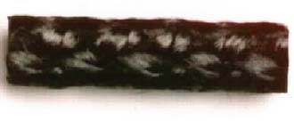
FIGURE 4B-2 Carbon pad, approximately 4 mm thick in discs up to 22 mm in diameter. Porosity is about 85%.
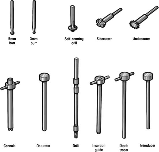
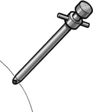
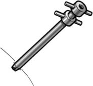
The cannula is positioned in the defect, on the bony surface (Fig. 4B-4).
The obturator is removed. Through the cannula, a 3.2-mm drill bit is put and a hole is drilled until depth- stop (Fig. 4B-5).
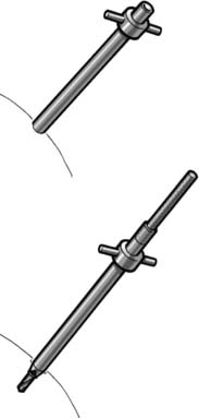
The drill is withdrawn and an insertion guide is put into the cannula. The guide should fit to the level of the stop (Fig. 4B-6)
< div class='tao-gold-member'>
Related posts:


Stay updated, free articles. Join our Telegram channel

Full access? Get Clinical Tree


