Benign Fibrous Histiocytoma
A variety of benign and malignant neoplasms in the soft tissues are considered to be of histiocytic origin. Many of these lesions are not histiocytic in origin. Nevertheless, they do have some common histologic features and fall into distinct clinicopathologic entities. The following features are commonly used to diagnose a fibrohistiocytic neoplasm in soft tissue: a spindle cell lesion with a storiform pattern and the presence of histiocytic-appearing cells, giant cells, foam cells, and a polymorphic-appearing infiltrate. These are also the features used to diagnose fibrohistiocytic neoplasms in bone.
The concept of benign fibrous histiocytoma of bone is complicated by the fact that authors have included several different entities in this terminology. Schajowicz, for instance, preferred the term histiocytic xanthogranuloma for the more commonly known metaphyseal fibrous defect, suggesting that this lesion is of histiocytic origin. However, he mentioned that it probably does not represent a true neoplasm. Fechner and Mills included xanthomas of bone under the rubric fibrohistiocytic neoplasm. They thought that a lesion containing cholesterol crystals and lipid-laden histiocytes and commonly called xanthoma probably represents an end stage of a fibrohistiocytic lesion.
In the fourth edition of this book, there were 10 examples called benign and atypical fibrous histiocytomas. There were only nine examples in the tabulations prepared for the fifth edition. One case that was previously considered a benign fibrous histiocytoma of the thoracic spine had been reclassified as a giant cell tumor. The patient developed a recurrence 12 years later, and at this time, the histology examination showed an osteosarcoma. Matsuno has also referred to the problem of differentiating benign fibrous histiocytoma from giant cell tumor when the lesions occur at the ends of long bones. As mentioned in the description of giant cell tumors, some giant cell tumors show spindling of the mononuclear cells and even a storiform pattern. A radiographically typical giant cell tumor may be so altered that only small and insignificant appearing zones of classic giant cell tumor may be found. We have added only one additional example of benign fibrous histiocytoma in the intervening 10 years.
INCIDENCE
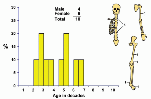
Only 10 of the 10,139 tumors in the Mayo Clinic series (Fig. 14.1) were considered to be benign fibrous histiocytoma.
SEX
There were four males and six females in this small series.
AGE
All patients were adults, with ages ranging from 17 to 60 years at the time of diagnosis.
LOCALIZATION
Four tumors involved the ilium and one the sacrum. Two tumors involved the femur: one the mid portion and one the lower end. One tumor each involved the distal portion of the tibia, the mid portion of the humerus, and a metatarsal.
SYMPTOMS
Local pain was the primary symptom of 8 of the 10 patients. One patient presented with osteomalacia, which was apparently related to this tumor. The osteomalacia recurred when the patient developed metastasis in the lung. The patient is alive and recently had a pulmonary metastasis, approximately 26 years after the original diagnosis was made.
Stay updated, free articles. Join our Telegram channel

Full access? Get Clinical Tree


