6
Balance and Coordination
1. Define and contrast balance and coordination.
2. Discuss the mechanoreceptor system and define four mechanoreceptors.
3. List static and dynamic balance and coordination tests and activities.
4. Define proprioception and kinesthetic awareness.
5. Discuss several factors that contribute to balance dysfunction.
6. Identify functional closed kinetic chain proprioceptive exercises.
7. Discuss the rationale for proprioceptive training for the upper extremity.
DEFINITIONS OF BALANCE, PROPRIOCEPTION, NEUROMUSCULAR CONTROL, AND COORDINATION
Balance is often considered as the ability to maintain the center of mass (COM) over the base of support.12 This definition is only appropriate, however, when the base of support is fixed, such as standing in a constant location on two feet. The base of support is defined as the area contained within the parts of the body making physical contact with the external environment (Fig. 6-1).9 During dynamic situations, such as gait and functional activity, the base of support does not remain fixed to a constant location. Rather, as part of locomotion, the base of support moves, increasing the challenge to the elements responsible for maintaining balance. For this reason, the concept of balance also needs to include consideration to these circumstances. Postural equilibrium is a broader term that refers to balancing all forces acting on the body’s COM to maintain COM within the limits of stability with optimal joint segment alignment.1 Forces that challenge postural equilibrium arise from gravity, unexpected perturbations (i.e., stumbling over an unforeseen obstacle), or performance of voluntary motor activities (i.e., picking up a bag of groceries). Maintaining postural equilibrium is accomplished by the postural control system, the collection of sensory sources (somatosensory, vision, and vestibular), central nervous system, and the musculoskeletal system, all serving to maintain postural equilibrium. The somatosensory sources relevant to postural equilibrium are the mechanoreceptor populations residing in joint, muscle, connective, and ligamentous tissues. Because these tissues are often damaged during orthopedic injury, postural equilibrium may be disturbed following injury because of sensory disruptions, musculoskeletal disruptions, or both.9

Performing motor tasks effectively and efficiently requires not only postural equilibrium, but also effective coordination of the many muscles serving to move and stabilize the joints upon which they cross. Coordination has been defined as the ability to produce patterns of body and limb motions in the context of environmental objects and events.13 For example, picking up an object from a table requires coordinating the shoulder, elbow, and wrists joints to put the hand and fingers into position so the object can be grasped. Essential to coordinating joint positions is sufficient sensory (afferent) information regarding joint position, movement (kinesthesia), and movement resistance/tension. The afferent information contributing to these three elements, joint position, movement (kinesthesia), and movement resistance/tension, is referred to as proprioception.12 When the proprioception elements are consciously perceived, they are referred to as the conscious perceptions of proprioception.12 Proprioception is vital for neuromuscular control. From a joint stability perspective, neuromuscular control refers to the subconscious activation of muscles occurring in preparation for and in response to joint motion and loading.12
Mechanoreceptors
Mechanoreceptors are the sensory receptors that are responsible for converting mechanical events (e.g., movement, tension) into neural signals that can be conveyed to the central nervous system.7 As mentioned previously, mechanoreceptors are located in muscle, tendon, ligament, joint capsules, and in skin and connective (fascial) tissues. Each mechanoreceptor has specific stimuli (e.g., light touch versus tissue lengthening) and thresholds (e.g., magnitude of stimuli required) to which it will respond.6 Mechanoreceptors most susceptible to disruption during orthopedic injury include the receptors located in the musculotendinous, ligaments, and joint capsules. Mechanoreceptors located in the musculotendinous tissues include the muscle spindles and Golgi tendon organs. Muscle spindles are responsible for conveying information regarding muscle length and rate of length change. Unique to muscle spindles is their adjustable sensitivity via the gamma motor neurons. Golgi tendon organs, located across a musculotendinous junction are responsible for conveying information regarding muscle tension. Located in the ligaments and joint capsules are Ruffini receptors, Pacinian corpuscles, Golgi tendon-like endings and free nerve endings. Collectively, based on their threshold and adaptation characteristics, these four mechanoreceptors provide the central nervous system with information regarding speed of joint position and movement and host tissue load levels.
BALANCE AND COORDINATION TESTS
To prescribe appropriate balance and coordination exercises, it is essential to have data related to present balance and coordination status. Most often, coordination is evaluated by using the simple tests such as those outlined in Box 6-1. Although quantifying a patient’s coordination abilities can be easily accomplished by counting the number of repetitions completed in a given time frame or the number or percentage of successes per number of attempts, qualitatively examining and describing the patient’s abilities and difficulties (e.g., steadiness, control, speed) can also be useful.
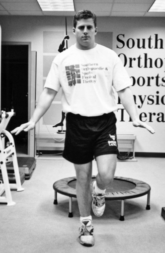
Interestingly, balance tests and specific balance treatment activities are rarely separated, and the same movements are used for fundamental balance exercises and clinically relevant balance tests. Recall that three sensory sources, somatosensory, visual, and vestibular, contribute afferent information to the central nervous system so that appropriate muscle actions can be selected. By manipulating the conditions in which balance tasks are conducted, different aspects of the postural control system may be more selectively challenged.9 For example, having a patient stand with eyes closed heightens their reliance on somatosensory and vestibular information. In addition, manipulating the base of support and support surface characteristics can also change the challenge imposed upon the postural control system. For example, compared to double-leg stance, single-leg stance requires that the postural control system reorganize itself over a narrow and short base of support, with the additional advantage that bilateral comparisons can be made. Functionally, periods of single-leg stance are often interspersed in many activities of daily living, such as walking, turning, climbing stairs, and putting on a pair of pants. Further, during activities of daily living, one does not usually solely concentrate on maintaining balance, but rather on the details of the task (e.g., reaching up to remove the correct book from a shelf). Additionally, during activities of daily living, situations arise where unexpected challenges (perturbations) to postural equilibrium occur. Thus comprehensive balance assessment and training frequently call for a progressive battery of specific tasks of incremental difficulty and should include not only static stances with varying bases of support and support surface characteristics, but also tasks that involve voluntary movement and task completion and unexpected perturbations.9 Close observation of the patient’s protective reactions during loss of balance is a critical component of all balance tests and training activities. Immediate corrective action by the patient to maintain balance is necessary to move the patient from low-level balance activities to more challenging, complex maneuvers.
Stay updated, free articles. Join our Telegram channel

Full access? Get Clinical Tree


