Chapter 9E Autologous Chondrocyte Implantation
Matrix-Induced Autologous Chondrocyte Implantation (MACI)
Introduction
Matrix-induced autologous chondrocyte implantation (MACI) is a method of cartilage regeneration that combines autologous cells with a type1/111 porcine membrane.1–6 MACI was developed from collagen-covered autologous chondrocyte implantation (CACI), but cells are seeded onto the membrane 3 days before implantation. The graft is implanted “cells down” and secured in the cartilage defect with fibrin glue.
Indications and Contraindications
Surgical indications and contraindications are similar to those described by the International Cartilage Research Society for other autologous chondrocyte implantation (ACI) techniques (see also Chapters 9C and 9D):
Some Key Questions Are Addressed
Cartilage Biopsy
Instruments
Notchplasty gouge, pituitary rongeurs, and ring curettes are useful additions to the standard arthroscopy set (see also Chapters 9C and 9D).
Surgical Technique
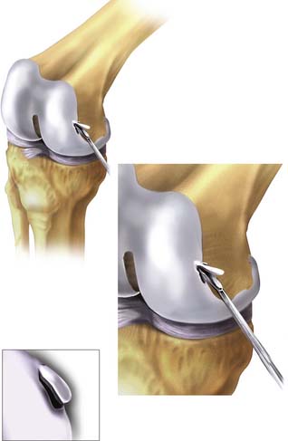
The biopsy for MACI is similar to other ACI techniques and may be taken from the median ridge at the level of the sulcus terminalis, the distal trochlea in the intercondylar region, or the surface of a fresh osteochondral fragment (see also Chapters 9C and 9D).
Using the small notchplasty gouge, a suitable-sized volume of cartilage is “scored” as deep as the subchondral plate. Using the gouge or ring curette, the biopsy is partially levered away from the subchondral plate of bone and finally grasped with pituitary rongeurs, immediately placed in culture medium (Fig. 9E-1), and transported to the laboratory for cell multiplication preparation.
Briggs et al.7 have demonstrated that surface cartilage of osteochondral fragments produces chondrocytes of good quality.
MACI Implantation
For open MACI implantation, the patient is positioned supine with a midthigh tourniquet and foot braces to position the knee in 90 and 120 degrees of flexion, respectively. A side support helps to control the leg in flexion.
< div class='tao-gold-member'>
Stay updated, free articles. Join our Telegram channel

Full access? Get Clinical Tree


