Chapter 9C Autologous Chondrocyte Implantation
ACI First and Second Generation
Introduction
Autologous chondrocyte implantation (ACI) is another means of repairing damaged cartilage.1 It is based on harvest of 200 to 300 mg of cartilage from a less loaded area in the knee. The cartilage is sent to a lab for processing. The cartilage is digested; the isolated chondrocytes are expanded in vitro during 2 to 3 weeks. The expanded final amount of cells is re-sent to the doctor as a suspension. The cells are to be injected into the defect covered with a membrane, periosteum (first generation), or collagen membrane (second generation).
Technical Overview
First-Stage Operation
The knee joint is examined by normal arthroscopy. A cartilage lesion is detected, and a decision is made to treat the defect by ACI. Often such a decision is made based on an earlier treatment that failed.1,2,3
Cartilage Harvest
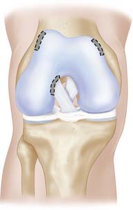
The cartilage is harvested from a less loaded weight-bearing area such as the upper medial femoral condylar area or similar on the lateral upper femoral trochlear area. Also the notch area is easy to use for harvest (Fig. 9C-1).
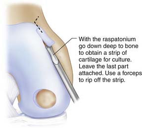
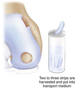
Use a ring curette or sharp gouge like a Wiberg raspatorium. Go deep down to cartilage-bone junction and leave a thin attachment of the strip. This strip may then be grasped with a forceps and ripped off with a gentle movement and taken through the portal to be put into a tube with saline. You may need two to three similar strips (Figs. 9C-2 and 9C-3).
Blood test tubes and harvested cartilage are sent to the cell laboratory that the hospital has a contract with.
Second-Stage Operation
The knee joint is opened by a miniarthrotomy.The cartilage defect is debrided (see Chapter 3). The debridement should result in a well-delineated defect, slightly oval.
< div class='tao-gold-member'>
Stay updated, free articles. Join our Telegram channel

Full access? Get Clinical Tree


