Autologous Chondrocyte Implantation

Chapter 35 Autologous Chondrocyte Implantation


INTRODUCTION


The ancient Greeks acknowledged ulcerated cartilage as troublesome to treat, with little to no natural healing capacity. Articular cartilage lesions have for a long time been undiagnosed and their severity underestimated. The increased surgical approach to ligament injuries, along with the introduction of arthroscopy and magnetic resonance imaging (MRI), has increased the diagnostic possibilities for articular cartilage lesions and activated the search for an improved treatment. Articular cartilage lesions are much more common than earlier recognized.


Anterior cruciate ligament (ACL) and meniscus injuries are often combined with articular cartilage injuries, in which the cartilage injury is the most serious and most difficult to treat in the end. Noyes and coworkers18,19 and Shelbourne and associates24 found that acute and chronic ACL injuries were associated with articular cartilage injuries in 40% to 70% of knees. There are reports that in cases of meniscus injury, 40% to 50% of patients also have cartilage damage.25 Hjelle and colleagues11 reported on 1000 consecutive patients with symptoms requiring arthroscopy. Chondral or osteochondral lesions of any type were found in 61% of the patients. Curl and coworkers5 found that in 31,516 patients who underwent arthroscopy, 63% had a cartilage lesion and 19% had an Outerbridge grade IV cartilage lesion. Bone bruises have been reported in acute knee injuries12 and in combination with ACL injuries in 80%.17 The damage to the trabecular bone may heal, but the overlying cartilage may degrade over time.


Since the early 1980s different treatments for full-thickness cartilage lesions have been developed that show promise. Some treatments have been discarded whereas others have been further developed. However, no “gold standard” is currently established and injuries to articular cartilage are still difficult to treat. Without proper management, articular cartilage injuries may degrade through enzymatic and mechanical breakdown into post-traumatic osteoarthritis.


Autologous chondrocyte implantation (ACI) has been in clinical practice since 1987, with over 25,000 patients treated worldwide. ACI was approved by the U.S. Food and Drug Administration in 1997.


INDICATIONS


ACI is indicated for full-thickness cartilage lesions or osteochondral lesions, including osteochondritis dissecans (OCD) in the knee (International Cartilage Repair Society [ICRS] or Outerbridge classification grade III or IV). The size of the lesion should be between 2 and 16 cm2, but larger lesions and multiple lesions can be treated in the same joint. The lesions should be situated on either the patella, the femur, or the tibia, and the opposing articular surface should be undamaged or have only superficial cartilage damage. Bone-to-bone conditions between the tibia and the femur or between the trochlea and the patella can be tried as a salvage procedure in young patients. The age recommendation is between 15 and 55 years, but there is no definite limit. Ligamentous instability, varus or valgus deformities, patella malalignment or instability, meniscus deficiency, and bone pathology or defect are not contraindications, but these must be addressed in the treatment.


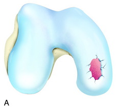
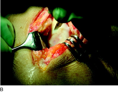
The recognition of serious articular cartilage injuries, including bipolar lesions in young and middle-aged individuals, has along with increased experience widened the indications for ACI (Fig. 35-1).


CLINICAL EVALUATION


INTRAOPERATIVE EVALUATION


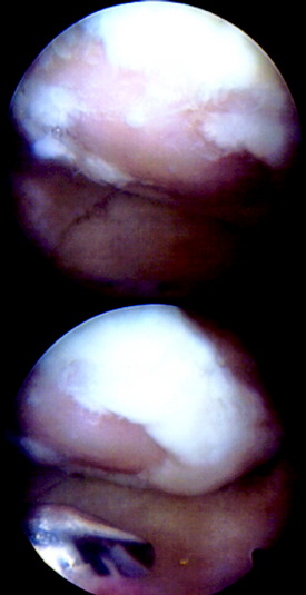
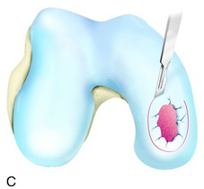
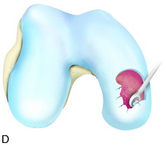
During the first-stage arthroscopy, knee stability is reassessed under anesthesia. The lesion is examined to decide whether it meets the indications for ACI. The lesion should be surrounded by healthy cartilage (but uncontainment is not a contraindication) and the opposing articular surface should be undamaged or have only minor superficial cartilage injury (Fig. 35-2). All intra-articular structures should be examined. The location, size, and depth should be noted using, for instance, the ICRS Knee Evaluation Package. Slices of cartilage with subchondral bone are harvested with a curet. The recommended harvest sites are the medial and lateral proximal rim of the femoral trochlea and the lateral aspect of the intercondylar notch in the knee joint. In more than 1400 patients who have had cartilage removed from the proximal medial trochlea for cell culturing, no complications or late symptoms have occurred from the donor site area. An optimal harvesting of cartilage is of great importance for the success of the cell culturing. Optimal cell quality is necessary for a successful result of this procedure and should be performed according to good laboratory practice or national regulations if present.


Other pathology within the knee should also be noted. If present, loose bodies are removed before the cartilage harvest. Meniscal tears are treated after the cartilage biopsy has been harvested. Always consider the possibility of meniscus repair, especially in young patients.


OPERATIVE TECHNIQUE


Either general or spinal anesthesia is used, and a tourniquet-controlled blood field is optional. A central straight skin incision is used. If previous incisions are present, these are used if possible to avoid further scar formation. The cartilage injury is approached through either a medial or a lateral arthrotomy depending on the location of the lesion and concomitant procedures, and the arthrotomy is adjusted for adequate access to the lesion. When the lesion is difficult to reach, the patella may have to be dislocated. In posterior femoral and tibial lesions, the anterior meniscus attachment has to be released or the medial or lateral collateral ligament femoral insertion detached with a bone block. The injured area is excised with vertical edges including all damaged cartilage and débrided without causing any bleeding from the subchondral bone. This avoids possible contamination with fibroblasts and undifferentiated stem cells from the bone marrow. Intralesional osteophytes may be present as a result of an intrinsic healing attempt or a previous microfracture or drilling procedure. Smaller osteophytes are carefully tapped down to the level of the subchondral bone plate, whereas larger and prominent osteophytes may be carefully curetted down to the bone plate. Surprisingly, there is very little bleeding from the curettage. If bleeding is present, use an epinephrine sponge or a drop of fibrin glue to stop the bleeding. Avoid electrocautery to prevent any necrosis in the bone.


The length and width of the defect is measured, and a template of the lesion is made with sterile paper or aluminum foil. Through a separate incision on the upper medial tibia, below the pes anserinus and the medial collateral ligament insertion, the periosteum is dissected free from overlying fascia, fat, fibrous tissue, and crossing vessels. A periosteal flap of the correct size and form is excised using a template, but because of shrinkage and room for suturing, the flap is oversized with 1 to 2 mm in the periphery. The flap with intact cambium layer is gently dissected from the cortical bone with a periosteal elevator. The periosteal flap should be as thin as possible and transparent, which will give more volume in the defect, allowing diffusion of synovial fluid and the cells to spread and expand and produce hyaline matrix. A thin flap also reduces the risk of periosteal complications. With increased age and inactivity, the periosteum atrophies and can become so thin that it is impossible to harvest. In smokers and obese patients, the quality of the periosteum is also decreased. The periosteum is thicker and more fibrous on the medial and lateral femoral condyle, proximal to the articular surface, and covered with a rich vascular network with risk for bleeding. This can lead to postoperative hematoma with swelling, adhesions, and arthrofibrosis. Therefore, the distal shaft of the femur is a better option as a second region for periosteal flap harvesting.


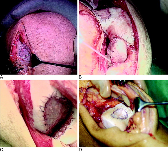
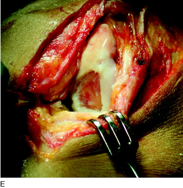
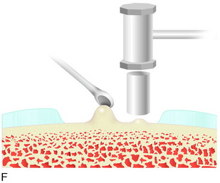
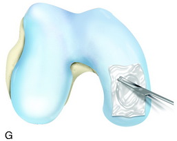
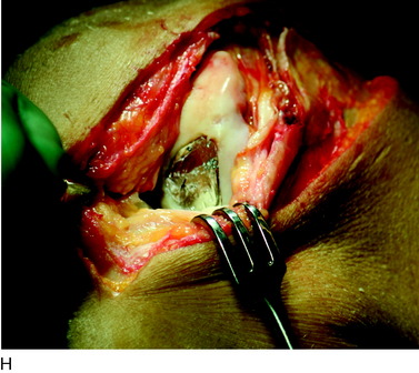
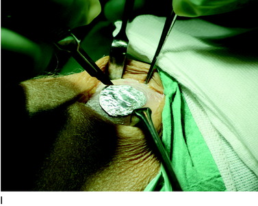
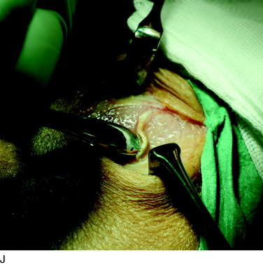
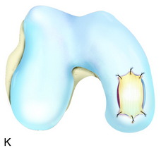
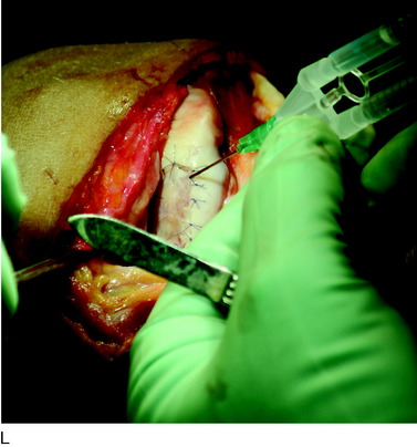
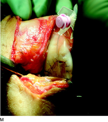
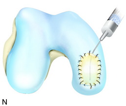
The flap is sutured to the cartilage rim of the defect at the level of the surrounding cartilage and the periosteum-cartilage border is sealed with fibrin glue. A gentle saline injection under the flap reveals any leakage along the cartilage rim, which must be sealed. After aspiration of the saline, the cultured chondrocytes are injected underneath the periosteal cover and the injection site is sutured and sealed (Fig. 35-3). The arthrotomy is closed in separate layers. Bandaging including the foot, lower leg, and knee is applied.


< div class='tao-gold-member'>

Stay updated, free articles. Join our Telegram channel

Jun 22, 2016 | Posted by in PHYSICAL MEDICINE & REHABILITATION | Comments Off on Autologous Chondrocyte Implantation

Full access? Get Clinical Tree

Get Clinical Tree app for offline access