Assorted Cases
WESTERN WISDOM
A diamond is a chunk of coal that made good under pressure.
CASE 1: ANTEROINFERIOR GLENOID FRACTURE WITH LARGE BONE FRAGMENT
History:
- 45-year-old man
- Fell 6 feet from ladder
- Taken to local ER
- Anterior dislocation reduced, but shoulder repeatedly redislocates
Exam:
- Neurovascular status intact
- Pain with any range of motion of shoulder
Imaging:
- X-rays show large anteroinferior glenoid fracture.
- 3-D CT images show a large main fracture fragment with some surrounding comminution.
Arthroscopic Findings:
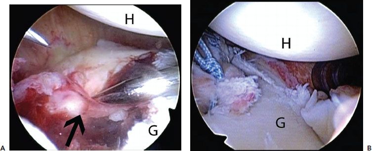
- Depressed fracture fragment that can be elevated to anatomic position (Fig. 28.1A)
Procedures Performed:
- Arthroscopic reduction and internal fixation with two screws
- Arthroscopic labral repair (Fig. 28.1B)
Key Points:
- To elevate the fracture fragment, one must partially detach the labrum from the margins of the fragment. In addition, the labral detachment must be medialized enough to allow for insertion of the screws. However, enough soft tissue attachments should be left intact so that the fragment is not devascularized.
- If the screw heads are “proud,” advance the capsulolabral sleeve to the main glenoid to “pad” the screw heads, and schedule arthroscopic removal of the screws at 6 to 9 months post-op (after the fracture has healed).
CASE 2: LOOSE AND FRACTURED GLENOID COMPONENT IN TOTAL SHOULDER REPLACEMENT
History:
- 61-year-old male self-employed machinist and lathe operator
- Had total shoulder replacement 21 years earlier for posttraumatic arthritis following a glenoid fracture (sustained in a motor vehicle accident)
- Had some mild aching pain the past 2 years
- Felt a painful pop in his shoulder 2 days ago while lifting a steel rod
- Owns his own machine-shop business and says he cannot afford to take off work more than 3 days
Exam:
- Pain with any shoulder motion
- Unable to elevate the arm >60° due to pain

Figure 28.1 A: Right shoulder, anterosuperolateral viewing portal, demonstrates a large bony Bankart fracture (blackarrow). B: Appearance postscrew fixation and capsulolabral repair. G, glenoid; H, humerus.
Imaging:
- X-rays show loosening and fragmentation of polyethylene and cement of glenoid component; humeral component is intact.
- CT scan confirms the x-ray impression.
Arthroscopic Findings:
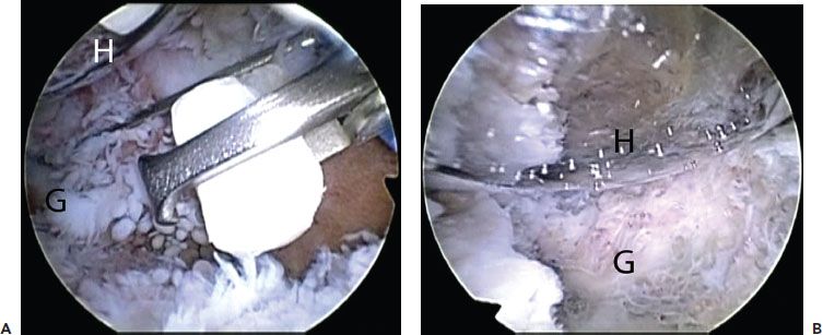
- Multiple loose fragments of polyethylene and methyl methacrylate cement (Fig. 28.2A)
Procedures Performed:
- Arthroscopic excision of loose fragments, debridement of fibrous interface, and contouring of glenoid (Fig. 28.2B)
Key Points:
- Large fragments of polyethylene can be reduced in size by using a ¼ or ⅜ ̋ osteotome, thereby facilitating removal through arthroscopic portals.
- Glenoid component removal is the ultimate salvage procedure for a failed total shoulder replacement, so it seemed to be the only alternative in this patient whose main goal was to return to his heavy occupation as soon as possible; he returned to regular activity as a machinist 2 days post-op.

Figure 28.2 A: Left shoulder, anterosuperolateral viewing portal, demonstrates removal of a loose polyethylene fragment in an individual with a previous total shoulder replacement. B: Appearance postarthroscopic removal of polyethylene component and debris. G, glenoid; H, humerus.
Stay updated, free articles. Join our Telegram channel

Full access? Get Clinical Tree


