Assessment of Gait
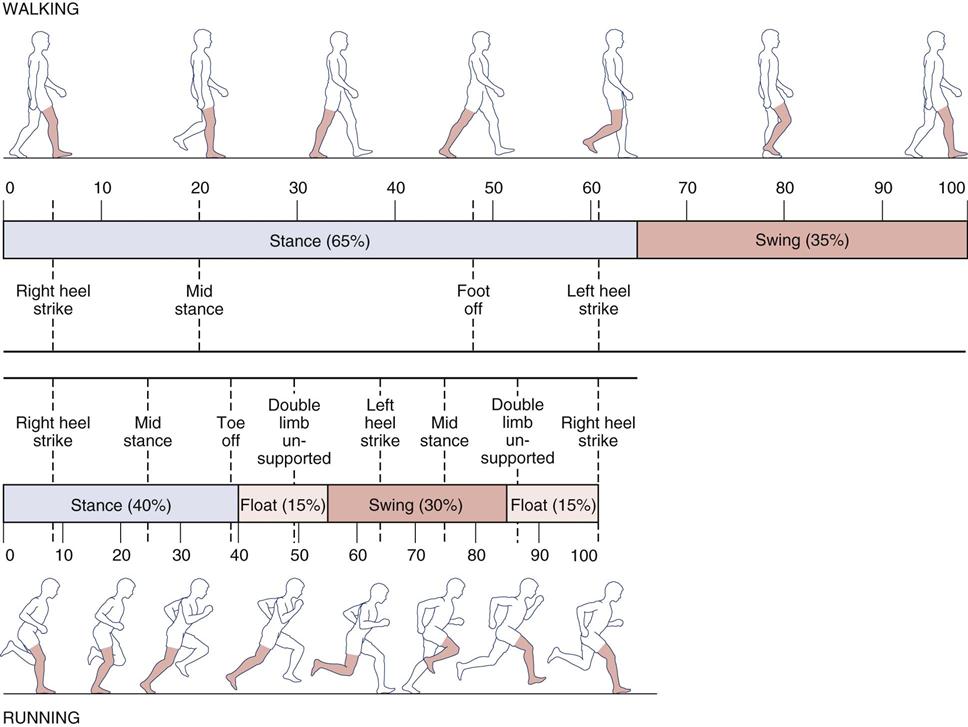
Walking is the simple act of falling forward and catching oneself. One foot is always in contact with the ground, and within a cycle, there are two periods of single-leg support and two periods of double-leg support. With running, there is a period of time during which neither foot is in contact with the ground, a period called “double float.”
Winter1 felt walking gait performs five main functions. First, it helps to support of the head, arms, and trunk by maintaining a semirigid lower limb. Second, it helps to maintain upright posture and balance. Third, it controls the foot to allow it to clear obstacles and enables gentle heel or toe landing through eccentric muscle action. Fourth, it generates mechanical energy by concentric muscle contraction to initiate, maintain, and, if desired, increase forward velocity. Finally, through eccentric action of the muscles, it provides shock absorption and stability and decreases forward velocity of the body.
The locomotion pattern tends to be variable and irregular until about the age of 7 years.2 Several functional tasks are involved in gait, including forward progression, which is executed in a stepping movement in a wide range of rapid and comfortable walking speeds. Second, the body must be balanced alternately on one limb and then the other; this is accompanied by repeated adjustments of limb length. Finally, there is support of the upright body.
Gait assessment or analysis takes a great deal of time, practice, and technical skill combined with standardization for the clinician to develop the necessary skills.3–5 Most gait analysis today is performed with force platforms to measure ground reaction forces, electromyography to measure muscle activity, and high-speed video motion analysis systems to measure movement. Discussion of these techniques, however, is beyond the scope of this book. This chapter gives only a brief overview of a complex task, assessment of normal and pathological gait; detailed assessment of gait is left to other authors.6–15 The various terms commonly used to describe gait, the normal pattern of gait, the assessment of gait, and common abnormal gaits are reviewed.
Definitions5–10
Gait Cycle
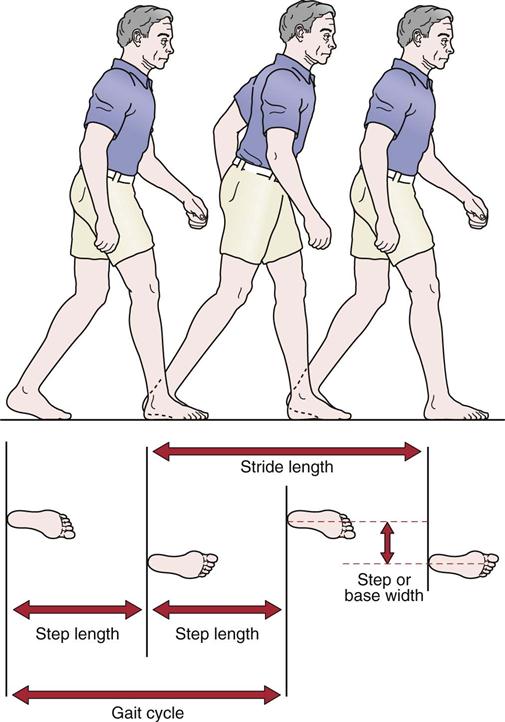
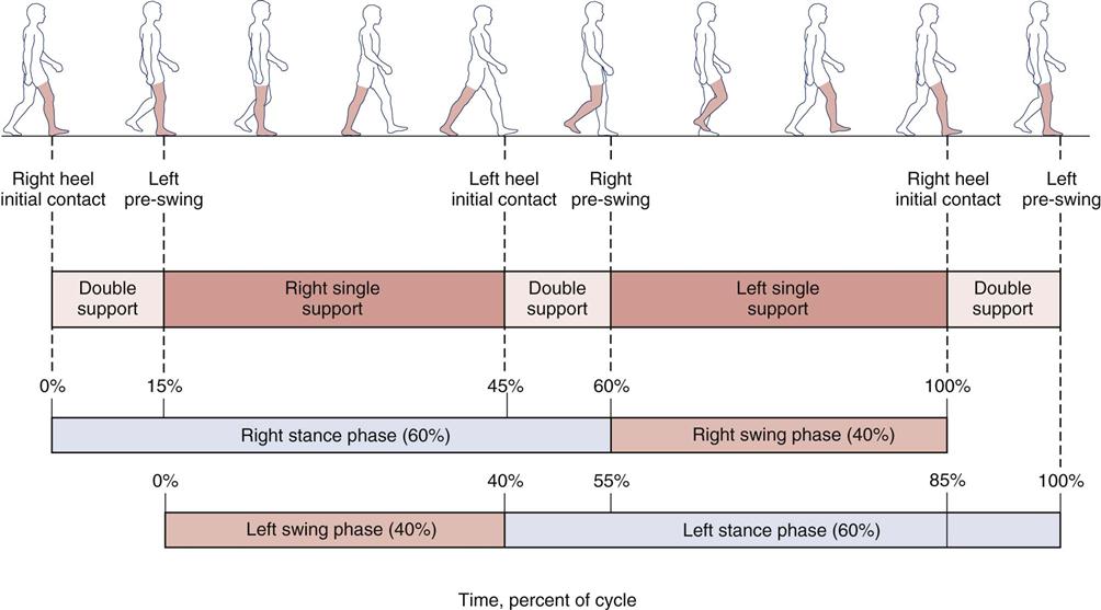
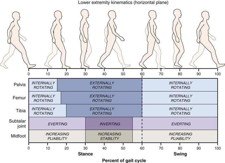
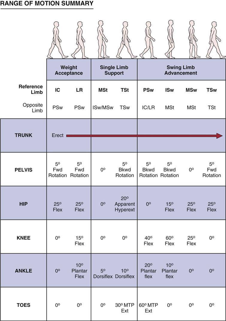
The gait cycle is the time interval or sequence of motions occurring between two consecutive initial contacts of the same foot (Figure 14-1). It is synonymous with the stride length. For example, if heel strike is the initial contact, the gait cycle for the right leg is from one heel strike to the next heel strike on the same foot. The gait cycle is a description of what happens in one leg. The same sequence of events is repeated with the other leg, but it is 180° out of phase.8 There are spatial descriptors of gait, such as stride length, step length and step width; time or temporal descriptors, such as cadence, stride time and step time; and, descriptors that involve time and space, such as walking speed.16 Another spatial descriptor that is sometimes discussed with gait is foot angle (Fick angle; see Figure 14-14). Each of these descriptors can and should be very similar for both limbs. For example, osteoarthritis in one hip can change many of the descriptors and the examiner should watch for these changes. Simoneau17 clearly described the terminology that applies to the gait cycle events (Figure 14-2). Table 14-1 demonstrates the periods or phases of the gait cycle, the function of each phase, and what is happening in the opposite limb.8 The gait cycle consists of two phases for each foot: stance phase, which makes up 60% to 65% of the walking cycle, and swing phase, which makes up 35% to 40% of the walking cycle. In addition, there are two periods of double support and one period of single-leg stance during the gait cycle.
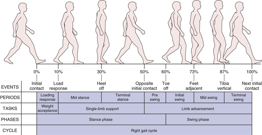
Initial contact corresponds to the beginning of stance when the foot first contacts the ground at 0% of gait cycle. Load response occurs when the contralateral foot leaves the ground at 10% of gait cycle. Heel off corresponds to the heel lifting from the ground and occurs at approximately 30% of gait cycle. Opposite initial contact corresponds to the foot contact of the opposite limb, typically at 50% of gait cycle. Toe off occurs when the foot leaves the ground at 60% of gait cycle. Feet adjacent takes place when the foot of the swing leg is next to the foot of the stance leg at 73% of gait cycle. Tibia vertical corresponds to the tibia of the swing leg being oriented in the vertical direction at 87% of gait cycle. The final event is, again, initial contact, which in fact is the start of the next gait cycle. These eight events divide the gait cycle into seven periods. Loading response, between initial contact and opposite toe off, corresponds to the time when the weight is accepted by the lower extremity, initiating contact with the ground. Midstance is from opposite toe off to heel rise (10% to 30% of gait cycle). Terminal stance begins when the heel rises and ends when the contralateral lower extremity touches the ground, from 30% to 50% of gait cycle. Preswing takes place from foot contact of the contralateral limb to toe off of the ipsilateral foot, which is the time corresponding to the second double-limb support period of the gait cycle (50% to 60% of gait cycle). Initial swing is from toe off to feet adjacent, when the foot of the swing leg is next to the foot of the stance leg (60% to 73% of gait cycle). Midswing is from feet adjacent to when the tibia of the swing leg is vertical (73% to 87% of gait cycle). Terminal swing is from a vertical position of the tibia to immediately before heel contact (87% to 100% of the gait cycle). The first 10% of the gait cycle corresponds to a task of weight acceptance—when body mass is transferred from one lower extremity to the other. Single-limb support, from 10% to 50% of the gait cycle, bears the weight of the body as the opposite limb swings forward. The last 10% of stance phase and the entire swing phase advance the limb forward to a new location. (Modified from Simoneau GG: Kinesiology of walking. In Neumann DA, editor: Kinesiology of the musculoskeletal system: foundations of physical rehabilitation, ed 2, St Louis, 2010, Mosby, p. 636.)
TABLE 14-1
Gait Cycle: Periods and Functions
| Period | Percentage of Cycle | Function | Contralateral Limb |
| Initial double limb support | 0 to 12 | Loading, weight transfer | Unloading and preparing for swing (preswing) |
| Single limb support | 12 to 50 | Support of entire body weight: center of mass moving forward | Swing |
| Second double limb support | 50 to 62 | Unloading and preparing for swing (preswing) | Loading, weight transfer |
| Initial swing | 62 to 75 | Foot clearance | Single limb support |
| Midswing | 75 to 85 | Limb advances in front of body | Single limb support |
| Terminal swing | 85 to 100 | Limb deceleration, preparation for weight transfer | Single limb support |
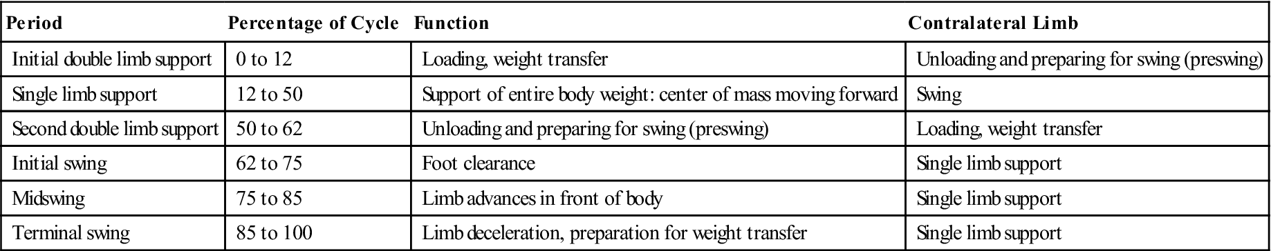
From Sutherland DH, et al: Kinematics of normal human walking. In Rose J, Gamble JG, editors: Human locomotion, Baltimore, 1994, Williams & Wilkins, p. 27.
As the velocity of the cycle increases, the cycle length or stride length decreases. For example, in jogging, the gait cycle is 70% of the walking cycle, and in running, the gait cycle is 60% that of walking.18 In addition, as the speed of movement increases, the function of the muscles changes somewhat, and their electromyographic activity may increase or decrease. Generally, gait velocity decreases with age.19 Montero-Odasso et al.20 found the gait velocity (less than 0.8 m/sec) could be used to determine mobility impairment in the elderly.
Stance Phase
The stance phase of gait occurs when the foot is on the ground and bearing weight (Figure 14-3). It allows the lower leg to support the weight of the body and, by so doing, acts as a shock absorber while allowing the body to advance over the supporting limb.18 Normally, this phase makes up 60% of the gait cycle and consists of five subphases, or instants.
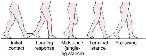
The initial contact instant is the weight-loading or weight acceptance period of the stance leg, which accounts for the first 10% of the gait cycle. During this period, one foot is coming off the floor while the other foot is accepting body weight and absorbing the shock of initial contact. Because both feet are in contact with the floor, it is a period of double support or double-leg stance.
The load response and midstance instants consist of the single support or single-leg stance, which accounts for the next 40% of the gait cycle. During this period, one leg alone carries the body weight while the other leg goes through its swing phase. The stance leg must be able to hold the weight of the body, and the body must be able to balance on the one leg. In addition, lateral hip stability must be exhibited to maintain balance, and the tibia of the stance leg must advance over the stationary foot.
The terminal stance and preswing instants make up the weight-unloading period, which accounts for the next 10% of the gait cycle. During this period, the stance leg is unloading the body weight to the contralateral limb and preparing the leg for the swing phase. As with the first two instants, both feet are in contact, and so double support occurs for the second time during the gait cycle.
Swing Phase
The swing phase of gait occurs when the foot is not bearing weight and is moving forward (Figure 14-4). The swing phase allows the toes of the swing leg to clear the floor and allows for leg length adjustments. In addition, it allows the swing leg to advance forward. It makes up approximately 40% of the gait cycle and consists of three subphases.
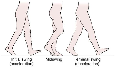
Acceleration occurs when the foot is lifted off the floor. During normal gait, rapid knee flexion and ankle dorsiflexion occur to allow the swing limb to accelerate forward. In some pathological conditions, loss or alteration of knee flexion and ankle dorsiflexion leads to alterations in gait.
The midswing instant occurs when the swing leg is adjacent to the weight-bearing leg, which is in midstance.
During the final instant (terminal swing or deceleration), the swinging leg slows down in preparation for initial contact with the floor. With normal gait, active quadriceps and hamstring muscle actions are required. The quadriceps muscles control knee extension, and the hamstrings control the amount of hip flexion.
During running or with increased velocity, the stance phase decreases and a float phase or double unsupported phase occurs while the double support phase disappears (Figure 14-5).18,21 Although the single-leg stance phase decreases, the load increases two or three times.22 The motion occurring at each of the joints (pelvis, hip, knee, ankle) is similar for walking and for running, but the required range of motion (ROM) increases with the speed of the activity. For example, hip flexion in walking is about 40° to 45°, whereas in running it is 60° to 75°.23
Double-Leg Stance
Double-leg stance is that phase of gait in which parts of both feet are on the ground. In normal gait, it occurs twice during the gait cycle and represents about 25% of the cycle. This percentage increases the more slowly one walks; it becomes shorter as walking speed increases (Figure 14-6) and disappears in running.
Single-Leg Stance
The single-leg stance phase of gait occurs when only one leg is on the ground; this occurs twice during the normal gait cycle and takes up approximately 30% of the cycle.
Normal Parameters of Gait7–11,24
The parameters that follow and their values are considered normal for a population between the ages of 8 and 45 years. It should be pointed out, however, that a relatively normal gait pattern is seen in persons as young as 3 years of age.2 There are, however, differences between individuals of the same sex and between men and women.25 For the majority of the population outside of these ages, there are alterations caused by neurological development, balance control, aging, changes in limb length, and maturation.2 For example, with maturity, walking velocity and step length increase, and cadence decreases.26 It is also important to evaluate gait on the basis of normal gait for someone the same age. This is especially true for children.
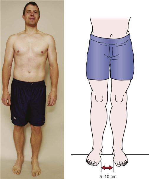
Base (Step) Width
The normal base width, which is the distance between the two feet, is 5 to 10 cm (2 to 4 inches; Figure 14-7). If the base is wider, the examiner may suspect some pathology (e.g., cerebellar or inner ear problems) that results in poor balance, a condition such as diabetes or peripheral neuropathy that may indicate a loss of sensation, or a musculoskeletal problem (e.g., tight hip abductors). In the first two cases, the patient tends to have a wider base to maintain balance. With increased speed, the base width normally decreases to zero, and in some cases, crossover occurs, in which one foot lands where the other should and vice versa. Such crossover can lead to gait alterations and other problems.27
Step Length
Step length, or gait length, is the distance between successive contact points on opposite feet (see Figure 14-1). Normally, this distance is about 72 cm (28 inches) being relatively constant for each individual (i.e., step length is commonly related to preferred walking speed)17,28 and should be equal for both legs. It varies with age and sex with children taking smaller steps than adults and females taking smaller steps than males.22 Height also has an effect: a taller person takes larger steps. Step length tends to decrease with age, fatigue, pain, and disease. If step length is normal for both legs, the rhythm of walking is smooth. If there is pain in one limb, the patient attempts to take weight off that limb as quickly as possible, altering the rhythm.
Stride Length
Stride length is the linear distance in the plane of progression between successive points of foot-to-floor contact of the same foot. The stride length is normally about 144 cm (56 inches) and in reality is one gait cycle.17 Stride length, like step length, decreases with age, pain, disease, and fatigue.19,29 The age changes are often the result of decreased walking pace or speed.29,30
Lateral Pelvic Shift (Pelvic List)
Lateral pelvic shift, or pelvic list, is the side-to-side movement of the pelvis during walking. It is necessary to center the weight of the body over the stance leg for balance (Figure 14-8). The lateral pelvic shift is normally 2.5 to 5 cm (1 to 2 inches). It increases if the feet are farther apart. The pelvic list causes relative adduction of the weight-bearing limb, facilitating the action of the hip adductors. If the abductor muscles are weak, a Trendelenburg gait results (see Figure 14-21).
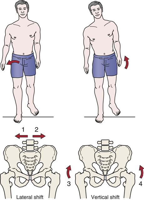
Vertical Pelvic Shift
Vertical pelvic shift keeps the center of gravity from moving up and down more than 5 cm (2 inches) during normal gait. By means of a vertical pelvic shift, the high point occurs during midstance and the low point during initial contact; the height of these points may increase during the swing phase if the knee is fused or does not bend because of protective spasm or swelling. The head is never higher during normal gait than it is when the person is standing on both feet. Therefore, if a person can stand in an opening, he or she should be able to move through the opening without hitting the head.7 On the swing phase, the hip is lower on the swing side, and the patient must flex the knee and dorsiflex the foot to clear the toe. This action shortens the extremity length at midstance and decreases the center of gravity rise.
Pelvic Rotation
Pelvic rotation is necessary to lessen the angle of the femur with the floor, and, in so doing, it lengthens the femur (Figure 14-9). The rotation decreases the amplitude of displacement along the path traveled by the center of gravity and thereby decreases the center-of-gravity dip. There is a total of 8° pelvic rotation with 4° forward on the swing leg and 4° posteriorly on the stance leg. To maintain balance, the thorax rotates in the opposite direction. When the pelvis rotates clockwise, the thorax rotates counterclockwise, and vice versa. These concurrent rotations provide counter-rotation forces and help regulate the speed of walking.
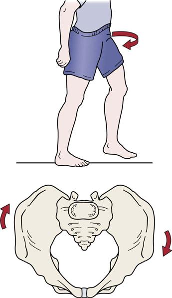
In the lower limb, rotation is evident at each joint (Figure 14-10). The farther the joint is from the trunk, the greater the amount of rotation. For example, rotation in the tibia is three times greater than rotation in the pelvis.7
Center of Gravity
Normally, in the standing position, the center of gravity is 5 cm (2 inches) anterior to the second sacral vertebra; it tends to be slightly higher in men than in women because men tend to have a greater body mass in the shoulder area. The vertical and horizontal displacements of the center of gravity describe a figure-eight, occupying a 5-cm (2-inch) square within the pelvis during walking. The vertical displacement, which describes a smooth sinusoidal curve during walking, can be observed from the side. The patient’s head descends during weight-loading and weight-unloading periods and rises during single-leg stance.
Normal Cadence
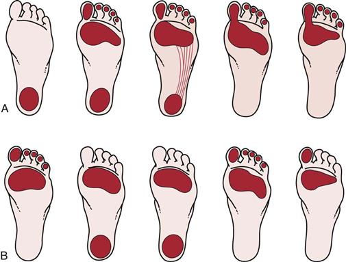
The normal cadence is between 90 and 120 steps per minute which varies, in part, because of the height of the individual.31–34 The cadence of women is usually six to nine steps per minute higher than that of men.33 With age, the cadence decreases. Figure 14-11, A, illustrates the cadence of normal gait from heel strike to toe off showing the changing weight distribution. With pathology or deformity (e.g., a cavus foot [Figure 14-11, B]), this weight-bearing pattern may be altered. As the pace of walking increases, the stride width increases, and the toeing-out angle decreases. Gait speed is about 1.4 m/sec (3 mph).17
Normal Pattern of Gait6–11,17,31,35,36
Stance Phase
As previously mentioned, there are five instants involved during the stance phase of gait. These are now described in order of occurrence. This phase is the closed kinetic chain phase of gait. The action occurring at the various joints causes a chain reaction because of the stresses put on the joints and supporting structures with weight-bearing. The foot becomes the fixed stable segment, and alterations occur from the foot up with the joints of the foot adapting first, followed by those of the ankle, knee, hip, pelvis, spine, and finally the upper limb, which acts as a counterbalance to movement in the lower limb.37 The relations between the joints are constantly changing. Table 14-2 summarizes the movement at the hip, knee, ankle, and foot during the stance phase.38
TABLE 14-2
| Hip | |||
| Kinematic Motion | KINETIC MOTION | ||
| Phase | Hip | External Forces | Internal Forces |
| Heel strike | 20° to 40° of hip flexion moving toward extension; slight adduction and lateral rotation | Reaction force in front of joint; flexion moment moving toward extension; forward pelvic rotation | Gluteus maximus and hamstrings working eccentrically to resist flexion moment; erector spinae working eccentrically to resist forward bend |
| Foot flat | Hip moving into extension, adduction, medial rotation | Flexion moment | Gluteus maximus and hamstrings contracting concentrically to bring hip into extension; erector spinae resisting trunk flexion |
| Midstance | Moving through neutral position; pelvis rotating posteriorly | Reaction force posterior to hip joint; extension moment | Iliopsoas working eccentrically to resist extension; gluteus medius contracting in reverse action to stabilize opposite pelvis; iliopsoas activity continuing |
| Heel off | 10° to 15° extension of hip abduction, lateral rotation | Extension moment decreasing after double-limb support begins | |
| Toe off | Moving toward 10° extension, abduction, lateral rotation | Decrease of extension moment | Adductor magnus working eccentrically to control or stabilize pelvis; iliopsoas activity continuing |
| Knee and Tibia | ||||
| KINEMATIC MOTION | KINETIC MOTION | |||
| Phase | Knee | Tibia | External Forces | Internal Forces |
| Heel strike | In full extension before heel contact; flexing as heel strikes floor | Slight lateral rotation | Rapidly increasing reaction forces behind knee joint causing flexion moment | Quadriceps femoris contracting eccentrically to control rapid knee flexion and to prevent buckling |
| Foot flat | In 20° flexion moving toward extension | Medial rotation | Flexion moment | After foot is flat, quadriceps femoris activity becoming concentric to bring femur over tibia |
| Midstance | In 15° flexion moving toward extension | Neutral | Maximum flexion moment | Quadriceps femoris activity decreasing; gastrocnemius working eccentrically to control excessive knee extension |
| Heel off | In 4° flexion moving toward extension | Lateral rotation | Reaction forces moving anterior to joint; extension moment | Gastrocnemius beginning to work concentrically to start knee flexion |
| Toe off | Moving from near full extension to 40° flexion | Lateral rotation | Reaction forces moving posterior to joint as knee flexes; flexion moment | Quadriceps femoris contracting eccentrically |
| Foot and Ankle | ||||
| KINEMATIC MOTION | KINETIC MOTION | |||
| Phase | Foot | Ankle | External Forces | Internal Forces |
| Heel strike | Supination (rigid) at heel contact | Moving into plantar flexion | Reaction forces behind joint axis; plantar flexion moment at heel strike | Dorsiflexors (tibialis anterior, extensor digitorum longus, and extensor hallucis longus) contracting eccentrically to slow plantar flexion |
| Foot flat | Pronation, adapting to support surface | Plantar flexion to dorsiflexion over a fixed foot | Maximum plantar flexion moment; reaction forces beginning to shift anterior, producing a dorsiflexion moment | Dorsiflexion activity decreasing; tibialis posterior, flexor hallucis longus, and flexor digitorum longus working eccentrically to control pronation |
| Midstance | Neutral | 3° of dorsiflexion | Slight dorsiflexion moment | Plantar flexor muscles (gastrocsoleus and peroneal muscles), activated to control dorsiflexion of the tibia and fibula over a fixed foot, contracting eccentrically |
| Heel off | Supination as foot becomes rigid for push-off | 15° dorsiflexion toward plantar flexion | Maximal dorsiflexion moment | Plantar flexor muscles beginning to contract concentrically to prepare for push-off |
| Toe off | Supination | 20° plantar flexion | Dorsiflexion moment | Plantar flexor muscles at peak activity but becoming inactive as foot leaves ground |
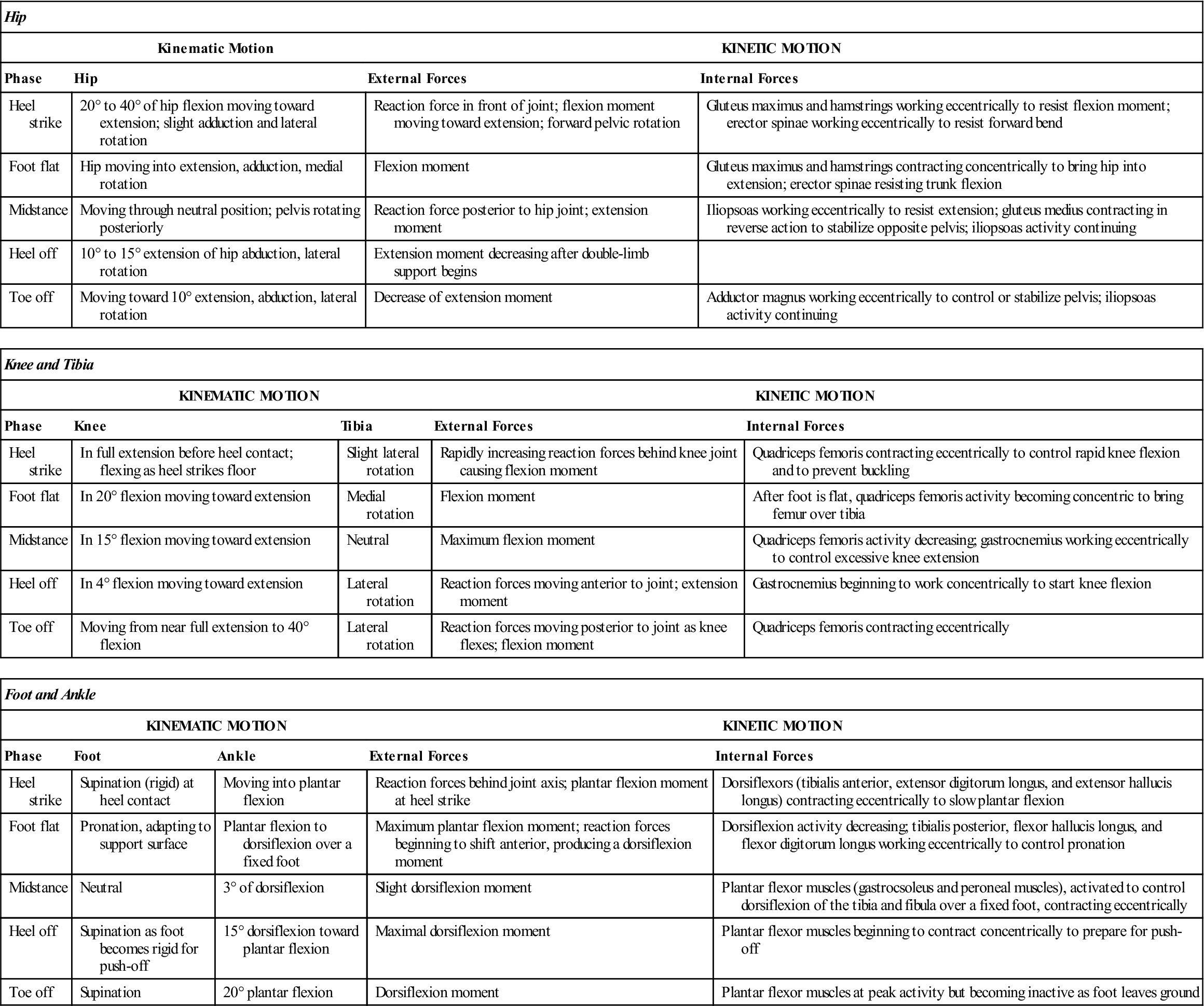
Modified from Giallonardo LM: Gait. In Myers RS, editor: Saunders manual of physical therapy practice, Philadelphia, 1995, WB Saunders, pp. 1108–1109.
Initial Contact (Heel Strike)
Initial contact occurs when the limb first strikes the ground. Normally, this occurs when the heel strikes and the limb is being prepared to take weight. During the initial contact, the pelvis is level and medially rotated on the side of initial contact, whereas the trunk is aligned between the two lower limbs. The hip is flexed 30° to 49° and is medially rotated; the knee is slightly flexed or extended; the tibia is laterally rotated; the ankle is at 90° with the foot supinated; and the hindfoot is everted. At this instant, there is little force going through the limb.
If pain occurs in the heel at this time, it may be caused by a heel spur, bone bruise, heel fat-pad bruise, or bursitis. This pain may cause increased flexion of the knee with early plantar flexion to relieve the stress or pressure on the painful tissues. If the knee is weak, the patient may extend the knee by using the hand or may hit the heel hard on the ground to whip the knee into extension. A patient may do this because of weakness of the muscles (e.g., reflex inhibition, poliomyelitis, an internal derangement of the knee, a nerve root lesion [L2, L3, or L4], femoral neuropathy). In the past, this instant was referred to as “heel strike;” however, with some pathological gaits, heel strike may not be the first instant. Instead, the toes, the forefoot, or the entire foot may initially contact the ground. If the dorsiflexor muscles are weak, the foot drops, slaps, or flops down. The weakness may be caused by a peroneal neuropathy or nerve root lesion (L4). A knee flexion contracture or spasticity may cause the same alteration.
Load Response (Weight Acceptance or Foot Flat)
Load response is a critical event in that the person subconsciously decides whether the limb is able to bear the weight of the body. The trunk is aligned with the stance leg. The pelvis drops slightly on the swing leg side and medially rotates on the same side. The flexed and laterally rotated hip moves into extension, and the knee flexes 15° to 25°. The tibia is medially rotated and begins to move forward over the fixed foot as the body swings over the foot. The ankle is plantar flexed, and the hindfoot is inverted. The foot moves into pronation, because this position unlocks the foot and enables it to adapt to different terrains and postures. The forefoot is pronated, unlocking the subtalar and metatarsal joints to enable them to absorb the shock more effectively, and the plantar aspect is in contact with the floor.
Abnormal responses include excessive or no knee motion as a result of weak quadriceps, plantar flexor contractures, or spasticity.9
Midstance (Single-Leg Support)
The midstance instant is a period of stationary foot support. Normally, the weight of the foot is evenly distributed over the entire foot. The trunk is aligned over the stance leg, and the pelvis shows a slight drop to the swing leg side.
During this stage, there is maximum extension of the hip (10° to 15°) with lateral rotation, and the greatest force is on the hip. Painful hip, knee, or ankle conditions cause this phase to be shortened as the patient hurries through the phase to decrease the pain. If the gluteus medius (L5 nerve root) is weak, Trendelenburg sign is present. The knee flexes, and the ankle is locked at 5° to 8° of dorsiflexion, rolling forward on the forefoot (roll-off). The foot is in contact with the floor; the forefoot is pronated, and the hindfoot is inverted. This instant is a critical event for the ankle. If pain is elicited during this period, the phase is shortened and the heel may lift off early. This pain is commonly caused by conditions such as arthritis, rigid pes planus, fallen metatarsal or longitudinal arches, plantar fasciitis, or Morton metatarsalgia. Therefore, pathology at the hip, ankle, or knee can modify the gait in this phase.
Terminal Stance (Heel Off)
In the final stages, the trunk is initially aligned over the lower limbs and moves toward the stance leg. The pelvis is initially level and posteriorly rotated and then dips to the swing leg side, remaining posteriorly rotated. The heel is in neutral and slight medial rotation; the knee is extended with the tibia laterally rotated. At the ankle, plantar flexion occurs as the critical event. This action helps to smooth the pathway of the center of gravity. The forefoot is initially in contact with the floor, and then the weight on the foot moves forward with plantar flexion so that only the big toe is in contact with the floor. At the same time, the forefoot moves from inversion to eversion.
Preswing (Toe Off)
The preswing phase is the acceleration phase as the toe pushes the leg forward. The trunk remains erect, the pelvis remains posteriorly rotated, and the hip is extended and slightly medially rotated. The knee flexes to 30° to 35° (critical event), and the ankle is plantar flexed. Because the center of gravity is anterior to the hip, the hip can be accelerated forward in initial swing.
If pain is elicited during this instant, it may be caused by a hallux rigidus, turf toe, or any other pathology involving the great toe (hallux), especially the metatarsophalangeal joint of the hallux. With injury to the joint, the patient is unable to push off on the medial aspect of the foot; instead, the patient pushes off on the lateral aspect of the foot to compensate for the painful metatarsophalangeal joint or, in some cases, a painful metatarsal arch resulting from increased pressure on the metatarsal heads. If the plantar flexors are weak (e.g., S1–S2 nerve root pathology), push-off may be absent. During this phase, the foot pronates so that there is a rigid base for better push-off.
During walking, a cane can be used to decrease the load on the limb. Lyu and associates39 have shown that using a cane in the contralateral upper limb, if the cane tip touches the ground at the same time as the heel, can reduce the force at heel strike by 34%, by 25% at midstance, and about 30% at toe off.
Swing Phase
The swing phase of gait involves the lower limb in an open kinetic chain; the foot is not fixed on the ground, and the stresses on the limb are therefore less and easier to dissipate. During this phase, alterations occur from the spine down through the pelvis, hip, ankle, and foot. The pelvis and hip provide the most stability in the lower limb during the non–weight-bearing phase. Table 14-3 summarizes the motions occurring in the lower limb during the swing phase.
TABLE 14-3
| ACCELERATION TO MIDSWING | MIDSWING TO DECELERATION | |||
| Joint | Kinematic Motion | Kinetic Motion | Kinematic Motion | Kinetic Motion |
| Hip | Slight flexion (0° to 15°) moving to 30° flexion and lateral rotation to neutral | Hip flexors working concentrically to bring limb through; contralateral gluteus medius concentrically contracting to maintain pelvis position | Continued flexion at about 30° to 40° | Gluteus maximus contracting eccentrically to slow hip flexion |
| Knee | 30° to 60° knee flexion and lateral rotation of tibia moving toward neutral | Hamstrings concentrically contracting | Moving to near full extension and slight lateral tibial rotation | Quadriceps femoris contracting concentrically and hamstrings contracting eccentrically |
| Ankle and foot | 20° dorsiflexion and slight pronation | Dorsiflexors contracting concentrically | Ankle in neutral; foot in slight supination | Dorsiflexors contracting isometrically |
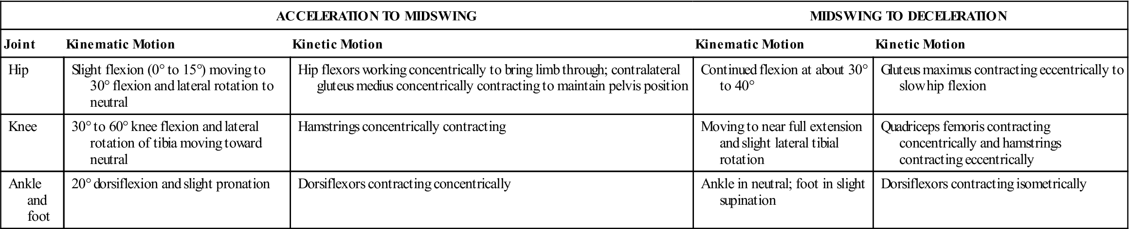
From Giallonardo LM: Gait. In Myers RS, editor: Saunders manual of physical therapy practice, Philadelphia, 1995, WB Saunders, p. 1110.
The three instants composing the swing phase of gait are now described in order of occurrence.
Initial Swing
During the first subphase of acceleration (Figure 14-12), flexion and medial rotation of the hip and flexion of the knee occur. The pelvis medially rotates and dips to the swing leg side. The trunk is aligned with the stance leg. In addition, the ankle continues to plantar flex. The foot is not in contact with the floor. The forefoot continues supinating, and the hindfoot continues everting. The dorsiflexor muscles of the ankle contract to allow the foot to clear the ground, and the knee exhibits its maximum flexion during gait of about 60°. If the quadriceps muscles are weak, the trunk muscles thrust the pelvis forward to provide forward momentum to the leg.
IC, Initial contact; ISw, initial swing; LR, load response; MSt, midstance; MSw, midswing; PSw, preswing; TSt, terminal stance; TSw, terminal swing. (Copyright 1991 LAREI, Rancho Los Amigos Medical Center, Downey, CA 90242; from The Pathokinesiology Service and The Physical Therapy Department, Rancho Los Amigos Medical Center: Observational Gait Analysis. Downey, CA, Los Amigos Research and Educational Institute, Inc., 1996, p. 30.)
Midswing
During the midswing instant, the hip continues to flex and medially rotate, and the knee continues to flex. The ankle is in the anatomical or plantigrade position (90°) for the first 25% of the stance phase to permit the foot and midtarsal joints to unlock so that the foot can adapt to uneven terrain when it begins weight-bearing. The forefoot is supinated, and the hindfoot is everted. The pelvis and trunk are in the same position as during the previous stage. If the ankle dorsiflexor muscles are weak (e.g., drop foot), the patient demonstrates a steppage gait (see Figure 14-27). In such a gait, the hip flexes excessively so that the toes can clear the ground.
Terminal Swing (Deceleration)
During the final subphase, the hip continues to flex and medially rotate, and the knee reaches its maximum extension. At the ankle, dorsiflexion has occurred. The forefoot is supinated, and the hindfoot is everted. The trunk and pelvis maintain the same position as before. The hamstring muscles contract during the terminal phase to slow the swing; if the hamstrings are weak (e.g., S1–S2 nerve root lesion), heel strike may be excessively harsh to lock the knee in extension.
Joint Motion During Normal Gait
Although there is a tendency to talk about gait as action around joints, the examiner must not forget that muscles play a significant role in what happens at the joints. Table 14-4 illustrates the actions of some of the muscles used during gait.40
TABLE 14-4
Muscle Actions During Gait Cycle
| Phase of Gait | Mechanical Goals | Active Muscle Groups | Examples |
| Stance Phase | |||
| Initial contact | Position foot, begin deceleration | Ankle dorsiflexors, hip extensors, knee flexors | Anterior tibialis, gluteus maximus, hamstrings |
| Loading response | Accept weight, stabilize pelvis, decelerate mass | Knee extensors, hip abductors, ankle plantar flexors | Vasti, gluteus medius, gastrocnemius, soleus |
| Midstance | Stabilize knee, preserve momentum | Ankle plantar flexors (isometric) | Gastrocnemius, soleus |
| Terminal stance | Accelerate mass | Ankle plantar flexors (concentric) |


