Arthroscopy for Symptomatic Hip Arthroplasty

CHAPTER 23 Arthroscopy for Symptomatic Hip Arthroplasty


Introduction


Arthroscopic surgery of the hip has gained immense popularity during recent years as a minimally invasive and low-risk surgical procedure for the treatment of many intra- and extra-articular conditions in the hip. A resurgence of interest in sports medicine and a proficiency in arthroscopic techniques has led to an expansion of the indications for—and, in some cases, perhaps a stretching the limits of—this procedure. The latest trends seem to be accessing the peripheral compartment on a regular basis; arthroscopic excision of the cam femoroacetabular impingement lesion; labral detachment; rim trimming and labral refixation for pincer impingement; and the treatment of several extra-articular conditions, such as snapping hip and iliopsoas tendonitis.


Alternatively, resurfacing arthroplasty of the hip is also increasingly being performed worldwide, and the early to mid-term results warrant optimism. Early complication rates have decreased, and this seems to have become the procedure of choice for the younger patient with end-stage arthritis and good bone stock. However, problems do occur, and assessing the radiologically well-fixed but symptomatic resurfacing arthroplasty can be a difficult task. Soft-tissue problems such as adhesions, iliopsoas tendonitis, trochanteric bursitis, synovitis, sports hernia, and metallosis can occur around the resurfacing; these are extremely difficult to diagnose initially. Secondary bony or soft-tissue impingement can also be fairly symptomatic, and these conditions do not always show up on the radiographs. Finally, septic or aseptic loosening can also result in a painful resurfacing with minimal or no radiologic changes.


Therefore, despite thoroughly investigating the patient, a definitive diagnosis is not always possible. Undertaking a revision of the resurfacing is perhaps the only solution, even when it is not warranted. However, an arthroscopy of the symptomatic joint could be useful in this scenario. Arthroscopy has already been reported to provide a diagnosis and possible treatment of symptomatic joint replacements of the knee, the elbow, the shoulder, and, in some cases, the hip. Larger series are available that have described the results of knee arthroscopy after total knee arthroplasty, with varying results. Although treatment options are often limited, obtaining a definite diagnosis with the use of a minimally invasive approach is undeniably advantageous, precluding further major surgery. On the basis of these reports and the large arthroscopic experience of our unit, we performed arthroscopies for patients with these symptomatic resurfacings, and we found that the procedures can be of great diagnostic and therapeutic value. This chapter outlines our experiences with these challenging but interesting patients.


History and physical examination


The assessment of a patient with a symptomatic resurfacing is quite challenging and should begin with a thorough history and physical examination. The patient typically presents with a history of pain in the buttock, groin, or thigh after the resurfacing. The character of the pain can provide some indication of the possible cause: a throbbing ache signifies underlying infection, whereas a sharp ache during weight bearing or start-up pain suggests possible implant loosening. The site of pain is also very helpful for determining the anatomic area of the possible pathology: pain in the buttock suggests problems with the acetabular component, whereas pain in the groin and the thigh suggests the femoral component. Patients who engage in strenuous physical or sports activity should be questioned about pain in and around the hernial orifices, because small sports hernias that are often missed can present with groin ache after a resurfacing. Patients with groin pain should also be questioned about the position or activity during which the pain is aggravated to elicit iliopsoas, adductor, or abductor tendonitis. Finally, referred pain from the lower back can also be a cause of pain in the buttock; a specific history that pertains to this pain should be elicited.


After a detailed history has been obtained, a physical examination that concentrates on the gait, the lower back, and the hip should be conducted. A limitation of the range of movement of the hip could be a result of soft-tissue or bony impingement lesions. The hernial orifices and the attachments of the iliopsoas, the adductors, the hamstrings, and the abductors should be palpated to ensure that pathology in these regions is not missed. Patients in whom there are clinical findings that are suggestive of sports hernias or muscle strains should be referred to a sports physician with an interest in groin pathology.


Surgical technique


The operation is performed as a day-care procedure with the patient under general anesthesia or with the use of a combination of a general anesthetic and a lumbar plexus block. We prefer to have the patient in the lateral position, but the supine position can also be used. The patient is placed in the lateral decubitus position, and appropriate abduction, flexion, and longitudinal traction are obtained with a distractor that can be attached to a standard Maquet table. The image intensifier is placed obliquely across the patient, and images of the resurfacing arthroplasty are obtained. It should be kept in mind that a large amount of longitudinal traction is not necessary for this procedure. Therefore, the hip is distracted to about 1 cm under fluoroscopic control, and a 17-gauge spinal needle is inserted into the joint. This allows for the negative intra-articular pressure to be equalized with the atmospheric pressure, and it also allows for the mandatory aspiration of the synovial fluid for culture before saline is instilled in the joint.


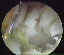
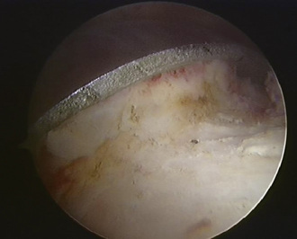
Standard lateral paratrochanteric and anterior paratrochanteric portals are made to access the central compartment; this allows for the visualization of the edge of the acetabular component, the bone–component interface (Figure 23-1), and the synovial membrane, and the surgeon is also able to look for any possible metallosis. If debridement is required, it is performed with an arthroscopic shaver or a radiofrequency probe. Synovial biopsy and further aspirates from the bone implant interface may also be obtained at this time.


< div class='tao-gold-member'>

Stay updated, free articles. Join our Telegram channel

Jul 24, 2016 | Posted by in MUSCULOSKELETAL MEDICINE | Comments Off on Arthroscopy for Symptomatic Hip Arthroplasty

Full access? Get Clinical Tree

Get Clinical Tree app for offline access