Fig. 42.1
Illustration showing a suprapatellar soft tissue mass, which results from sustained irritation of the quadriceps tendon on the upper edge of the intercondylar box. The soft tissue can be trapped in the box when the knee is extended from deep flexion

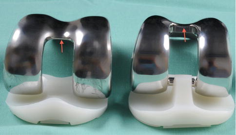
Fig. 42.2
The intercondylar box of the femoral TKR component is deeper (arrow) in the posterior stabilizing implant (right) than in the cruciate retaining one (left)
Patella clunk syndrome: catching or locking at about 40° flexion when extending the knee, which is suddenly released by audible popping. This phenomenon is due to a suprapatellar soft tissue mass on the back of the quadriceps tendon, which gets caught in the femoral box while the knee is moved from flexion to extension.
However, not all patients with patellofemoral problems after PS TKR develop these prominent fibrotic nodules and the typical clunk syndrome. Pollock et al. [7] described a number of cases of synovial entrapment in a series of 459 PS TKRs due to suprapatellar soft tissue interposition. The spectrum of symptoms includes crepitus, anterior knee pain, and difficulties rising from chairs or climbing stairs. In contrast to the patellar clunk syndrome, the typical symptoms occurred at 90° of flexion. As different models of PS TKR were used in this study, it could be shown that the soft tissue entrapment was related to certain design features such as a more superior and anterior transition from the intercondylar box into the distal trochlea.
Patellofemoral impingement can be subtler than the patella clunk syndrome. The symptoms include crepitus, anterior knee pain, and difficulties climbing stairs or rising from a chair.
Similar to the patellar clunk, an association with the intercondylar box was found. Certain design modifications were made on the first Insall-Burstein PS prosthesis to address the patellofemoral problems. The trochlea was deepened, the transition of the notch to the trochlea was smoothed, the box was narrowed, and the trochlea was angled differently. The goal of these modifications was to decrease the irritation of the suprapatellar extensor apparatus. Indeed, the introduction of the new design effectively decreased the occurrence of synovial entrapment problems [8, 9].
Nevertheless, other implants with a similar design were still prone to patellofemoral impingement. Schroer et al. [10] found that a patellar clunk syndrome occurred more frequently in patients who were operated on using a minimally invasive approach and who had a particular high-flexion ability. The effect was observed with one out of three femoral posterior stabilized TKR, a fact that indicates that other factors such as range of motion might also influence the occurrence of a synovial impingement.
42.2 Diagnostic Steps
The diagnosis is primarily a clinical one. The condition usually develops within months after the index procedure but can occur as late as several years after the implantation [6, 7, 10]. Soft tissue entrapment as a cause of patellofemoral symptoms is unlikely during the first postoperative weeks. Typically, the patient presents with anterior knee pain on active extension with or without a feeling of catching or snapping. Radiographs are rarely helpful in identifying soft tissue lesions. However, radiographs should be obtained to rule out other causes such as malpositioning of the TKR components or maltracking of the patella. A patella baja together with typical clinical symptoms raises the suspicion of a patellofemoral soft tissue impingement as the quadriceps tendon engages earlier with the femoral TKR component [11]. Due to metal artifacts, MRI is limited in visualization and identification of soft tissue lesions near the femoral component. Ultrasound is clearly of potential value but, considering the available literature [12], it seems not to be in widespread use.
A thorough history and clinical examination are the key steps to identifying patellofemoral soft tissue impingement. Typically, symptoms develop within months after primary TKR; it is unlikely during the first weeks. Patients with a posterior stabilized TKR design are more prone to it. Symptoms are anterior knee pain on active extension with or without catching or snapping. Radiographs are rarely helpful.
42.3 Treatment
In most cases treatment is initially conservative. If symptoms do not resolve within a few months, surgical debridement is the treatment of choice. While this was done in an open fashion in the early days of PS TKR, today arthroscopic treatment is indicated.
The primary treatment is nonsurgical. If symptoms do not resolve within a few months, surgical arthroscopic debridement is indicated.
42.3.1 Arthroscopic Procedure
The patient is positioned supine, and the knee is prepared as preferred by the surgeon (Fig. 42.3). However, full flexion should be possible in order to allow for verification of the impingement.


Fig. 42.3
Image of a right knee after TKR, prepared for arthroscopy. In addition to standard infrapatellar portals, a suprapatellar portal provides excellent access to the soft tissue to be resected. The arrows shows the area of depth
While standard anteromedial and anterolateral portals might be sufficient to enter the knee, atypical portals are often necessary to gain access to the region of interest. The author’s preferred technique is to start with an infrapatellar, periligamentous lateral portal.
Standard anteromedial and anterolateral portals might be sufficient, but atypical portals are often necessary to gain adequate access to the region of interest.
However, the common approach of advancing the arthroscope into the upper recess through or beside the patellofemoral joint should be done with all due care. In particular, in the case of extension deficit, forcing the arthroscope through the patellofemoral joint should be avoided. As the Hoffa fat pad is usually reduced, possibly resected, the arthroscope can be advanced medially toward the notch and, in most cases, adequate visualization of the TKR can be achieved. The patella height and possible soft tissue entrapment can be assessed in the flexed knee. If needed, a medial portal is created. It is strongly recommended that the second portal is done after a guiding cannula has been placed, as the shape of the knee differs from the normal knee and misplacement of the portal can easily happen. It has to be emphasized that the periarticular soft tissue usually is substantially thicker and less pliable than in healthy knees. Thus, the range of motion of the instruments used can be considerably limited. Contact of the arthroscopic instruments with the TKR components should be avoided in all cases (Fig. 42.4).


