CHAPTER 15  Arthroscopic treatment of glenoid bone loss—surgical technique
 Consider patient age, gender, and activity such as whether he or she is a collision/contact athlete or not.


Introduction
Significant bone loss, often characterized by an inverted-pear glenoid or engaging Hill-Sachs lesion, is believed to be a main cause of failures after arthroscopic stabilization.1–3 According to a three-dimensionally (3-D) reconstructed computed tomography (CT) study, the prevalence of glenoid bone defect has been reported as high as 90% in shoulders with chronic recurrent traumatic anterior instability, and an associated bony fragment is present in about half of shoulders with glenoid bone loss.4 Further, bone loss in shoulders associated with a bony fragment is relatively significant compared with that in shoulders with attritional glenoid without bony fragment.4,5 Therefore, the majority of shoulders with a significant bone loss can be treated through arthroscopic bony Bankart repair.5
Preoperative history, examination, and radiographic findings
Preoperative history
The diagnosis of recurrent traumatic anterior glenohumeral instability is usually made easily on the basis of the history of distinct dislocation or subluxation and the positive apprehension sign. However, with collision athletes, care should be taken because they may not experience clear dislocation or subluxation and only complain of pain or weakness when they bring their arm to maximum external rotation in abduction.6
Examination findings
The anterior apprehension test is done with the patient in the supine position. In this test, the shoulder is moved passively into maximum external rotation with the arm at side; at 30, 60, 90, 120, and 150 degrees of abduction, and maximum flexion.6 At the same time, the posterior apprehension test is done with the arm at maximum internal rotation in 90 degrees of abduction. The feeling of apprehension is reported in each arm position. However, the most important and reliable physical examination usually can be done with the patient under anesthesia, comparing stability testing to the contralateral extremity.
Radiographic findings
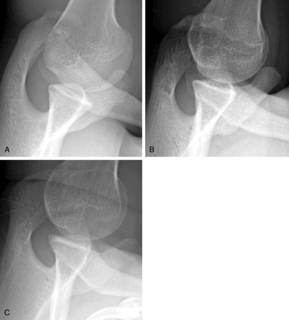
X-ray images are sometimes helpful in detecting the Hill-Sachs lesion and the anterior glenoid rim lesion, especially during the first patient visit. Bernageau described a unique method for detecting an anterior glenoid rim lesion with the patients in the standing position.7 However, this technique requires fluoroscopic control in order to obtain optimal diagnosable images and, therefore, radiation exposure is an unignorable issue.8 We have developed a modified Bernageau method with the patient lying on their axilla in their most relaxed position (Fig. 15-1).6 In this method, clear Xx-ray images can be obtained more easily with a high probability of ascertaining bony pathology without using fluoroscopic imaging (Fig. 15-2).6
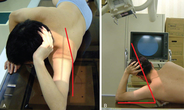
(From Sugaya H: Instability with bone loss. In Angelo R, et al, editors: AANA advanced shoulder arthroscopy, Philadelphia, 2010, Elsevier.)
(From Sugaya H: Instability with bone loss. In Angelo R, et al, editors: AANA Advanced shoulder arthroscopy, Philadelphia, Elsevier, 2010.)
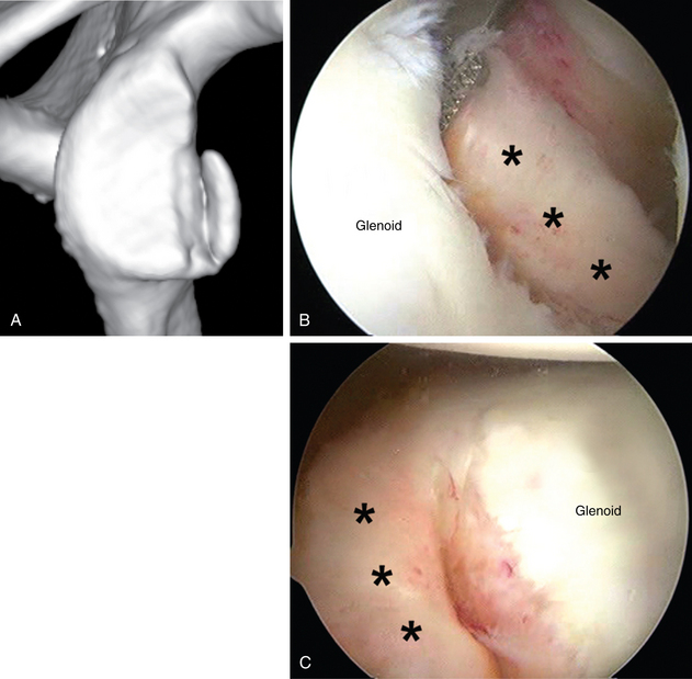
Three-dimensional CT is the most important imaging study in assessing glenoid morphology accurately.4 In a shoulder with a bony Bankart, detecting accurate configuration of the bony fragment is not easy because the bone fragment is covered by the surrounding soft tissue. Through preoperative 3-D CT, surgeons can determine whether the glenoid is attritional or if a bony Bankart lesion is present. They also can quantify bone loss with attritional glenoids and detect the size and shape of the bony fragment in shoulders with a bony Bankart lesion (Fig. 15-3).4,5,9
Description of techniques
If a bone fragment is present with 3-D CT, an arthroscopic bony Bankart repair, in which the fragment is incorporated into the Bankart repair, is indicated regardless of the severity of glenoid bone loss.5,10,11 Therefore, a majority of shoulders with a large glenoid bone loss can be treated arthroscopically using this technique.6 For shoulders with significant bone loss associated with the attritional glenoids in young and active patients, although the number of such patients is limited, open or arthroscopic bone grafting procedures are indicated. The author’s preference is an arthroscopic iliac bone block grafting in combination with an anterior-inferior capsulolabral repair, which is described later in detail.12,13
Arthroscopic bony bankart repair
Because most of the bony Bankart lesions are displaced and partly malunited chronic avulsion type fractures, the bony fragment is firmly connected to the adjacent labrum or soft tissue. These characteristics of chronic bony Bankart lesions make arthroscopic bony reconstruction feasible.5,10 Normally, bony Bankart lesion is never completely healed to the glenoid neck because the fragment is displaced from the original place, and the adjacent glenohumeral ligament is not functioning. Therefore, most of the bony fragment associated with a bony Bankart lesion can be separated easily from the glenoid neck, using a standard straight rasp. Although the gap between the fragment and original glenoid is well demarcated in most shoulders, if otherwise, careful palpation or preoperative 3-D CT greatly helps surgeons to delineate the gap.6
Procedure (see video 15-1)
Related posts:
 Open surgical solutions for posterior instability of the shoulder
 Clinical history, examination, arthroscopic findings, and treatment of multidirectional instability
 Recognition and management of combined instability and rotator cuff tears
 Rehabilitation after posterior instability repair—open vs. arthroscopic
 Humeral head defects—biomechanics, measurements, and treatments
 Nonoperative rehabilitation for traumatic and atraumatic glenohumeral instability
Stay updated, free articles. Join our Telegram channel

Full access? Get Clinical Tree


