Arthroscopic Subscapularis Repair

Chapter 22


Arthroscopic Subscapularis Repair


As arthroscopic techniques in shoulder surgery have improved, recognition of subscapularis tendon injuries has increased. Although isolated subscapularis tears continue to be rare, estimated at 5% of rotator cuff tears,1 they are increasingly being seen in association with other supraspinatus, infraspinatus, and teres minor tears, with an incidence estimated at 27% to 43%.2 Because of this, there has been increased interest in arthroscopic subscapularis tendon repairs. The main advantages of an arthroscopic repair of the subscapularis tendon are smaller incisions, less postoperative pain, and the ability to better visualize and address coexisting pathologic processes, including labral tears, long head biceps injury, and superior and posterior rotator cuff tears. This chapter addresses the preoperative considerations and the techniques involved in performing an arthroscopic subscapularis repair.


Preoperative Considerations


History


Often there is a history of trauma, which may include an abduction and external rotation moment, direct blow, heavy lifting, or traction injury.3 For degenerative tears, the patient may report a gradual worsening of anterior shoulder pain without specific trauma. Patients may note difficulty with internal rotation—for example, in tucking in a shirttail. Consider a full differential diagnosis for anterior shoulder pain, including acromioclavicular joint arthrosis or dislocation, biceps tendon tears or inflammation, anterior capsulolabral damage, and fractures of the lesser tuberosity.


Physical Examination


A complete shoulder examination should begin with observation, range of motion, and strength testing. The most common physical examination findings associated with a subscapularis tear are weakness and pain with isolation of internal rotation. External rotation is evaluated with the arm at the side and compared with the contralateral extremity.


It is important to isolate the subscapularis muscle from other internal rotators during examination. Owing to the high association with biceps tendon pathology, careful examination with the Speed and O’Brien tests is also important. The belly press test is performed by asking the patient to press the ipsilateral hand on the abdomen, maintaining the elbow anterior to the body. The belly press test result is considered positive if the patient is not able to keep the elbow anterior to the trunk or if the wrist is flexed in attempting to press into the abdomen. The lift-off test requires the patient to be able to place the ipsilateral hand behind the back. The patient is asked to lift the hand off the back; if the patient is unable to do so, the test result is considered positive. The most sensitive test is the bear hug test.4 The patient places his or her ipsilateral hand on the contralateral shoulder with the elbow elevated forward. The test result is positive if the surgeon is able to lift the patient’s hand off the shoulder.


Imaging


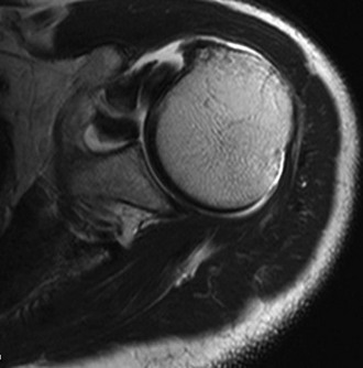
A standard shoulder imaging series including an axillary view is obtained to assess for alternative pathologic changes, such as fractures and glenohumeral arthritis. Magnetic resonance imaging is the gold standard imaging modality for diagnosis of subscapularis tendon tears and evaluation of muscle belly quality, fatty infiltration, and displacement of the long head of the biceps tendon (Fig. 22-1). Magnetic resonance arthrography improves the sensitivity of diagnosis of partial tears and labral and biceps pathology. In addition, the coracohumeral distance is measured from the tip of the coracoid to the humerus on an axial cut with the smallest distance.2 A distance of 6.5 mm is considered narrowed. The tendon should be assessed on both axial and sagittal images, with verification of its insertion on the lesser tuberosity.


Indications and Contraindications


Indications for subscapularis repair are pain and weakness nonresponsive to conservative management. Important secondary indications are restoration of shoulder strength and function and treatment of recurrent shoulder instability.


Relative contraindications to arthroscopic repair include lack of pain, severe atrophy, retraction or significant fatty degeneration on magnetic resonance imaging, and rotator cuff tear arthropathy. For patients with massive tears, significant retraction, or atrophy, we recommend open surgery including pectoralis major tendon transfer and possible tissue augmentation. Absolute contraindications are severe medical illness that precludes anesthesia, and active infection. The decision to perform an arthroscopic or open procedure should be based on the surgeon’s individual comfort level with the chosen technique.


Surgical Technique


Anesthesia and Positioning


Most patients are placed under general anesthesia. We prefer to supplement every case with an interscalene block or nerve catheter depending on the skill and comfort of the anesthesiologist. This reduces the amount of anesthetic required during the case and improves postoperative pain control. If the patient’s medical condition does not allow general anesthesia, regional anesthesia can be used along with sedation.


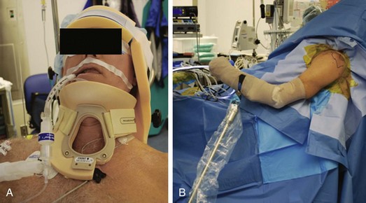
We prefer the beach chair position for arthroscopic repairs of the subscapularis (Fig. 22-2). The upper extremity can be easily moved and rotated to better visualize the subscapularis and its insertion. Furthermore, the beach chair position allows the surgeon to convert easily to an open procedure if necessary.


The patient is aligned on the edge of the table so that the affected shoulder and scapula are exposed. The head is secured to the operating table with a headrest, and a Philadelphia collar (Philadelphia Cervical Collar Company, Thorofare, NJ) is used to prevent excessive motion in the neck. We use the Spider Limb Positioner (Smith & Nephew, London) to aid in arm positioning during the case.


Surgical Landmarks, Incisions, and Portals


We begin every case by outlining the bony landmarks on the skin, including posterior and anterior corners of the acromion, and the soft spot between the posterior clavicle and anterior scapular spine. A line is drawn between the two corners of the acromion. The anterior and posterior edges of the clavicle and scapular spine are marked next. The acromioclavicular joint is palpated and marked. A circle is drawn over the prominence of the coracoid.


The posterior portal is the first portal to be established. With use of a three-finger shuck, the index finger of the same hand as the shoulder being operated on is placed in the soft spot between the clavicle and scapular spine. The middle finger is placed on the coracoid, and the thumb feels the interval between the infraspinatus and teres minor.


The anterior portal is generally placed just lateral to the coracoid and below the coracoacromial ligament. This portal can easily be established with an outside-in technique localizing with a spinal needle or an inside-out technique over a Wissinger rod. An accessory anterolateral portal is made in all subscapularis repairs. This portal is located in the rotator interval anterior and medial to the anterolateral corner of the acromion. This places it about 1 to 2 cm superior and 2 cm lateral to the standard anterior portal. A spinal needle is used to localize this portal, with an intra-articular entrance site just posterior to the native biceps tendon. After the portal is made, it is enlarged to allow the placement of a threaded 8-mm clear cannula. It is important not to place the two anterior portals too close to each other. When there is also an associated superior rotator cuff tear, this portal may be placed through the defect.

Stay updated, free articles. Join our Telegram channel

Sep 11, 2016 | Posted by in SPORT MEDICINE | Comments Off on Arthroscopic Subscapularis Repair

Full access? Get Clinical Tree

Get Clinical Tree app for offline access