Arthroscopic Subacromial Decompression, Distal Clavicle Resection, and Coracoplasty
Steven B. Singleton MD
James M. Bothwell MD
John E. Conway MD
Arthroscopic Subacromial Decompression
History of the Technique
Subacromial decompression as a treatment for impingement syndrome was first described by Neer in 1972.1,2,3,4 The goal of the procedure was to reduce extrinsic compression on the rotator cuff musculature by increasing the volume of the subacromial space. The original description entailed the open exposure of the anterior acromion and subacromial space to resect the coracoacromial ligament, the anterior lip of the acromion, and the subacromial bursa. Over the past 30 years, multiple investigators have noted satisfactory results with this procedure when performed in the proper setting.3,4,5
The advent of shoulder arthroscopy has furthered the evolution of the subacromial decompression. Neer’s description of the open technique has been the gold standard by which arthroscopic techniques have been measured. Many, including Ellman,6 have proposed various techniques for arthroscopic subacromial decompression that differ slightly in their means, but have the very same end point: that is, to increase the volume of the subacromial space. The main advantage of arthroscopic subacromial decompression is sparing of the deltoid muscle origin and, therefore, an expedited recovery. In general, most authors report good to excellent results in the majority (73% to 88%) of their patients.3,4,5,6,7,8,9,10
Indications and Contraindications
The main indications for arthroscopic subacromial decompression include the following: (a) Symptomatic subacromial impingement refractory to a comprehensive rehabilitation program usually lasting 6 to 12 months, where the impingement is either classical extrinsic or chronic secondary with adaptive changes; (b) when rotator cuff repair is performed.
The contraindications for arthroscopic subacromial decompression include the following: (a) Shoulder stiffness, (b) massive or irreparable rotator cuff tear, (c) inaccurate diagnosis: be aware of instability.
Preoperatively, consider the amount of anterior acromion to be resected. The supraspinatus outlet view enables one to determine the acromial morphology and may prove helpful in determining the appropriate volume of bone removal for adequate acromioplasty. Similarly, postoperative radiographic evaluation utilizing the supraspinatus outlet view to evaluate the decompression may be useful, especially for those learning the arthroscopic technique.
Surgical Technique
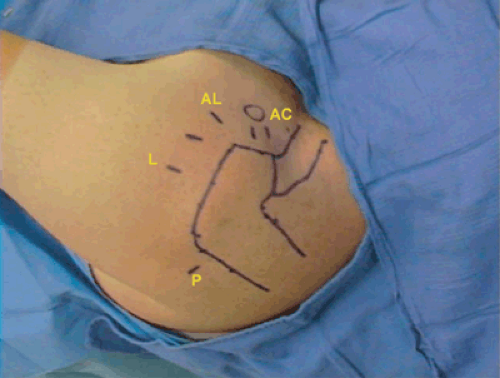
Anesthesia and patient positioning are the same for the three procedures described in this chapter. General anesthetic or interscalene regional block is used. Hypotensive anesthesia maintaining systolic blood pressure at or below 95 mm Hg will assist visualization by minimizing intraoperative bleeding. Our preference in performing arthroscopic shoulder procedures is the lateral decubitus position. The arm is suspended with 10 to 12 lb of arm traction in 30 degrees of abduction, and in 20 degrees to 30 degrees of forward flexion. The spine of the scapula, lateral acromion, and anterior and posterior borders of the clavicle are marked on the skin. When indicated, the location and angle of obliquity of the acromioclavicular (AC) joint is also noted on the skin (Fig. 7-1).
Procedure
A 30-degree arthroscope is placed in the standard posterior portal. This portal is located approximately 2 cm inferior
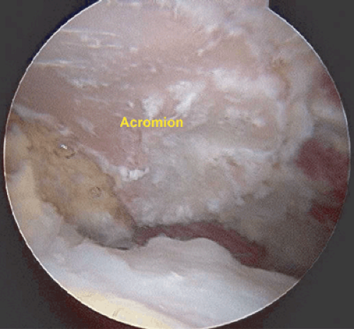
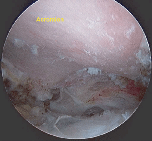
and 1.5 cm medial to the posterior angle of the acromion in the “soft spot.” An anterior portal is established approximately 1 cm anterior to the anterior edge of the acromion. If this procedure is done in conjunction with an arthroscopic distal clavicle excision, an AC joint portal, as described below, can be utilized. A thorough diagnostic examination of the glenohumeral joint is performed. Any indicated intra-articular procedures may be accomplished. In the same posterior portal, the arthroscope is removed from the glenohumeral joint and reintroduced into the subacromial space. The anterior cannula is redirected into the subacromial space and is utilized primarily as an inflow portal. A midlateral portal (working portal) is established approximately 4.0 cm lateral to the midportion of the lateral acromion, in line with the posterior border of the clavicle. A complete bursectomy of the subacromial space is performed using an arthroscopic shaver. Cautery or a radiofrequency device is used for hemostasis. This is an essential step because visualization of the complete undersurface of the acromion is necessary for this procedure. After completion of the subacromial bursectomy, the cautery or radiofrequency device is used to outline the anterior and lateral aspects of the acromion. The coracoacromial ligament is detached in its entirety from the anterior aspect of the acromion. Care should be taken to coagulate the acromial branch of the thoracoacromial artery because this can be a significant source of bleeding as the ligament is detached. The ligament should be further resected until the anterior deltoid fascia is visualized. The undersurface of the acromion should be visualized in its entirety from its posterior border to its anterior border such that its morphology is visualized and confirmed based on preoperative radiographs (Fig. 7-2). An arthroscopic burr is inserted via the midlateral working portal and used to remove the appropriate amount of anterior acromion to produce a flat undersurface from its lateral to medial borders. Caution should be exercised to not violate the capsule of the acromioclavicular joint or to excessively detach the deltoid fascia from the acromion. The anterior border of the acromion should be resected to be coplanar with the anterior edge of the clavicle (usually a resection of 5 to 8 mm from the anterior border). It is essential for adequate decompression that the resection of the acromion be flat from its lateral to medial borders, including the acromioclavicular joint (Fig. 7-3A). An arthroscopic rasp may be utilized to smooth the resection of the undersurface of the acromion. Final evaluation and visualization of the acromion after resection is performed by tilting the angle of the lens parallel to acromion (medial or lateral) (Fig. 7-3B). Also, one may visualize the undersurface of the acromion from the midlateral portal and place a switching stick from the posterior border to the anterior border of the acromion to confirm that the decompression is adequate. A last option is to use digital palpation to evaluate the acromial undersurface. Portals are reapproximated and dressings applied. The patient’s extremity is placed in an immobilizer for comfort; however, passive motion is begun immediately and full range of motion is the short-term immediate goal, followed by strengthening once full range of motion is encountered.
and 1.5 cm medial to the posterior angle of the acromion in the “soft spot.” An anterior portal is established approximately 1 cm anterior to the anterior edge of the acromion. If this procedure is done in conjunction with an arthroscopic distal clavicle excision, an AC joint portal, as described below, can be utilized. A thorough diagnostic examination of the glenohumeral joint is performed. Any indicated intra-articular procedures may be accomplished. In the same posterior portal, the arthroscope is removed from the glenohumeral joint and reintroduced into the subacromial space. The anterior cannula is redirected into the subacromial space and is utilized primarily as an inflow portal. A midlateral portal (working portal) is established approximately 4.0 cm lateral to the midportion of the lateral acromion, in line with the posterior border of the clavicle. A complete bursectomy of the subacromial space is performed using an arthroscopic shaver. Cautery or a radiofrequency device is used for hemostasis. This is an essential step because visualization of the complete undersurface of the acromion is necessary for this procedure. After completion of the subacromial bursectomy, the cautery or radiofrequency device is used to outline the anterior and lateral aspects of the acromion. The coracoacromial ligament is detached in its entirety from the anterior aspect of the acromion. Care should be taken to coagulate the acromial branch of the thoracoacromial artery because this can be a significant source of bleeding as the ligament is detached. The ligament should be further resected until the anterior deltoid fascia is visualized. The undersurface of the acromion should be visualized in its entirety from its posterior border to its anterior border such that its morphology is visualized and confirmed based on preoperative radiographs (Fig. 7-2). An arthroscopic burr is inserted via the midlateral working portal and used to remove the appropriate amount of anterior acromion to produce a flat undersurface from its lateral to medial borders. Caution should be exercised to not violate the capsule of the acromioclavicular joint or to excessively detach the deltoid fascia from the acromion. The anterior border of the acromion should be resected to be coplanar with the anterior edge of the clavicle (usually a resection of 5 to 8 mm from the anterior border). It is essential for adequate decompression that the resection of the acromion be flat from its lateral to medial borders, including the acromioclavicular joint (Fig. 7-3A). An arthroscopic rasp may be utilized to smooth the resection of the undersurface of the acromion. Final evaluation and visualization of the acromion after resection is performed by tilting the angle of the lens parallel to acromion (medial or lateral) (Fig. 7-3B). Also, one may visualize the undersurface of the acromion from the midlateral portal and place a switching stick from the posterior border to the anterior border of the acromion to confirm that the decompression is adequate. A last option is to use digital palpation to evaluate the acromial undersurface. Portals are reapproximated and dressings applied. The patient’s extremity is placed in an immobilizer for comfort; however, passive motion is begun immediately and full range of motion is the short-term immediate goal, followed by strengthening once full range of motion is encountered.
Pitfalls of the Technique
There are several potential complications to be aware of and to avoid. It is important to choose a burr for the resection that is able to smoothly resect the appropriate amount of bone from the undersurface without being too aggressive. Our personal choice is a 12-fluted barrel burr that is able to
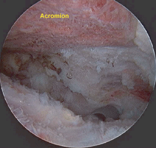
spin at 9,000 revolutions per minute. A lack of familiarity with an arthroscopic burr can result in a burr that is overly aggressive and resecting excessive amounts of bone in inappropriate areas of the acromion. This may result in an acromion that is not well contoured and rough upon completion of the acromioplasty. Excessive resection of the anterior acromion can lead to a fracture of the acromion. Preoperative planning and templating with correlation of the postoperative supraspinatus outlet view is an important part of evaluation of the technique utilized for the arthroscopic acromioplasty. Lateral acromionectomy should be avoided due to detachment of the deltoid insertion; loss of acromial fulcrum and development of adhesions between the undersurface of the rotator cuff and the deltoid can all lead to loss of deltoid function and pain. Inadequacy of the decompression is a relatively common cause of failure. Resection of the coracoacromial ligament may be important part of this procedure. Retention or regeneration of the coracoacromial ligament has been implicated as a frequent cause of failures.11,12 Also, residual anterior or medial acromial spurs can also be a cause for failure of this procedure13,14 (Figs. 7-4, 7-5).
spin at 9,000 revolutions per minute. A lack of familiarity with an arthroscopic burr can result in a burr that is overly aggressive and resecting excessive amounts of bone in inappropriate areas of the acromion. This may result in an acromion that is not well contoured and rough upon completion of the acromioplasty. Excessive resection of the anterior acromion can lead to a fracture of the acromion. Preoperative planning and templating with correlation of the postoperative supraspinatus outlet view is an important part of evaluation of the technique utilized for the arthroscopic acromioplasty. Lateral acromionectomy should be avoided due to detachment of the deltoid insertion; loss of acromial fulcrum and development of adhesions between the undersurface of the rotator cuff and the deltoid can all lead to loss of deltoid function and pain. Inadequacy of the decompression is a relatively common cause of failure. Resection of the coracoacromial ligament may be important part of this procedure. Retention or regeneration of the coracoacromial ligament has been implicated as a frequent cause of failures.11,12 Also, residual anterior or medial acromial spurs can also be a cause for failure of this procedure13,14 (Figs. 7-4, 7-5).
Alternative Techniques: Posterior Cutting Block Technique
Utilizing the previously outlined portals, after completion of a thorough bursectomy and coracoacromial resection, the arthroscope is positioned in the midlateral portal and the shaver is inserted in the posterior portal. The midhalf of the acromion is marked and the burr is inserted from the posterior portal. The shaft of the burr is laid upon the posterior one half of the acromion. The burr is advanced anteriorly while sweeping medially to laterally removing the bone that is posterior to this line. The anterior acromion is resected to be coplanar with the bone removed from the anterior one half of the acromion. Final evaluation of the resection can be made by inserting the arthroscope into the posterior portal to ensure that the acromion is flat in a medial to lateral
plane. Portals are closed and dressings applied in a similar manner to the method described above.
plane. Portals are closed and dressings applied in a similar manner to the method described above.
Rehabilitation
Immediate passive and active assist range of motion can be initiated on postoperative day 1 following the procedure. Active range of motion can be initiated on postoperative day 7 to 10. Emphasis is placed on achieving full range of motion prior to instituting strengthening. Similar to the rehabilitation program discussed in the other sections of this chapter, emphasis is placed on scapular stabilization and control of the trunk. As the patient progresses, the return to full activity and sports may occur as early as 6 weeks. The return to play for the overhead athlete may require a more prolonged rehabilitation.
Stay updated, free articles. Join our Telegram channel

Full access? Get Clinical Tree


