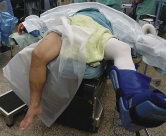
Chapter 58 1. Arthroscopic technique allows for anatomic placement of both meniscal horns, which minimizes meniscal extrusion and maximizes hoop stresses, presumably leading to enhanced knee kinematics. 2. Bone plugs can be adapted to a patient’s individual anatomy. 3. Concomitant surgical procedures can be performed at the time of MAT to address ligamentous instability, mechanical malalignment, and cartilaginous defects. 4. An all-arthroscopic technique is minimally invasive and can be used in an outpatient setting. The patient is placed supine on the surgical bed with a thigh tourniquet applied, though rarely inflated. A lateral post allows for application of a valgus force to open the medial compartment. The foot of the bed is flexed maximally with enough room behind the passively flexed knee to allow a fist to be placed. Two rolled sheets placed under the pad of the bed prevent hyperextension at the hip. The contralateral lower extremity is safely cradled in a well leg holder with the hip and knee slightly flexed in an abducted position (Fig. 58-1). The graft is checked by the surgeon at least 1 day before the scheduled surgery to verify it is for the correct limb and for the correct compartment. While the patient is being brought into the room and positioned for surgery, the graft is thawed and prepared for transplantation on the back table. The graft is usually ready for implantation as the leg is being prepared and draped. Anesthesia routinely includes a regional block performed in the preoperative area once the patient’s site is signed by the surgeon. A general anesthetic is used as well to help expedite the surgery.
Arthroscopic Meniscus Transplantation
Bone Plug
Surgical Technique
Anesthesia and Positioning


