Arthroscopic Management of Lateral Epicondylitis
Champ L. Baker III
Champ L. Baker Jr
In 1873, Runge first described the pathologic entity of lateral humeral epicondylitis. Ten years later, Morris noted an association between lawn tennis and lateral epicondylitis, leading to its common designation as tennis elbow. Since these original descriptions, various etiologies regarding the pathogenesis of lateral epicondylitis have been proposed. Advancing the works of Cyriax (1), Goldie (2), and Coonrad and Hooper (3), Nirschl and associates (4, 5) noted the basic underlying lesion is in the origin of the extensor carpi radialis brevis (ECRB) tendon. Repetitive overuse leads to microtears in the ECRB origin. Subsequent failed tendon healing and replacement with immature reparative tissue follows. Histologic examination of the essential lesion reveals a degenerative, noninflammatory process characterized with fibroblasts, disorganized collagen, and vascular hyperplasia. These findings have been termed angiofibroblastic hyperplasia with later modification to angiofibroblastic tendinosis. Despite advances in our understanding of the pathoanatomy of lateral epicondylitis, controversy remains regarding its optimal treatment. Various nonoperative and operative interventions have been proposed with most yielding short-term success. For patients who require surgery for recalcitrant symptoms, the authors have had high rates of clinical success with arthroscopic resection of pathologic tissue at both short- and long-term follow-up (6, 7).
CLINICAL EVALUATION
The most common presenting complaint is pain about the lateral aspect of the elbow. The pain may extend distally into the dorsal forearm or radiate proximally. Typically, the pain is of insidious onset with a history of repetitive activity. Patients often report a decrease in grip strength and difficulty holding or lifting objects, especially away from their body with their arms extended. On clinical examination, the patient may give minimal effort or wince with a handshake. Evaluation of the elbow reveals characteristic point tenderness to palpation at an area just anterior and distal to the lateral epicondyle. Reproducible pain localized to the lateral epicondyle is found with resisted wrist extension with the elbow fully extended. Passive wrist flexion, again with the elbow extended, places the ECRB on stretch and can reproduce pain. Evaluation of the cervical spine and upper extremity can help to differentiate lateral epicondylitis from other causes of lateral elbow pain, such as cervical radiculopathy, radial tunnel syndrome, osteochondritis dissecans of the capitellum, radiocapitellar arthrosis, posterolateral rotatory instability of the elbow, and posterolateral elbow plica.
Although lateral epicondylitis is a clinical diagnosis, we routinely obtain plain anteroposterior, lateral, and axial elbow radiographs as part of the initial evaluation in the patient presenting with elbow pain. Radiographs may demonstrate soft tissue calcification adjacent to the lateral epicondyle, which is present in approximately 25% of patients, especially if the patient has had previous steroid injections. MRI can provide additional information regarding suspected intra-articular disorders, the extent of extensor tendon involvement, the presence of associated tendon tears, and the integrity of the lateral collateral ligamentous complex.
TREATMENT
Currently, no consensus exists regarding the optimal treatment for lateral epicondylitis. Various nonoperative measures have been recommended, including activity modification, physical therapy, nonsteroidal anti-inflammatory medications, counterforce bracing, and corticosteroid injections. More recent approaches include injections of botulinum toxin, buffered platelet-rich plasma, and application of extracorporeal shock-wave therapy. Most patients respond successfully to various conservative methods. In the reported studies of Coonrad and Hooper (3), Nirschl and Pettrone (4), and Boyd and McLeod (8), only 4% to 11% of patients required operative intervention for recalcitrant symptoms.
Indications for surgical treatment include persistent lateral elbow pain and dysfunction that interferes with the patient’s activities despite appropriate nonoperative treatment. We recommend a minimum of 6 months of nonoperative treatment before considering surgery. Although rare, we consider earlier surgical intervention in the patient who sustained a direct trauma, resulting in a partial tear of the ECRB. Many different operative procedures have been reported: percutaneous, endoscopic, or open extensor tendon release; open techniques for excision of abnormal degenerative tissue with either simple suture repair or formal repair of the extensor tendons back to the lateral epicondyle; and arthroscopic release or resection of degenerative tissue.
We prefer arthroscopic management of lateral epicondylitis because we have found it to be safe and reproducible with proven high rates of clinical success at long-term follow-up. In addition, it affords the opportunity to evaluate and treat for coexistent intra-articular pathoanatomy and offers a potentially shorter postoperative rehabilitation with expedited return to work and unrestricted activities. Relative contraindications to the arthroscopic technique include previous medial elbow surgery with ulnar nerve transposition or with a subluxating ulnar nerve.
AUTHORS’ PREFERRED TREATMENT
For the arthroscopic procedure, patients can be placed in the supine, lateral, or prone position. We prefer the prone position with the use of a general anesthetic (Fig. 36.1). This position allows for an accurate postoperative neurovascular assessment. A well-padded tourniquet is placed about the upper arm. For larger individuals, a sterile tourniquet can be used. The arm is placed into a commercially available arm holder and is prepared and draped in the standard sterile fashion. The following anatomic landmarks are identified and outlined on the skin to safely create the arthroscopic portals: medial epicondyle and intermuscular septum, ulnar nerve, olecranon tip, lateral epicondyle, and radial head (Figs. 36.2 and 36.3). Next, we mark the lateral soft spot in the center of the triangle created by the radial head, olecranon, and lateral epicondyle. We wrap the forearm with a compressive dressing to prevent leakage of fluid into the distal soft tissues. The limb is exsanguinated, and the tourniquet insufflated to approximately 250 mm Hg.
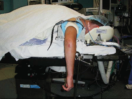 FIGURE 36.1. Patient is positioned prone for surgery with arm placed in arm holder. |
Before we create the portals, the elbow joint is distended with approximately 20 to 25 mL of normal saline through the lateral soft spot (Fig. 36.4). Joint distention displaces the neurovascular structures anteriorly to help protect against iatrogenic injury during portal creation and introduction of the instruments. In particular, the ulnar nerve should be identified and protected. First, we establish the proximal medial portal approximately 2 cm proximal and 1 cm anterior to the palpable medial epicondyle (Fig. 36.5). For larger individuals, we place this portal slightly more proximal and anterior. After incising the skin, we utilize a straight hemostat to spread the subcutaneous tissues (Fig. 36.6). This “nick-and-spread” technique helps prevent injury to the sensory nerves. A blunt trocar and cannula without side portals is inserted through this portal aiming toward the center of the joint while maintaining contact with the anterior humeral
border. Backflow of fluid confirms intra-articular placement (Fig. 36.7). Stothers et al. (9) found the ulnar nerve to be located, on average, 12 mm away from this proximal medial portal posterior to and protected by the medial intermuscular septum. This portal is also an average of 2.3 mm from the medial antebrachial cutaneous nerve, 7.6 mm from the median nerve, and 18 mm from the brachial artery with the elbow in flexion (Fig. 36.8).
border. Backflow of fluid confirms intra-articular placement (Fig. 36.7). Stothers et al. (9) found the ulnar nerve to be located, on average, 12 mm away from this proximal medial portal posterior to and protected by the medial intermuscular septum. This portal is also an average of 2.3 mm from the medial antebrachial cutaneous nerve, 7.6 mm from the median nerve, and 18 mm from the brachial artery with the elbow in flexion (Fig. 36.8).
Related posts:
 Anthroscopic Cuff Repair: Tissue Graft Applications
 Arthroscopic Management of Osteochondritis Dissecans of The Elbow
 Arthroscopic Treatment of Dorsal and Volar Ganglions
 Arthroscopic Treatment of Anterior Glenoid Bone Loss: Latarjet Techniques
 Double-Bundle Acl Reconstruction
 Arthroscopy and Management of Ankle Fractures
Stay updated, free articles. Join our Telegram channel

Full access? Get Clinical Tree


