Arthroscopic Management of Glenohumeral Arthritis

Chapter 29


Arthroscopic Management of Glenohumeral Arthritis


Chapter Synopsis


• Glenohumeral degenerative disease is a growing problem in the active patient population. Patient discomfort may be related to isolated articular cartilage disease, but may also result from other glenohumeral abnormalities, including labral, rotator cuff, and/or long head of the biceps tendon pathology. Although shoulder arthroplasty remains the gold standard for reducing pain in patients with advanced shoulder arthritis, it is typically not the most appropriate option for the relatively young patient who wishes to remain active without postoperative activity limitations. Therefore shoulder arthroscopy has become an increasingly popular treatment option for this challenging patient population, with a growing body of literature supporting its ability to improve pain and function while potentially delaying the need for arthroplasty.


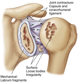
Glenohumeral degenerative disease is a growing problem in the relatively young, active patient population. Patients with shoulder arthritis have increased pain and decreased function compared with patients with normal shoulder joints,1 and such symptoms can certainly have a significant impact on quality of life, particularly at a younger age. However, given the complexity of the shoulder joint, it can be difficult to determine the exact pain generator (Fig. 29-1). Articular cartilage degradation can contribute to the patient’s symptoms, as can other shoulder pathologies including synovitis and capsulitis, labral tears, rotator cuff pathology, and long head of the biceps (LHB) tenosynovitis. Thus a comprehensive diagnostic and therapeutic approach is necessary.


Nonoperative treatment techniques including activity modification, physical therapy, nonsteroidal anti-inflammatory drugs (NSAIDs), and intra-articular injections of corticosteroids and/or hyaluronic acid solutions (off-label use) are typically effective as first-line options; however, their effects are often temporary.2,3 Surgical options are varied and range from simple arthroscopic debridement to total shoulder arthroplasty. Arthroplasty has certainly shown favorable outcomes with regard to pain relief; however, postoperative limitations and risks can be significant in an active patient.


Nonarthroplasty alternatives, including arthroscopic debridement, are increasingly attractive options in contemporary orthopedics.47 Shoulder arthroscopy and debridement is a commonly used initial surgical technique in the approach to treating patients with early glenohumeral degeneration, and several authors have demonstrated its potential for decreasing pain, improving function, and potentially delaying the need for eventual arthroplasty.815 These debridement and arthroscopic procedures often include a combination of debridement, chondroplasty, synovectomy, capsular release, and subacromial decompression (SAD). However, it is important to keep in mind that the goal of arthroscopy at this stage is not to cure the underlying disease process (arthritis) but rather to provide symptomatic improvement and maintenance of function for as long as possible.


Preoperative Considerations


History


Classically, patients with shoulder arthritis will have decreased range of motion (ROM) and global pain associated with activities or motion, most commonly in the older patient. A history of trauma or weightlifting should be solicited in a younger patient. As with most shoulder conditions, night pain may be a prominent component of the patient’s complaints. The onset is usually insidious and has been progressively affecting the patient’s activity level. A history of prior treatments such as injections and NSAIDs should be solicited. Such conservative measures may provide temporary relief and improved function, but with time the pain may be refractory to these modalities. However, the key in the history is not diagnosing the “degenerative condition,” but rather determining associated and contributing pathologies and developing a treatment algorithm.


The activity level of the patient should be determined, as well as the postoperative goals. Is the patient a sedentary household ambulator who wants to be able to reach things without pain, or is he or she an avid tennis player who hopes to return to some level of sport? This initial fact will guide decision making as to whether the patient is a good candidate for arthroplasty, or whether a less invasive technique such as arthroscopy should be tried first. The onset of pain is also important, as are the characteristics of the discomfort. An acute onset suggests an exacerbation of a concomitant pathology. Pain at the extremes of motion may be the result of decreased motion and capsulitis rather than the underlying degeneration, which may also cause pain in mid-ROM. A sensation of “popping” or “clicking” can be attributed to loose bodies or chondral irregularity, and pain isolated to the anterior aspect of the shoulder may occur secondary to biceps tendonitis. Nonetheless, a complete history related to the shoulder joint should be completed, and the recognition of concomitant pathologies is essential.


Furthermore, it is imperative for the orthopedic surgeon to ask about previous shoulder injuries and procedure(s), as certain implants and techniques have well-documented correlations with the development of arthritis. For example:


Physical Examination


A complete physical examination of both shoulders should be performed for every patient with possible glenohumeral arthritis. After visualization of the shoulder, standard range-of-motion, strength, sensation, and stability tests should be performed, followed by any special tests deemed appropriate. The physical examination should be completed with the mindset that the patient has an underlying degenerative condition and that accompanying pathology can be treated to improve the symptomatology. The cervical spine examination must also be performed to rule out referred pain. All examination findings must be compared with the contralateral shoulder to determine what is abnormal versus a normal variant for that patient. Typical physical examination findings include the following:


• Decreased ROM—specific documentation with preoperative ROM is essential


• Sensation of crepitus, clicking, catching, or locking


• Limited ROM compared with normal shoulder


• Possible point tenderness


• Provocative testing such as LHB tension signs


• Normal strength, though examination can be limited by pain


• Neurovascular examination to document normal status


Imaging


Imaging is useful in the evaluation of the patient with glenohumeral degenerative joint disease (DJD), and the typical modalities include radiographs, magnetic resonance imaging (MRI), and computed tomography (CT).


• Plain radiographs


• MRI (without contrast) can show the following:


Stay updated, free articles. Join our Telegram channel

Sep 11, 2016 | Posted by in SPORT MEDICINE | Comments Off on Arthroscopic Management of Glenohumeral Arthritis

Full access? Get Clinical Tree

Get Clinical Tree app for offline access