CHAPTER 12 Arthroscopic and Open Radial Ulnohumeral Ligament Reconstruction for Posterolateral Rotatory Instability of the Elbow
Dysfunction of the lateral ligamentous complex of the elbow may produce considerable dysfunction in the activities of daily living.1 Unlike the medial ulnar collateral ligament, whose injury results in disability during athletic activities, the radial ulnohumeral ligament (RUHL) complex is involved in even the simplest activities of the upper extremity. There has been a growing interest in the diagnosis and treatment of posterolateral rotatory instability (PLRI) of the elbow since the original description by O’Driscoll and colleagues in 1991.1 Because the RUHL complex stabilizes the elbow during supination and extension activities, even mild injury to this area can cause difficulty in lifting and twisting activities, such as turning a key or opening a door.
ANATOMY
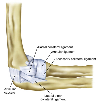
The RUHL complex as described by O’Driscoll and coworkers1 is formed by three separate components that may have a variable expression. The radial collateral ligament is adjacent to the capsule and courses from the lateral epicondyle to the annular ligament and then down to the ulna. The RUHL begins at a variable point on the posterolateral aspect of the lateral epicondyle and courses distally to the crest of the ulna while sending fibers to the annular ligament and blending with the lateral collateral ligament (Fig. 12-1). The annular ligament originates and inserts on the ulna while following a course around the radial neck.
Anatomic studies have attempted to define the involved tissue. Dunning and colleagues2 stated that the RUHL and the radial collateral ligament must be sectioned to achieve PLRI. They also found that they could not visually differentiate the two ligaments at their humeral origin. They could differentiate the RUHL from the radial collateral ligament only by identifying the distal extent of the RUHL at the supinator crest of the ulna.2 Seki and associates3 were able to show that sectioning just the anterior band of the lateral collateral complex induced instability. This suggests that an intact RUHL cannot stabilize the elbow.3 These data demonstrate that the cause of PLRI is a spectrum of injury. Although originally described as sequelae of an elbow dislocation, these anatomic studies and a report by Kalainov and Cohen4 support our own experience that there is a continuum of injury between PLRI and frank elbow dislocation.1,5
Instability findings may coexist with the standard examination findings of lateral epicondylitis, radial tunnel, and posterolateral plica syndrome. Kalainov and Cohen4 posit that PLRI may be a cause of these problems of the elbow. Twenty-five percent of patients in their study had previous surgery for chronic, recurrent lateral epicondylitis. We think that uncorrected posterolateral instability of the elbow may result in increased tension on the lateral musculature as it attempts to stabilize the elbow, thereby producing a secondary lateral epicondylitis. Other tertiary findings, such as an inflamed posterolateral plica and inflammation of the posterior interosseous nerve in or near the radial tunnel, may also occur with the instability. Physicians must look for the instability and fully evaluate the elbow of patients with all of these findings. The clinical examination recommended by O’Driscoll and colleagues1 and by Regan and Lapner6 can assist in the determination of a coexisting instability as a base cause of these problems in the elbow.
Smith and colleagues5 initially described the role of arthroscopy in treating PLRI, and there have been various anecdotal reports since then.7 In this chapter, we update and summarize the current information about the diagnosis and management of PLRI.
PATIENT EVALUATION
History and Physical Examination
Instability is best demonstrated clinically with the pivot shift test of the elbow. As first described by O’Driscoll and colleagues,1 this test with the patient in the supine position may elicit gross instability or pain and apprehension.1 Two other clinical tests assess (1) pain when pushing up from an arm chair with the palms facing inward and (2) having the patient push up from a prone or wall-leaning position first with the forearms maximally pronated and then repeating the test with the forearms supinated, reproducing pain or instability, or both.6,8
Diagnostic Imaging
Imaging studies for PLRI can be helpful. Radiographs may reveal an avulsion fragment from the posterior humeral lateral epicondyle in acute cases. However, radiographic findings often are normal. A stress radiograph or fluoroscopic scan while performing the pivot shift test may show the radial head and proximal ulna moving together in a subluxated and posterolaterally rotated position. Magnetic resonance imaging (MRI) of the elbow can identify a lesion in the RUHL.9 It has been our experience that MRI is most helpful when contrast is added. This can be done for formal arthrography or, in the case of office MRI, an injection of 20 to 30 mL of sterile normal saline with or without gadolinium delivered into the olecranon fossa just before the scan can greatly enhance the effectiveness of the test.
TREATMENT
Indications and Contraindications
The indications for treatment of PLRI are the same as for any other injury: pain and functional impairment. Although much has been written about the pathologic anatomy and biomechanics of the lesion, little has been reported on the surgical treatment of these patients. Consequently, no level 1 studies comparing the effectiveness of operative or nonoperative management of this disorder have been published, nor are there any large published series describing the outcomes of the surgical treatment of PLRI. In the following sections, we review the outcomes of our experiences with arthroscopic repair, plication, and open grafting techniques.5
Arthroscopic Technique
The surgical treatment of posterolateral instability may be divided into distinct subgroups based on cause: acute dislocations, recurrent dislocations, and PLRI. The procedures used may also be divided into subgroups based on available tissue at the time of reconstruction: repair of ligamentous avulsion, plication of the RUHL complex with or without repair to bone, and tendon graft reconstruction.


