Arthrogryposis
CASE PRESENTATION
A 2-year-old female presents for evaluation of a stiff elbow. Prenatal diagnosis was made by prenatal ultrasound examination and pediatric orthopaedic consultation of arthrogryposis multiplex congenita (amyoplasia), and this was confirmed on postnatal genetic evaluation. She has had prior treatment of neurogenic clubfeet but is independently ambulatory and otherwise healthy. She has had elbows fixed in extension since birth, with considerable limitations in wrist and hand function as well. Radiographs of the elbow appear normal (Figure 23-1). The family now seeks treatment options to improve both function and aesthetics of the upper limbs.
CLINICAL QUESTIONS
What is arthrogryposis?
What causes arthrogryposis?
How is it classified?
What are the principles of treatment?
What surgical procedures can be performed to improve hand and upper limb function in patients with arthrogryposis?
THE FUNDAMENTALS
Few conditions present as many clinical challenges for the pediatric hand and upper limb surgeon as arthrogryposis. The combination of functionally limiting joint contractures, hypoplastic or absent skeletal muscle, and normal to high intelligence motivate the patient and family to seek aggressive treatment. Unfortunately, to date, imperfect surgical solutions exist. Despite these challenges, a number of interventions may provide improved upper limb and hand function in selected patients. The ultimate goals of any musculoskeletal treatment are improved functional independence as an adult and as healthy and happy a childhood as feasible. Making the hand and upper limb “normal” is not possible and needs to be stressed early on in life, no matter how difficult it is for the family and patient to hear. As challenging as it may be, admitting our present limits can be very helpful for patients and families to focus their energy where it can do the most good.
Etiology and Epidemiology
Motion is life.
—Robert Salter
Arthrogryposis is a descriptive term used to describe a host of clinical conditions resulting in multiple congenital joint contractures.1 The most common type is amyoplasia (also known as arthrogryposis multiplex congenita or classic arthrogryposis).2, 3 and 4 Other variants include distal arthrogryposis, which is characterized by hand and foot involvement with relative sparing of the proximal limb segments.5,6 Arthrogryposis is also associated with a number of systemic or generalized conditions (syndromic arthrogryposis), including Freeman-Sheldon (so-called whistling face) syndrome.5,7,8
Although the primary condition involves failure of skeletal muscle formation and development, arthrogryposis manifests as multiple joint contractures. Indeed, “arthrogryposis” loosely translates into “abnormally curved” or “stiff joints.” While the pathoanatomy and pathophysiology vary and continue to be investigated, it appears that the joints initially have full developmental potential but fail to form mobile articulations secondary to the absence of movement in utero. This theory has been supported by a number of chick embryo studies, in which paralytic agents were administered during development, resulting in abnormal joint morphology and stiffness.9, 10, 11 and 12 The lack of joint motion results in articular cartilage abnormalities, failure of joint cavitation, and secondary fusions.
Amyoplasia is seen in approximately 1:3,000 live births.13 It is thought to be sporadic, with no genetic or hereditary predisposition. In contrast, distal arthrogryposis is comprised of a number of autosomal dominant disorders, all of which are due to mutations in genes responsible for myofiber function, including TNNI2, TNNT3, TPM2, MYH3, and MYH8.14, 15 and 16
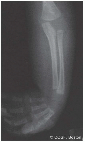 FIGURE 23-1 Radiograph of the elbow, forearm, wrist, and hand of a patient with arthrogryposis. Note is made of the elbow extension and wrist flexion contractures. The osseous elements and joint spaces appear normal. |
Clinical Evaluation
If you ain’t looking at the big picture, you just ain’t looking…
—Stephen Root
Amyoplasia is characterized by multiple, often symmetric joint contractures associated with marked muscle underdevelopment. The upper limbs will typically demonstrate posturing and/or contractures of shoulder internal rotation and adduction, elbow extension, wrist flexion and ulnar deviation, digital metacarpophalangeal (MCP) flexion, interphalangeal (IP) extension, and thumb-in-palm deformity. Associated musculoskeletal conditions include hip dysplasia, knee contractures, and clubfeet. Up to 10% of patients will have abdominal abnormalities.17 Intelligence and cognitive abilities are normal or above average in distal arthrogryposis, amyoplasia, and many patients with syndromic arthrogryposis.
Careful history and physical examination are performed to assess the overall functional abilities and needs of the patient, particularly with regard to upper extremity and hand use. Passive range-of-motion measurements are made of the shoulder, elbow, forearm, wrist, and hand. The presence of active joint motion and strength, if any, is similarly noted. The functional status of the patient is very important to assess. These children will use head tilt, trunk sway, table stabilization, and leg and opposite arm assistance to improve functional independence. Do not just get lost in the specifics of each joint and muscle; see the child as a whole for what he or she can accomplish. If surgery is considered, radiographs are obtained of the affected joint(s), particularly in the older patient, to rule out joint fusions and/or secondary bony deformities.
Surgical Indications
We try to stress the little things because little things lead to big things.
—Steve Alford
Goals of treatment are to improve upper limb mobility and function in order to increase independence with activities of daily living and ambulation/locomotion. Occupational and physical therapy are essential components of treatment for these children and begin at a very young age. Surgical strategies for arthrogryposis focus on functional improvement and are performed in up to 25% of patients.18 Indications include limited joint motion and upper extremity function refractory to nonoperative care in the motivated patient.1,19, 20 and 21 As with all systemic conditions in children, nonoperative and surgical treatment must be tailored to each individual patient.
Ezaki19 has previously outlined the guiding principles of surgical treatment. First, any joint motion that is present should be preserved. Second, passive motion must be obtained before active motion is surgically provided. Third, the potential for bimanual hand/upper limb use must be prioritized, due to the relative weak grasp and associated wrist and digital stiffness. Finally, upper limb function should be maximized for use at the tabletop level.
The optimal timing of surgical intervention is unclear and remains individualized according to the overall needs and status of the individual patient. Considerations are made to (1) maximize upper limb mobility during the development of gross and fine motor activities; (2) optimize upper limb function by the time the patient is of school age; (3) prevent irreversible bone and joint changes; and (4) perform upper limb surgery in conjunction with other (lower extremity) surgical procedures whenever possible. Surgical treatment in these children most commonly addresses contractures of elbow extension, wrist palmar flexion and ulnar deviation, and thumb-in-palm deformity.
SURGICAL PROCEDURES
• Splinting, Occupational Therapy
Occupational and physical therapy, including stretching and splinting, are critical components of upper extremity care in patients with arthrogryposis.22 Indeed, the vast majority of patients will continue to undergo therapy and splinting treatments well into the second decade of life.23 Modest gains in elbow, wrist, and digital motion will go a long way in improving function. The younger the age at which therapy is initiated, the more likely it is to be successful. However, it may take 18 to 24 months to achieve maximum outcome with therapy or to be truly determined as ineffective. For example, aggressive elbow flexion therapy can result in up to 90 degrees of passive motion in up to 80% of patients if consistently performed until 18 months of age, a huge advantage for these patients. It enables them, with functional adaptations such as table stabilization, trunk sway, and head tilt, to feed themselves and perform independent facial hygiene.
In addition, adaptive devices may further increase independence of feeding, dressing, personal hygiene, and other activities of daily living with considerable benefits to the patient.24 This is particularly important as these patients approach school age, where adaptive tools may assist with keyboarding and tabletop activities, and independent hygiene and feeding are very important. The computer age has brought many technologic advances for these children, including voice-activated software and memory-recognition programs.
▪ Elbow Release, V-Y Tricepsplasty
Given that the most important purpose of the elbow is to move the hand to and from the face, correction of elbow extension contractures remains a primary goal of surgical treatment.19 Elbow surgery is indicated in roughly 20% of patients who fail passive motion therapy, with posterior capsulectomy and V-Y lengthening of the triceps being the procedure of choice in patients with amyoplasia (Figure 23-2). Preoperative radiographs must confirm the presence of sufficient joint space to allow for correction. Remember, passive motion must be achieved through therapy or surgery before consideration of active transfers.
Under general anesthesia and tourniquet control, a posterior midline incision is created, curved radially over the olecranon process. Skin flaps are raised and hemostasis obtained. Despite the apparently normal girth of the brachium, there is typically an abundance of subcutaneous fat with very thin and fibrotic triceps muscle mass. The ulnar nerve is identified, released, and protected at all times. It too will be atrophic and at times, almost fibrotic.
The medial and lateral margins of the triceps are identified, and the plane between the capsule and triceps developed. A long V-shaped incision in the triceps is created through the tendinous region, with the base distal and apex proximal. Care is made to make this “V” as long as possible to allow for adequate lengthening. The triceps flaps are then reflected proximally and distally, exposing the posterior elbow capsule. With release of the triceps alone, there is usually very little improvement in passive elbow flexion.
Related posts:

Stay updated, free articles. Join our Telegram channel

Full access? Get Clinical Tree


