Arthrodesis for the Chronically Infected Total Knee Arthroplasty
Stephen J. Incavo
Michael R. Dayton
INDICATIONS
Knee arthrodesis is a salvage operation used when staged revision has failed to resolve infection after total knee arthroplasty (TKA), when the extensor mechanism is unreconstructable, or when soft tissue coverage is inadequate for further revision arthroplasty. Its goals are the eradication of infection and a successful fusion resulting in a stable and pain-free knee.
Arthrodesis does not guarantee resolution of infection. In fact, given the chronicity of infection in some patients, the risk of post-fusion sepsis or osteomyelitis likely remains elevated (supporting the role of chronic suppressive antibiotic therapy in many of these patients). Even if successful arthrodesis is achieved, recurrent local infection may cause chronic pain and pose a systemic threat.
CONTRAINDICATIONS
Successful fusion requires not only bone healing but also reasonable soft tissue coverage. Therefore, a fusion may not be feasible in patients who have compromised and unreconstructable soft tissue coverage. Plastic surgery consultation should be sought early to determine the feasibility of flap coverage in the setting of chronic knee infection with wound breakdown.
Arthrodesis is also contraindicated in most nonambulatory patients who might be served better with resection arthroplasty or above-knee amputation. Fusion may be contraindicated in the presence of overwhelming or persistent sepsis, particularly when arthroplasties are present in other joints, because they would be at increased risk for metachronous infection.
Patients with unreconstructable severe vascular insufficiency may be better candidates for an above-knee amputation.
In the presence of severe bone deficiency, successful arthrodesis may be untenable, although emerging fusion devices or future biologic options may expand the indications for fusion. Chronically infected knees treated previously with hinged implants with metaphyseal-filling or segmental-replacing metal augments may not be amenable to fusion; in such cases, above-knee amputation may be a more appropriate solution.
There are unusual situations that may render the patient an unsuitable candidate for knee arthrodesis, although it is unclear whether these criteria are as appropriate in patients requiring salvage of a chronically infected TKA as they are in patients considering arthrodesis for primary or secondary arthritis of the knee. These include patients with a contralateral above-knee amputation or knee or hip arthrodesis, or those with ipsilateral hip or ankle arthritis.
FIXATION OPTIONS
A variety of fixation options exist for knee arthrodesis. These include either cephalomedullary or modular intramedullary nails, external fixation, and internal plate fixation. Traditionally, external fixation with circular frames or multiplanar fixation and internal fixation using plates and screws have had lower fusion rates than intramedullary techniques. This statistic may, in fact, be changing with the use of modern locking plates, which provide improved stability. External fixators have the unique risks of pin tract problems and a relatively limited capacity for compression. Fusion rates are relatively low with external fixation compared to alternative methods and is therefore used less frequently than the other methods of fusion.
Plate fixation and external fixators have the advantage of limiting the fusion procedure to the knee, and intramedullary techniques theoretically carry a potential risk of spreading a residual infection up and down the femoral or tibial intramedullary canals. Despite this theoretical concern regarding intramedullary fixation, however, the meticulous attention placed on the eradication of infection prior to knee arthrodesis surgery has limited its occurrence in most contemporary reports on intramedullary techniques of knee fusion (1). Intramedullary techniques are also facilitated with the use of improved intramedullary nail systems. Long intramedullary nails are easily removed should further surgery be necessary. On the other hand, although short intramedullary fusion nails are appealing because they are relatively easily inserted and assembled through the knee, they can be very difficult to remove if infection recurs once the joint is successfully fused, unless an osteotomy or large bony window is made in the knee.
In the absence of ipsilateral hip problems or “obstacles” (such as a total hip arthroplasty), we prefer to use intramedullary nail fixation; therefore, the remainder of this chapter focuses on the insertion technique of long cephalomedullary nails, with a brief discussion on double plating.
PREOPERATIVE PREPARATION
Arthrodesis for salvage of the chronically infected TKA should be preceded by aggressive treatment of the infection, including removal of implants, cement debris and necrotic tissue, an appropriate course of antibiotics (perhaps even chronic antibiotic suppressive therapy), and a period of treatment with antibiotic-impregnated spacers (Fig. 34-1). Ideally, the serum C-reactive protein and erythrocyte sedimentation rate should be normalized, and there should be negative cultures of fluid from a joint aspiration or tissue biopsy. If evidence exists of residual infection or necrotic tissue, further debridement and attempts at eradication of the infection should be performed before attempting arthrodesis.
The history should determine whether the patient has ipsilateral hip, ankle, or foot problems that would influence the decision to proceed with arthrodesis and the choice of technique for fusion. Determine whether the patient has a hip replacement, previous hip fracture, or hardware in place. Examine the ipsilateral hip and ankle to make sure range of motion is adequate to sustain the patient following the arthrodesis. Determine if there are other problems related to the ipsilateral hip or femur, such as deformity, which would have an impact on knee arthrodesis. Document the neurovascular status of the limb. Make sure the patient has an adequate vascular supply to heal skin incisions at the knee. Examine the skin integrity to determine whether muscle flaps are present from previous reconstructions that need special consideration during surgical exposure.
Anteroposterior and lateral radiographs of the affected knee and entire lower extremity from hip to ankle are necessary to identify existing deformity, assess bone stock, and ensure that no unexpected findings are present through the entire length of the femur and tibia (such as extra-articular deformity or retained hardware) that would preclude the use of intramedullary fixation. Apparent obstructions, such as fracture malunion, callus or deformity, ipsilateral hip arthroplasty, or retained hardware require an alternative to intramedullary fixation (such as plating or external fixation). A long, standing radiograph from hips to ankles with radiographic magnification markers is helpful to determine the overall limb alignment, to identify whether there are bony deformities of the femur or tibia, and to plan for fixation device dimensions. If fusion with a long antegrade intramedullary nail is anticipated, calculate the proper nail length and diameter; this calculation needs to take into account the radiographic magnification. The measurement should extend from the tip of the greater trochanter to within 8 cm from the tibial plafond. The calculation should also account for the gap between the distal femur and the proximal tibia that will be closed at arthrodesis and the bone that will be removed as the distal femur and proximal tibia are trimmed to optimize apposition during
arthrodesis. Lateral radiographs of the femur and tibia with radiographic markers are helpful to determine the proper diameters of the femoral and tibial portions of the rod. Restoring limb length after failed treatment of infection after TKA is less important than maximizing bone apposition and achieving successful fusion. Therefore, use of structural graft to restore lost bone mass and length is not generally advised.
arthrodesis. Lateral radiographs of the femur and tibia with radiographic markers are helpful to determine the proper diameters of the femoral and tibial portions of the rod. Restoring limb length after failed treatment of infection after TKA is less important than maximizing bone apposition and achieving successful fusion. Therefore, use of structural graft to restore lost bone mass and length is not generally advised.
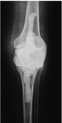 FIGURE 34-1 Preoperative x-ray showing the presence of a spacer block in a patient’s knee after multiple attempts at salvage of a chronically infected total knee arthroplasty. |
TECHNIQUE
Setup
Placement of an intramedullary device for knee arthrodesis requires a radiolucent operating room table with a single C-arm fluoroscopic unit. The C-arm is best positioned over the patient (not under the operating room table). The patient is placed in a lateral decubitus position with the affected limb up (Fig. 34-2). Alternatively, the patient could be positioned semi-supine with a bump under the ipsilateral hip. The affected limb should be prepared and draped free from the gluteal region, torso, anterior hemipelvis, and buttock down to the ankle. This optimizes positioning, access to the
hip area, and maneuverability while the guide, reamer, and nail are passed antegrade through the hip. Do not use an ipsilateral arm board, as this will get in the way during passage of the guidewire, reamers, and the long nail. A sterile tourniquet may be placed on the thigh during knee exposure. Prior to starting surgery, confirm that the starting point for antegrade intramedullary nailing is accessible. Also confirm that the distal part of the leg is accessible where a distal interlocking screw may be inserted.
hip area, and maneuverability while the guide, reamer, and nail are passed antegrade through the hip. Do not use an ipsilateral arm board, as this will get in the way during passage of the guidewire, reamers, and the long nail. A sterile tourniquet may be placed on the thigh during knee exposure. Prior to starting surgery, confirm that the starting point for antegrade intramedullary nailing is accessible. Also confirm that the distal part of the leg is accessible where a distal interlocking screw may be inserted.
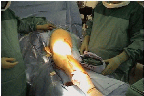 FIGURE 34-2 Patient positioned in the lateral decubitus position. |
Procedure
The knee is approached through the standard anterior arthrotomy. If that is not possible, use the same criteria for choosing incisions as one would for revision knee arthroplasty. Be sure to maintain full-thickness skin flaps and avoid devascularization of the skin. If the patella is still present, perform either a straight midline or a medial parapatellar arthrotomy, and elliptically excise the patella. Obtain adequate exposure to débride the knee. Remove antibiotic-impregnated cement spacer blocks if present and débride soft tissue from the metaphyseal bone ends. Remove only the minimum amount of bone necessary to obtain adequate contact between the surfaces of the femur and tibia. The goal is to cut the femur and tibia perpendicular to the long axis of the medullary canal of each with no posterior slope. The distal femoral and proximal tibial canals are identified (Fig. 34-3A). The tibia is reamed in an antegrade fashion under direct vision with flexible intramedullary reamers, the last of which matches the size of the tibial portion of the nail (Fig. 34-3B


Related posts:


Stay updated, free articles. Join our Telegram channel
Full access? Get Clinical Tree
