Chapter 7 Arm and Hand
1. Understand the musculoskeletal design of the arm.
2. Explain the design of the elbow joint.
3. List and describe the ligament of the elbow joint.
4. Describe and identify common locations of nerve impingements in the elbow.
5. List and describe the nerves of the arm.
6. Explain the differences between tendonitis and tendinosis.
7. Demonstrate massage applications for arm massage.
lateral collateral (radial) ligament
medial collateral (ulnar) ligament
proximal ulnar (cubital) tunnel
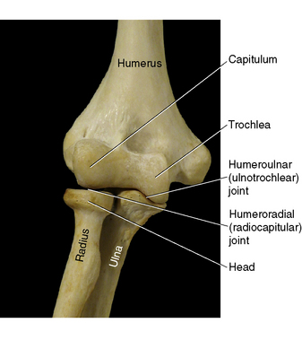
Although classified as a hinge joint, the elbow is unique in that it has three articulations located within one joint capsule. Both the humeroradial and the humeroulnar joints work together to allow flexion and extension of the forearm. The proximal radioulnar joint allows for pronation and supination of the wrist (Figure 7-1).

Figure 7-1 Elbow joint.
(From Muscolino JE: Kinesiology: the skeletal system and muscle function, ed 2, St Louis, 2011, Mosby.)
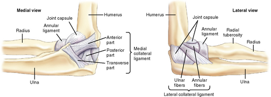
The elbow has a complex design of ligaments to help support and protect the joint consisting of the three bones and three articulation structures. These ligaments are the medial collateral (ulnar) ligament, the lateral collateral (radial) ligament, the annular ligament, and the fibrous joint capsule (Figure 7-2).

Figure 7-2 Elbow ligaments.
(From Muscolino JE: Kinesiology: the skeletal system and muscle function, ed 2, St Louis, 2011, Mosby.)


