Anterior Cruciate Ligament Reconstruction in The Pediatric Patient


Anterior Cruciate Ligament Reconstruction in The Pediatric Patient


Craig Finlayson

Adam Nasreddine

Mininder S. Kocher


The knee is the most common site of injury in the skeletally immature athlete (1). The incidence of anterior cruciate ligament (ACL) tears appears to be on the rise. The treatment of these injuries is controversial. Nonoperative management can lead to functional instability and difficulty with cutting and pivoting sports. In addition, the pathologic shear forces are associated with meniscal and chondral damage over time. ACL reconstruction in children and adolescents risks iatrogenic injury to the physis. This chapter reviews the historic perspective of ACL injuries in the young patient, clinical and diagnostic findings in children, treatment options, and results of treatment.


HISTORICAL NOTE

The ACL is the principal intraarticular stabilizer of the knee. As in adults, an ACL injury in a child or adolescent is usually a noncontact valgus injury. Before the 1980s, these injuries were thought to be rare in the pediatric athlete. Advances in diagnostic imaging and improved clinical acumen have allowed physicians to identify midsubstance ACL tears in patients with open physis (2, 3 and 4).

The results of nonoperative management in children are consistently associated with poor outcomes (5, 6). Aichroth et al. reported on 23 children that were treated nonoperatively between 1980 and 1990. At final followup, meniscal tears were present in 15 knees, three osteochondral fractures occurred, and osteoarthritic changes developed in 10 knees. From 1980 to 1985, McCarroll followed 16 patients younger than 14 years with open physes and midsubstance tears of the ACL treated without reconstruction. Six patients underwent arthroscopy for meniscal tears. Only seven patients returned to sports, all experiencing recurrent episodes of giving way, effusions, and pain.

Attempts at primary repair of the ligament in children have resulted in poor outcomes (7). Engebretsen et al. presented eight adolescents that were followed 3 to 8 years after primary suture of a midsubstance rupture of the ACL. Only three patients had good function, and five were functionally unstable. Failure of primary repair has led to the development of various procedures to stabilize the knee. Surgical options include transphyseal, partial transphyseal, and physeal-sparing reconstructions.


TIBIAL SPINE FRACTURES AND PARTIAL ACL TEARS

It is important to understand the different types of injuries that can occur in the skeletally immature patient. Partial ACL tears and avulsion fractures of the tibial spine are more common in the pediatric population (8). Excellent functional results have been reported following arthroscopic reduction and internal fixation of tibial spine fractures, although long-term follow-up does demonstrate some residual laxity, indicative of associated intrasubstance injury to the ACL (2). Many partial tears can be treated nonoperatively (9). On the basis of a prospective study of arthroscopically confirmed partial ACL tears, failure of nonreconstructive treatment has been associated with tears greater than 50%, tears of the posterolateral bundle, older skeletal age, and presence of a pivot shift.


CLINICAL EVALUATION


History and Physical Examination

Important history questions are as follows:


  • 1. How did the injury occur?


    • a. Was there contact with another athlete?


    • b. Was there a fixed position of the foot and rotation or twisting movement?


  • 2. Were you able to continue to compete?


  • 3. Was there significant swelling directly after the injury?


  • 4. Have there been previous injuries to the knee?

Our understanding of ACL tears in the setting of younger athletes has changed considerably. The tibial spine fracture was once thought to be the pediatric equivalent of an ACL tear. Midsubstance ACL ruptures are now diagnosed more frequently in pediatric athletes
participating in cutting and contact sports. The typical presentation is a young athlete who has a decelerating, twisting injury. Approximately two-thirds of ACL injuries occur by noncontact mechanisms (10). The patient will often report a “pop” and the inability to return to the field. A large amount of swelling due to hemarthrosis is expected. The presentation is less dramatic in athletes who have had a prior partial tear of the ACL.

The findings on physical examination are dependent on the timing in relation to the injury. Directly after the injury, the stability of the knee can be tested on the sideline. The Lachman and pivot shift tests are positive before swelling and guarding occurs. When the patient presents for evaluation in the emergency department or clinic, the knee is typically swollen, compromising the ability to perform an accurate physical examination. Rates of ACL injury are reported between 10% and 65% in pediatric patients presenting with traumatic hemarthrosis of the knee; therefore, young athletes presenting with a hemarthrosis of the knee should raise suspicion for an ACL tear (11, 12). The differential diagnosis of hemarthrosis of the knee includes patellar dislocation, meniscal tear, osteochondral fracture, tibial spine fracture, and epiphyseal fracture of the femur or tibia.

A thorough examination of the knee must be performed to rule out concomitant injuries. Associated injuries include meniscal tears, posterior cruciate and/or collateral ligament tears, osteochondral fractures, and physeal fractures of the distal femur or proximal tibia. Given the higher prevalence of generalized ligamentous laxity in skeletally immature patients, a direct comparison to the contralateral knee should also be made. The Lachman and pivot shift maneuvers are used to test for ACL insufficiency.


Imaging

Evaluation of the knee by MRI is an important part of the assessment, particularly in children. The MRI is useful to distinguish between partial tears, avulsions, and midsubstance tears of the ACL. Secondary findings in an acute injury include hemarthrosis and the presence of a bone contusion at the posterior lateral tibial plateau and anterior lateral femoral condyle. The MRI is useful for confirming the diagnosis of ACL tear, ruling out associated injuries, and assisting in preoperative planning (Fig. 80.1).


INDICATIONS AND TIMING OF SURGERY

Indications for ACL reconstruction in a skeletally immature patient include complete ACL tear with functional instability, partial ACL tear that has failed nonoperative treatment, and ACL injury with associated repairable meniscal or chondral injury. Owing to higher rates of postoperative stiffness, acute ACL reconstruction is not recommended for isolated ACL tears (13). Surgery is typically delayed at least 3 weeks from the time of injury until adequate range of motion has been achieved and joint effusion minimized. Patients must be mature enough to participate in the extensive rehabilitation process following ACL reconstruction.


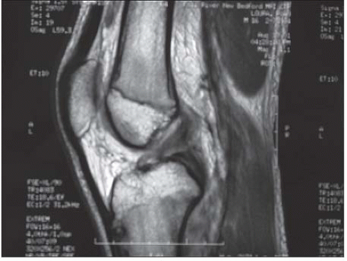
FIGURE 80.1. MRI demonstrating midsubstance ACL tear.


TREATMENT OPTIONS

The choice of surgical technique is dependent on the physiologic age of the patient and the amount of growth remaining. For prepubescent children, violation of the tibial and femoral physis presents a risk of significant growth disturbance that would require limb lengthening or osteotomy. Animal studies have demonstrated a risk of physeal arrest with transphyseal ACL reconstruction(14, 15). A number of growth disturbances following ACL reconstruction in this age group have been documented (16). Radiographs and developmental findings are used to determine the physiologic age. Referencing radiographs of the left wrist to the atlas of Greulich and Pyle (17) provides an efficient means to determine skeletal age. The physiologic age is based on the Tanner staging system (Fig. 80.2 and Table 80.1) (18).

The prepubescent child (Tanner stage I or II) with a midsubstance ACL tear presents a difficult problem. Because of the large amount of growth remaining, the consequences of iatrogenic physeal arrest are severe. Unfortunately, activity modification such as refraining from cutting sports is difficult in this age group, and nonreconstructive treatment has been associated with meniscal and chondral injury (19, 20, 21 and 22).

Surgical techniques include physeal-sparing, transphyseal, and partial transphyseal reconstructions. In theory the extraarticular reconstruction provides a method to restore stability and avoid risk of growth disturbance. At our institution, we use a modification of the MacIntosh ACL reconstruction to perform a physeal-sparing reconstruction with an extra- and intraarticular component that is described in detail later in the chapter.


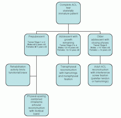
FIGURE 80.2. Algorithm for management of complete ACL injuries in skeletally immature patients.


Table 80.1 Tanner staging classification of secondary sexual characteristics


Tanner Stage


Male


Female


Stage I (prepubertal)


Growth development


5-6 cm/y


Testes <4 mL or <2.5 cm


No pubic hair


5-6 cm/y


No breast development


No pubic hair


Stage II


Growth development


5-6 cm/y


Testes 4 mL or 2.5-3.2 cm


Minimal pubic hair at base of penis


7-8 cm/y


Breast buds


Minimal pubic hair on labia


Stage III


Growth development


7-8 cm/y


Testes 12 mL or 3.6 cm


Pubic hair over pubis


Voice changes


Muscle mass increases


8 cm/y


Elevation of breast; areolae enlarge


Pubic hair of mons pubis


Axillary hair


Acne


Stage IV


Growth development


10 cm/y


Testes 4.1-4.5 cm


Pubic hair as adult


Axillary hair


Acne


7 cm/y


Areolae enlarge


Pubic hair as adult


Stage V


Growth development


No growth


Testes as adult


Pubic hair as adult


Facial hair as adult


Mature physique


No growth


Adult breast contour


Pubic hair as adult


Other


Peak height velocity: 13.5 y


Adrenarche: 6-8 y


Menarche 12.7 y


Peak height velocity: 11.5 y

Only gold members can continue reading. Log In or Register to continue

Stay updated, free articles. Join our Telegram channel

Jul 22, 2016 | Posted by in ORTHOPEDIC | Comments Off on Anterior Cruciate Ligament Reconstruction in The Pediatric Patient

Full access? Get Clinical Tree

Get Clinical Tree app for offline access