Ankle Replacement Arthroplasty


Ankle Replacement Arthroplasty


John M. Schuberth

Jerome K. Steck

Jeffrey C. Christensen


Over the past 50 years, tremendous knowledge has been accumulated regarding total joint arthroplasty technique, prosthetic materials science, and advanced engineering designs. Driven by this formative knowledge base, the science of total arthroplasty has been applied to other major joints including the ankle. Over the past decade, with these refinements and accumulated global experience, total ankle replacement (TAR) has emerged as a viable alternative in the surgical management of advanced ankle arthritis. In concept, ankle replacement is more attractive to many patients than fusion, but clearly not all are good candidates for implantation. Successful implantation requires a synchronization of sound component design, astute patient selection, precise prosthetic placement and sizing, and comprehensive patient education.

The authors’ goal is to provide a comprehensive chapter that has utility to all practicing foot and ankle surgeons, even those who choose not to perform TAR. Where necessary, due to the vast and rich literature on hip and knee total arthroplasty, references from non-ankle joint studies are included to provide perspective and means of extrapolation to the reader. With increasing popularity of TAR, it will be necessary for all foot and ankle specialists to have a fundamental knowledge of the procedure and to properly evaluate, troubleshoot, and discuss with patients the complexities of their condition.


PERTINENT BIOMECHANICS AND ANATOMY

Restoration of function to the diseased ankle joint requires a sound understanding of the joint kinematics of the hindfoot and ankle, surface geometry combined with ligamentous constraints, and periarticular mechanical properties of a normal articulation. The biomechanical environment and complexities of the ankle are more demanding that those found in the knee and hip, with a smaller working area combined with additional moving parts functioning under higher stresses. When treating a patient with an arthritic ankle, one needs to recognize and treat any prevailing biomechanical deficiencies superimposed with the end-stage arthritic joint to optimize the prosthetic ankle function.

The functioning ankle is one of the six determinants of gait and plays an essential role in the kinetic chain of the lower extremity (1,2). The ankle integrates with the other critical components of gait that allow for energy efficient ambulation. In normal walking, the ankle and foot functions to maintain dynamic limb length. Throughout the contact phase of gait, the majority of ankle movement occurs in the sagittal plane. At heel strike, the ankle plantarflexes in a controlled manner through eccentric contracture of the anterior leg musculature. With the foot plantigrade, the ankle then dorsiflexes as the triceps surae decelerates forward leg movement. Finally, at heel off, the motion of the ankle reverses and begins to plantarflex until toe off. Several factors affect ankle joint mechanics. It is the resultant movement and stability during gait and is a product of external and muscular loads, joint surface geometry, and ligamentous arrangement and constraints (3).


RANGE OF MOTION MECHANICS

The location of the ankle joint axis approximates the tips of the malleoli (3,4) (Fig. 52.1). The true range of motion (ROM) has been debated and is dependent on landmarks, measurement technique, and open or closed kinetic chain. Clinical ankle range of motion is a blending of ankle, subtalar, and midtarsal joints. Roaas and Andersson (5) measured total ROM at 55 degrees (dorsiflexion 15.3 degrees; plantarflexion 39.7 degrees), while Boone and Azen (6) measured a total of 68.8 degrees (dorsiflexion 12.6 degrees; plantarflexion 56.2 degrees). Tibiotalar ROM measured radiographically has been reported for dorsiflexion at 14 ± 7 degrees and plantarflexion at 32 ± 6 degrees (7). While these parameters are important in clinical examination of the ankle, none of this clinical information is useful in understanding the individual contributions of the subtalar and midtarsal joint complex in comparison to pure ankle motion three dimensionally. One must look at static and dynamic kinematic data to begin to understand the complexity of the rearfoot and ankle complex. More sophisticated investigations have analyzed ankle complex kinematics in weight-bearing and non-weight-bearing protocols under different input stresses that showed resultant triaxial motion between the ankle and surrounding intertarsal joints (3,8,9,10,11,12 and 13).

One difficult problem to resolve is defining the precise segmental motions during gait when closed kinetic chain influences are under greatest loads. Motions of the talus and other tarsal bones are particularly hard to measure accurately and are prone to external sensor errors in typical gait analysis investigations (14). In recent years, this issue has been addressed; two investigations have overcome these barriers by placing intraosseous pins with reflective markers into each segment and performed three-dimensional (3-D) gait analysis (15,16). These studies confirmed that the ankle and subtalar joints are working in all three planes during gait. The data by Lundgren and colleagues are the most complete, using six participants with average tibiotalar motion of 15.3 ± 2.0 degrees sagittal, 8.1 ± 3.8 degrees frontal, and 7.8 ± 2.7 degrees transverse. Simultaneous subtalar motion was 6.8 ± 1.4 degrees sagittal, 9.8 ± 1.8 degrees frontal, and 7.5 ± 2.0 degrees transverse.


SURFACE GEOMETRY AND LOAD CHARACTERISTICS

The ankle joint is a highly congruent joint and has a unique surface anatomy, which has been described in detail (17,18). The ankle mortise with the intact support of the lateral collateral, deltoid complex, and syndesmotic ligaments is able to
provide stability in all three planes. When considering joint replacement, there are certain anatomic stabilizing features that need to be considered. First, the curvature of the talar dome in the ankle mortise has stabilizing qualities in anterior to posterior shear loads. Additionally, frontal plane stability is a result of congruent medial and lateral talomalleolar articulations. In the clinical setting, any condition that alters the ankle geometry (i.e., surface incongruity) or through extra-articular malalignments (i.e., end-stage hindfoot pronation) may affect ankle stability under weight-bearing conditions (19).


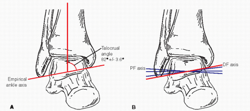
Figure 52.1 A: Diagram of ankle with empirical axis through the talocrural line. B: Diagram showing actual axes of plantar and dorsal flexion.

A normal ankle, including the talomalleolar facets, has a surface area of 12 cm2, which is larger than the hip or knee. However, the tibial plafond surface area is approximately 7 cm2 (20). Compressive loads across the normal ankle is about three times body weight from heel strike to foot flat, followed by additional compressive forces in late midstance to exceed five times body weight (20,21). Medial to lateral shear forces are less than one body weight, whereas forward and aft shear forces are calculated to be in excess of two body weights (21).

Load path analysis has been studied at the ankle, and it has been determined that body weight is shared by the talar dome, and both medial and lateral talomalleolar articulations (22,23). Lateral loads are diverted from the tibia and are transmitted by the fibula. Fibular loads have been determined to be 3.7% to 13% of axial forces depending on ankle position (23).


BONE STRENGTH

The mechanical strength of bone to support total ankle components is unknown. Based on finite element analysis, the simple act of resecting the joint surfaces from the ankle joint immediately will weaken the bone structurally on either side of the articulation (24). Hvid et al (25) evaluated trabecular bone strength of the talus and distal tibia in amputated limbs (25). On average, they demonstrated that tibial bone was 40% weaker than the talus and had an eccentrically positioned area of peak bone strength that was posterior medial. Additionally, in this study, it was confirmed that tibial metaphyseal bone strength is greatest at the subchondral bone level and significantly weakens at each deeper level away from the joint. A similar finding using finite element analysis modeling of a talus predicted a 500% increase in circumferential (hoop) stress with the same load applied (24).


LIGAMENTOUS RESTRAINT

Aside from the articular geometry of the ankle, ligamentous attachments collectively guide and control mobility and stability (19,26). The ligaments that control mobility are designed to maintain length throughout normal ROM (ligament isometry). These guiding ligaments are the calcaneofibular (CFL) laterally and tibiocalcaneal ligament medially (27). The remaining ligaments are stabilizing ligaments that resist movements from externally applied forces. These ligaments work in tandem with the joint surfaces to create a stable joint complex. As an external load is applied, there is an instantaneous response through increased tensile ligament and joint pressure loads that effectively counteract the external force (19). During weight-bearing activities, the lateral collateral and the deltoid ligaments function to keep aberrant joint motions in check. However, the integrity of the deep deltoid is arguably the most critical ligament for proper ankle function; it is the primary restraint for valgus rotation of the talus and has secondary restraint function against lateral and anterior talar excursion (Fig. 52.2). The syndesmotic ligaments are also critical for normal ankle function; chronic instability can lead to posterior migration of the fibula and rotational instability of the syndesmosis with external rotation loads (28,29).


VASCULAR ANATOMY AND SUPPLY

Since most incisional exposures for TAR are an anterior approach, understanding normal arterial anatomy and the incidence and patterns of variation is essential. The normal anatomy has been well studied and known to involve the anterior tibial (AT) artery that then continues as the dorsalis pedis artery distal to the inferior extensor retinaculum. This normal anatomy pattern has been reported to occur 95.7% of the time (30). In their study, they described three variable patterns of arterial flow. Occasionally (2% occurrence), the AT artery will
take a more lateral course deep to peroneus tertius muscle and pass anterior to lateral malleolus. In the second variation (˜1% occurrence), the perforating peroneal assumes the position of the AT artery. In the third variation (1% occurrence), the AT artery will give off a lateral branch to take over function of perforating peroneal. While the AT artery shows mild variation, the dorsalis pedis is consistent in its pattern.

Angiosomes: An angiosome is a 3-D anatomic unit of tissue that is fed by a source artery (31). Choke vessels link neighboring angiosomes to each other and are important conduits that provide blood flow to adjacent zones. The AT artery angiosome covers the anterior leg and ankle, while the dorsalis pedis artery angiosome covers the entire dorsum of the foot. Conceptually, the AT artery angiosome is important surgically to understand the fragility of the soft tissue envelope (see Complications: Wound Dehiscence and Focal Necrosis).


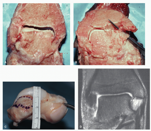
Figure 52.2 A: Frontal cadaveric section of ankle showing intact deltoid ligament including superficial and deep components. B: The superficial component of the deltoid has been removed. The deep portion has been isolated. C: Photo of talus demonstrating the deep deltoid insertion on the medial surface of the talus. D: MRI showing the course of the deep deltoid ligament.


SURGICAL RATIONALE

The surgical rationale for prosthetic replacement is simple: to achieve a durable and functional restoration of the ankle that permits return to normal activities of daily living with reduction or elimination of pain. However, the precise manner, conditions, and limitations at which replacement arthroplasty can accomplish these aims are not fully understood. Clearly, the ultimate expectation is that restoration or even preservation of motion of the ankle will be beneficial. While the rationale for functional restoration is intuitive, other factors that can compromise device survivorship have been appreciated through careful observations of previous failures. Recognition of these factors has shaped refinements in selection criteria, surgical instrumentation, device design, and material selection to better combat the stressful mechanical environment of the ankle.
The preoperative determination of ultimate outcome for the individual patient is an imperfect science to date since there are many contributing factors that are known and unknown that collectively determine clinical outcome. Yet as the collective experience with implant arthroplasty increases, more definitive and predictive parameters will emerge.


ETIOLOGY AND PATHOMECHANICS OF END-STAGE ANKLE ARTHRITIS

Patients with end-stage ankle arthritis often experience significant symptoms of progressive pain, joint immobility, deformity, and reduced activities. As a result, many are challenged to perform basic activities of daily living such as level walking, stair climbing, or stooping. The individual impairment of advanced arthritis of the ankle is severely disabling and is comparable to that of the hip (32). In bilateral disease, such as seen in rheumatoid arthritis, the impact can be even more devastating (33,34).

The pathomechanics of end-stage arthritis is poorly understood and minimally covered in the TAR literature. The majority of arthritis in the ankle is traumatically induced, unlike the hip and knee. Even though many of the patients with end-stage ankle arthritis have a common etiology, there are distinct patterns of disease and rates of degeneration (Fig. 52.3). The specific etiologies can often be explained by the patients’ history but in some instances can only be inferred from experimental data. It has been shown in the laboratory that sectioning of the CFL disconnects some of the kinematic coupling functions or the ankle joint (35) and destabilizes the joint complex at two levels. Clinically, this type of ligament insufficiency can lead to ankle joint incongruity and varus talar tilt. If instability persists, eventual degenerative changes develop from the varus talus alignment against the medial plafond (36). In these varus ankles, the deltoid ligament can also contract, making it difficult to rebalance the ligaments upon reconstruction (37). Valgus talar tilt due to impact injury to the lateral tibial plafond or compromise of the deltoid ligament complex can cause rapid degeneration of the ankle (Fig. 52.3). Degeneration can also occur in situations in which ligament insufficiency is not apparent; however, often these ankles have functional impairment with restricted tethered motion from scarified ligaments; thus, the motion guiding function may be impaired.

In nearly all of these traumatically induced cases, there is some form of ligamentous compromise. Even with intact ligaments, there is often scarification and dysfunction from loss of ligament elasticity (38). The capacity to restore physiologic ROM and maintain proper ligament balance in the setting of severe ligamentous compromise can often be the largest challenge for the surgeon during implant arthroplasty.


INFLAMMATORY ARTHROPATHY

The treatment of patients with end-stage inflammatory ankle arthritis is controversial (39). Ankle arthrodesis has been a primary surgical treatment; however, pain relief may be at the expense of minimal or lost functional improvement (40,41). The rationale for use of TAR in inflammatory arthritis is not as well defined as in posttraumatic arthritis. TAR historically has shown considerable failures in patients with inflammatory arthritis; these primarily involved first-generation devices with constrained prosthetic designs (42,43 and 44). Interest in TAR in inflammatory arthritis has been reawakened in secondgeneration prostheses that demonstrate encouraging survival rates that are equal to or better than patients with osteoarthritis (39,45,46 and 47).


CONSEQUENCE OF ANKLE FUSION

Ankle arthrodesis became a useful technique for providing stability in cases of postpolio paralysis as a result of multiple epidemics from the late 1800s and early 1900s (48,49). It was not until the 1930s that reports of ankle arthrodesis surfaced to treat late results of ankle fracture (50,51). Modern techniques have incorporated internal and external fixation methods to achieve more consistent pain relief and stability and are considered to be beneficial in the surgical treatment for end-stage ankle arthritis (52,53,54,55,56,57,58,59,60,61,62,63 and 64). Regretfully, the assorted types of ankle fusion procedures are burdened with complications and sequelae including wound problems, nerve entrapment, infection, nonunion, and malunion.

The long-term effects of ipsilateral adjacent joint arthrosis after ankle arthrodesis have been studied more recently. The mounting evidence of disappointing long-term results with ankle arthrodesis reflects the heightened pursuit for a reliable ankle prosthesis that is durable and functionally restorative (Fig. 52.4). It is further driven by the continued successes with knee and hip arthroplasty. While ankle arthrodesis techniques and outcomes have improved over time, there are still major complications ranging from 6% to 56% that have been reported in various clinical series (56,58,62,63,65,66,67 and 68). Malunion and pseudoarthrosis are problems that still plague the procedure. Frey et al reported a pseudoarthrosis rate of 41% (32 of 78 ankles). Haddad et al published a meta-analysis on intermediate and long-term outcomes of TAR and ankle arthrodesis involving 852 and 1,262 patients, respectively. TAR revision rates were 7% as compared with 9% in fusions (nonunions being the primary cause of revision in 65% of fusions). Similarly, amputation rates were 5% with ankle arthrodesis compared with 1% in the TAR group (69).

It is clear that the gait pattern after ankle arthrodesis is not normal. Some patients find it impossible to walk without a limp or climb stairs (70). Gait studies have consistently measured decrease in stride length, gait velocity, and cadence (58,61,71). Loss of ankle motion from fusion or advanced arthrosis has a profound impact on the orderly transmission of body weight in the gait cycle and increases energy utilization. Average walking speed of a patient with a unilateral arthrodesis of the ankle is 67 m/min, which is 84% of normal velocity. This results in 3% higher oxygen consumption and a gait efficiency of 90% of normal (72). In a gait analysis investigation analyzing the intermediate results of ankle arthrodesis with age/sex matched controls revealed, kinematic patterns that had significantly diminished triplane motion of the hindfoot and midfoot in the arthrodesis group (71).

It can be argued that patients that present with end-stage arthritis already have limited ROM of the ankle and that fusion would serve to relieve pain but not specifically precipitate adjacent joint arthritis. However, the net and longer-term effects of limited motion are the same regardless of whether the loss of motion is surgically induced or not. Compensatory mechanisms of the hindfoot are activated by the loss of motion at the ankle at the time of fusion or as the ROM decreases insidiously

from loss of joint space and contour. Motion demands are shifted to adjacent and distal joints of the foot. With the loss of ankle motion, these joints are subjected to abnormal forces including torque and excursions, which are at variance to their natural axes of motion. In a recent prospective study, there is a 10.8% increase in combined subtalar and medial column motion within 33 months after isolated ankle arthrodesis (73). In long-term follow-up, ipsilateral adjacent joint arthrosis has been documented to consume Lisfranc, midtarsal, and subtalar joints and not the ipsilateral knee (74,75 and 76). Functional
impairment of end-stage ankle arthritis that is severe has been measured using a Musculoskeletal Functional Assessment (MFA) outcomes measurement tool (77). This study evaluated 426 patients with ankle arthritis and compared them to a general population group, and an ankle/foot trauma group (9 to 12 months after surgery). The results showed MFA scores that were more than triple the scores in the general population, and dysfunction was more than double than patients having a recent history of trauma.


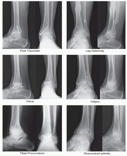
Figure 52.3 Patterns of ankle arthritis.


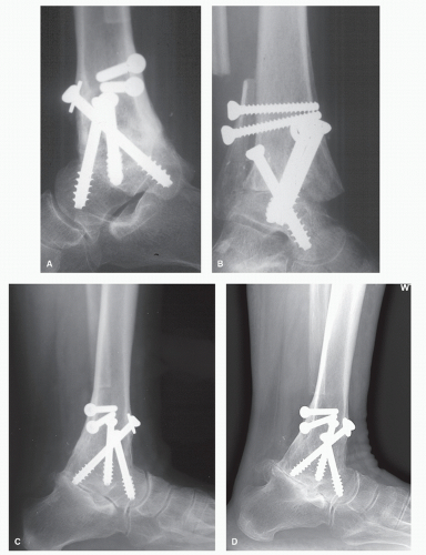
Figure 52.4 Hindfoot arthritis example 20 years after ankle fusion. A: Lateral view with fused ankle. B: Mortise view of ankle showing normal subtalar joint space. C: Lateral x-ray of same patient 8 years postoperatively with subtalar arthritis. D: A 19-year postoperative lateral x-ray with progression of subtalar disease.

Patients with multilevel midstage arthritis that involves the ankle and adjacent joint of the hindfoot and midfoot, have effectively lost their capacity to adequately compensate for lost ankle motion. With ankle fusion, these adjacent joints will rapidly become more symptomatic and progress to end-stage arthritis. This potentially will place the patient at risk for pantalar arthrodesis (78,79).

Patient satisfaction with ankle fusions is initially favorable but deteriorates as the arthritic process expands into the foot. Mazur et al (61) in a classic study evaluating gait in 12 patients post ankle fusion found that motion of the midfoot joints are necessary for patients to compensate for their fused ankle. Patients post ankle fusion will often demonstrate calf atrophy in the ipsilateral limb, which has ranged between 1 and 7 cm (mean 3 cm) (71). The same atrophic developments appear in patients who have had long-standing arthrosis and loss of ankle motion from any cause.


RESTORATION OF NORMAL GAIT PATTERNS

Preservation of movement in combination with enhanced functional locomotion after TAR is a meaningful ultimate goal. Unfortunately, gait laboratory analysis after first-generation implantation showed disappointing results with persistent pain, poor push-off, and slow cadence of gait (80). However, second-generation devices have been analyzed with gait laboratory analysis and have displayed normalization of gait parameters (81,82,83,84 and 85). Sequential 3-D kinematic-kinetic gait analysis performed preoperatively and 3, 6, 9, and 12 months postoperatively after TAR demonstrated a normalization of all gait parameters including improved functional ankle motion (84). While these gait parameters did not return to control levels, beneficial effects were clearly demonstrated postoperatively when compared with preoperative levels with the majority of improvement noted at 1 year. Corroborating this dynamic response, a more recent study evaluated the effects of TAR on gait disability (81). The authors found significant improvement in locomotor function between pre- and postoperative analysis, measuring a significant decrease in energy expenditure with patients functioning with a more propulsive gait pattern.


INDICATIONS, CONTRAINDICATIONS, AND PATIENT SELECTION

The indications for ankle arthroplasty essentially parallel those for ankle fusion (Table 52.1) and include severe, end-stage primary osteoarthritis, inflammatory arthritis (i.e., rheumatoid), and posttraumatic arthritis of the tibial-talar joint in patients who have failed conservative treatments.

Patients with end-stage arthrosis of the ankle can be considered candidates for implantation. There are numerous factors that the surgeon should consider, including age, smoking status, body mass index, overall alignment, quality of the soft tissue envelope, bone stock, neurovascular status, patient physical demands, ligamentous integrity, and medical comorbidities (86). These parameters should be analyzed by the surgeon when selecting a patient for TAR.


TABLE 52.1 Indications and Contraindications for Total Ankle Replacement (95,96,139)


Indications


  • Good bone stock


  • Normal vascular status


  • Good hindfoot alignment


  • Sufficient collateral ligament function


  • Low physical demands


  • Coexistent midfoot and hindfoot arthrosis


  • Bilateral arthritic ankles


Relative Contraindications


  • Previous severe trauma (i.e., open fracture of ankle, talar body dislocation, segmental bone loss)


  • AVN talus 25%-50% of body


  • Severe osteopenia/osteoporosis


  • Long-term use of steroids


  • Insulin dependent diabetes mellitus


  • Demanding sport activities


  • Obesity (especially with relatively small ankle sizes)


  • Younger patients (<50 years) with intact hindfoot joint function


Absolute Contraindications


  • Charcot arthropathy


  • Active or recent infection


  • AVN talus (>50% of talar body)


  • Severe benign hypermobile joint syndrome


  • Nonreconstructible malalignment of the ankle


  • Severe soft tissue problems around ankle


  • Progressive sensory or motor dysfunction of lower leg


  • High physical demand sports/work activities


GENERAL HEALTH

Since the stress of surgery and postoperative rehabilitation demands are nearly identical, the general health of the patient is not a primary determinant in deciding between fusion and implant arthroplasty. Since the goal of salvage surgery for ankle arthritis is to improve the quality of life of the patient and improve functional ambulatory capacity, patients that have substantial health problems that would preclude realization of the benefits of successful ankle surgery might be better served with a nonsurgical approach. Yet, segregation of the impact of end-stage arthritis from the patient’s overall health condition is difficult and subjective at best. Further, the balance of the risks and benefits in patients with superincumbent health problems is intricate. There are published studies that suggest that a patient who is under the management of immunosuppressive medications, diabetes, or rheumatoid arthritis or is malnourished should be considered a high-risk surgical patient for total joint replacement (87,88,89,90 and 91).


DIABETES/INSENSATE FOOT

Diabetes mellitus is a relative contraindication for TAR. Certainly, diabetics with insensate feet or who are morbidly obese are not candidates for TAR. However, diabetics with a proven history of excellent glycemic control, no severe weight related concerns, and no evidence of progressive neuropathy can be candidates for TAR in the right setting. However, the topic is controversial, with some authors listing type I diabetes as an absolute contraindication (86). Diabetics have routinely undergone total joint replacements of the knee and hip. As such, it has been shown that diabetes is a risk factor for wound complications in patients undergoing total knee replacements (92). As a group, outcomes have not been reported in TAR, but the potential for ankle joint instability or pedal collapse from neuroarthropathy is far more common in the diabetic patient even without any ankle surgery whatsoever. Because the hip or knee is rarely a target of neuroarthropathy, extrapolation of the results to total hip or knee replacements is spurious. Yet, it is unknown what the long-term effect of implant arthroplasty of the ankle would be on the potential genesis of neuroarthropathy, latent infection, or limb salvage should either of these situations evolve.


AGE AND ACTIVITY LEVEL

The optimum age for TAR has not been established and is controversial. There are concerns that the implant survivorship is threatened in younger patients (93). However, no difference in survivorship between age groups has also been reported (94). Younger patients are likely to be more active following successful implant arthroplasty. However, many younger patients with end-stage arthritis are not active because of their disease process and cannot necessarily expect to assume new activities that may cause premature failure. Secondly, the joint-sparing feature of implant arthroplasty is more appealing in the younger patient. Elderly patients may not live long enough to make the increased risk of implant arthroplasty appealing on the basis of adjacent joint protection alone. In larger clinical series, the age of TAR placement ranges from 18 to 90 years (Table 52.2). Nevertheless, the issue of age needs further clarification, even as the age threshold is decreasing.

The concept of activity level as it relates to the use of implant arthroplasty is synchronous with age and can be considered as a continuum. Those young very active patients are at higher risk for revision than those older, sedentary individuals. Young, sedentary patients and older, active patients may be considered within the same risk profile with regard to early failure, but no definitive guidelines exist.


BODY MASS

Surgeons must realize that there is an increase in obesity in the American population (97). This translates to an increased prevalence of overweight patients. It is unclear if this has an impact on the genesis or affects the progression of osteoarthritis in the ankle. Nevertheless, it is a concern when considering TAR in the obese patient. While body mass index does not discriminate between fat weight and lean body weight, it represents load that will be applied directly to the prosthesis. While it can be argued that increased BMI will induce a stronger bone construct in those patients, there is concern about proportionality of the size of the ankle relative to the bone mass of the patient (98). Thus, patient weight not only is an important consideration in the decision to offer implant arthroplasty but may also influence the type of device that is utilized. It can be argued that devices that accommodate a thicker polyethylene insert and/or maximize the surface area for biologic fixation would be preferable in the obese patient. However, there are no published reports that substantiate this notion. Obese patients in total hip replacement did not have any significant outcome differences with implant survival as compared with nonobese patients (99). The authors feel there may be a protective effect of patients having larger components and reduced activity level. However, there may be an increased relative risk of deep vein thrombosis in these patients.


SOFT TISSUE ENVELOPE

The integrity of the soft tissue envelope is considered one of the more critical factors when considering TAR (100). To date, all of the readily available implants utilize an extensive anterior exposure for insertion. Prior trauma, surgery, or other conditions that have resulted in poor skin turgor, scarring, previous skin grafts, or muscle flaps in the vicinity of the anterior surface of the ankle escalate the risk for wound problems with TAR.


OSTEOPOROSIS AND OSTEOPENIA

Diminished bone density is a factor of relative contraindication that presents along a continuum that is etiologically diverse. The severity of the clinical expression must be weighed by the surgeon when considering TAR especially when combined with other comorbidities (i.e., large hindfoot deformity). These patients are often less active and are projected to have less cumulative stress on the prosthesis than most patients undergoing TAR. Furthermore, the determination of actual bone density preoperatively is not easily accomplished. Although factors known to contribute to overall bone density can be identified, there is no useful correlation for the surgeon when trying to identify preoperative risk factors.

Implant survivability is a concern in an environment of weak bone. Critical unanswered questions remain:


  • Which is the better implant design for this patient type: a low-profile device that is placed in denser periarticular bone or a larger device that can maximize surface area for better load dispersion?


  • Is a low-profile two-component device going to be more susceptible to premature aseptic loosening or subsidence than a similar-sized mobile bearing device due to increased loads placed at the bone-implant interface?

In time, better guidelines for management of these patients will exist. Patients need to understand that TAR may have higher risks with lower bone mass.


SMOKING

The negative effects of smoking have been studied extensively relative to osteotomy, fusion, and fracture healing. The longterm success of a prosthetic joint replacement requires osseointegration of the implant with the surrounding bone, which is a process similar to fracture healing (101). There is a striking

paucity of high-quality evidence that relates specifically to the effects of smoking on the success of total joint replacements. Conflicting reports relate that smoking can cause implant loosening and affect device survivorship (102,103). Further research is needed to isolate and confirm the true effects of smoking on total joint replacement. There remains concern with smokers if any type of arthrodesis is part of the surgical plan. Furthermore, it is well known that smokers have more difficulties with wound healing as compared with nonsmokers and would likely carry more risk with delayed incisional healing in TAR.


TABLE 52.2 Second-Generation Total Ankle Replacement Clinical Results


Study


System


Time Frame


Mean Age (range)


n


Study Type; Follow-up Time; Other


Knect 2004 (193)


Agility


1983-1994


61 (27,28,29,30,31,32,33,34,35,36,37,38,39,40,41,42,43,44,45,46,47,48,49,50,51,52,53,54,55,56,57,58,59,60,61,62,63,64,65,66,67,68,69,70,71,72,73,74,75,76,77,78,79,80,81,82 and 83)


132


Prospective cohort; mean f/u 9 y; survivorship 63% at 10 y; 76% radiolucency, 14 subsidence


Vienne 2004 (226)


Agility


1999-2003


58


36


Prospective cohort; mean f/u 2.4 y; 2.8% failure rate


Spirtl 2004 (93)


Agility


1995-2001


53.5 (19,20,21,22,23,24,25,26,27,28,29,30,31,32,33,34,35,36,37,38,39,40,41,42,43,44,45,46,47,48,49,50,51,52,53,54,55,56,57,58,59,60,61,62,63,64,65,66,67,68,69,70,71,72,73,74,75,76,77,78,79,80,81,82,83,84 and 85)


306


Retrospective cohort; mean f/u 2.8 y; 10.8% failure rate


Kopp 2006 (191)


Agility


1998-2002


63 (32,33,34,35,36,37,38,39,40,41,42,43,44,45,46,47,48,49,50,51,52,53,54,55,56,57,58,59,60,61,62,63,64,65,66,67,68,69,70,71,72,73,74,75,76,77,78,79,80,81,82,83,84 and 85)


43


Retrospective cohort; mean f/u 3.7 y; 5% failure rate


Schuberth 2006 (104)


Agility


4.16 y


57.6 (23,24,25,26,27,28,29,30,31,32,33,34,35,36,37,38,39,40,41,42,43,44,45,46,47,48,49,50,51,52,53,54,55,56,57,58,59,60,61,62,63,64,65,66,67,68,69,70,71,72,73 and 74)


50


Retrospective cohort; mean f/u 2.0 y; 16% failure rate


Hurowitz 2007 (47)


Agility


1998-2002


54.5 (28,29,30,31,32,33,34,35,36,37,38,39,40,41,42,43,44,45,46,47,48,49,50,51,52,53,54,55,56,57,58,59,60,61,62,63,64,65,66,67,68,69,70,71,72,73,74,75,76 and 77)


65


Retrospective cohort; mean f/u 3.3 y; 32.3% failure rate; survivorship 67% at 6 y


Hosman et al 2007 (227)


Agility


2000-2005


?


117


National Joint Registry; mean f/u 2.8 y; 7.7% failure rate


Buechel 2004 (100)


BP


1981-1988


55


40


Prospective cohort, shallow sulcus; 27% failure rate


BP


1991-2000


49


75


Prospective cohort, deeper sulcus; 8% failure rate


Su 2004 (228)


BP


1994-2001


50


19


Retrospective cohort; 5.3% failure rate


Doets 2006 (39)


BP


1988-1999


57.6


93


Prospective cohort, 19 cases were with the LCS design; 83.6% survival at 8 y. Varus or valgus >10° had a survival of 48%


San Giovanni 2006 (229)


BP


1990-1997


61


31


Unknown study type; 93.4% at 8 y survivorship


Ali 2007 (230)


BP


1990-2005


69


35


Unknown study type; 2.9% failure at 5 y


Wood 2009 (231)


BP


2000-2003


64 (29,30,31,32,33,34,35,36,37,38,39,40,41,42,43,44,45,46,47,48,49,50,51,52,53,54,55,56,57,58,59,60,61,62,63,64,65,66,67,68,69,70,71,72,73,74,75,76,77,78,79,80,81,82,83 and 84)


100


Randomized controlled study; 79% 6-y survivorship


Rudigier 2005 (232)


ESKA


1990-2004


137


Prospective cohort; 5.8% failure rate; medium term; mean f/u not reported


Kofoed 2004 (233)


STAR


1990-1995


58 (29,30,31,32,33,34,35,36,37,38,39,40,41,42,43,44,45,46,47,48,49,50,51,52,53,54,55,56,57,58,59,60,61,62,63,64,65,66,67,68,69,70,71,72,73,74,75,76,77,78,79,80 and 81)


25


Prospective cohort; f/u 9.5 y; 4% failure rate; survivorship 95.4% at 12 y


Schill 1998 (234)


STAR


1998


22


Prospective cohort; 6% failure rate at 5 y


Carlsson 2006 (178)


STAR


1993-1999


57 (27,28,29,30,31,32,33,34,35,36,37,38,39,40,41,42,43,44,45,46,47,48,49,50,51,52,53,54,55,56,57,58,59,60,61,62,63,64,65,66,67,68,69,70,71,72,73,74,75 and 76)


51


Prospective cohort; 29.4% failure rate at 10 y


Carlsson et al 2006 (178)


STAR


1999-2005


56 (26,27,28,29,30,31,32,33,34,35,36,37,38,39,40,41,42,43,44,45,46,47,48,49,50,51,52,53,54,55,56,57,58,59,60,61,62,63,64,65,66,67,68,69,70,71,72,73,74,75,76,77,78,79,80,81,82 and 83)


58


Prospective cohort; 1.7% failure rate at 5 y


Anderson 2003 (161)


STAR


1993-1999


57 (27,28,29,30,31,32,33,34,35,36,37,38,39,40,41,42,43,44,45,46,47,48,49,50,51,52,53,54,55,56,57,58,59,60,61,62,63,64,65,66,67,68,69,70,71,72,73,74,75 and 76)


51


Prospective cohort; 23.5% failure rate at 5 y


Wood 2003 (105)


STAR


1993-2000


59.6 (18,19,20,21,22,23,24,25,26,27,28,29,30,31,32,33,34,35,36,37,38,39,40,41,42,43,44,45,46,47,48,49,50,51,52,53,54,55,56,57,58,59,60,61,62,63,64,65,66,67,68,69,70,71,72,73,74,75,76,77,78,79,80,81,82 and 83)


200


Prospective cohort; 92.7% survivorship at 5 y; 7% failure rate; mean f/u 46 mo


Woodl 2008 (235)


STAR


1993-2000


59.6 (18,19,20,21,22,23,24,25,26,27,28,29,30,31,32,33,34,35,36,37,38,39,40,41,42,43,44,45,46,47,48,49,50,51,52,53,54,55,56,57,58,59,60,61,62,63,64,65,66,67,68,69,70,71,72,73,74,75,76,77,78,79,80,81,82 and 83)


200


Prospective cohort; 80.3 survivorship at 10 y


Valderabano 2004 (236)


STAR


1996-1999


68


Prospective cohort; 13.2% failure rate at 3.7 y (same patient group as Wood and Deakin)


Lodhi 2004 (237)


STAR


1997-2001


73


30


Retrospective cohort; 3.3% failure; mean f/u not reported


Hagena 2003 (238)


STAR


1997-2003


58


147


Prospective cohort; mean f/u not reported


Murnaghan 2005 (239)


STAR


60 (31,32,33,34,35,36,37,38,39,40,41,42,43,44,45,46,47,48,49,50,51,52,53,54,55,56,57,58,59,60,61,62,63,64,65,66,67,68,69,70,71,72,73,74,75,76 and 77)


22


Retrospective cohort; 9.1% failure rate at 26 mo


Schutte 2008 (240)


STAR


1999-2004


57.1 (37,38,39,40,41,42,43,44,45,46,47,48,49,50,51,52,53,54,55,56,57,58,59,60,61,62,63,64,65,66,67,68,69,70,71,72,73,74,75,76,77,78,79,80 and 81)


49


Retrospective cohort; 24.5% radiographic loosening; 8.2% failure; f/u 2.3 y


Hosman 2007 (227)


STAR


2000-2005


45


New Zealand Joint Registry; 6.7% failure rate at 3.6 y


Fevang 2007 (241)


STAR


1994-2006


58


216


Joint Registry; 9.7% failure rate at 3.1 y


Henricson 2007 (242)


STAR


1993-2006


318


Swedish Joint Registry; 23.4% failure rate


Wood 2009 (231)


STAR


2000-2003


65 (23,24,25,26,27,28,29,30,31,32,33,34,35,36,37,38,39,40,41,42,43,44,45,46,47,48,49,50,51,52,53,54,55,56,57,58,59,60,61,62,63,64,65,66,67,68,69,70,71,72,73,74,75,76,77,78,79,80,81,82 and 83)


100


Randomized controlled study; 95% 6-y survivorship


Tanaka and Takakura 2006 (243)


TNK


1975-2000


160


Early ankles (30) had 23% failure rate and loosening. Second generation 50% loose at 5 y. Extensive tibial bone resection with high radiolucency


Hintermann 2004 (156)


Hintegra


2000-2002


56.4 (22,23,24,25,26,27,28,29,30,31,32,33,34,35,36,37,38,39,40,41,42,43,44,45,46,47,48,49,50,51,52,53,54,55,56,57,58,59,60,61,62,63,64,65,66,67,68,69,70,71,72,73,74,75,76,77,78,79,80,81,82,83,84 and 85)


122


Prospective cohort; failure rate 18%


Hintermann 2006 (244)


Hintegra


2000-2004


58.4 (25,26,27,28,29,30,31,32,33,34,35,36,37,38,39,40,41,42,43,44,45,46,47,48,49,50,51,52,53,54,55,56,57,58,59,60,61,62,63,64,65,66,67,68,69,70,71,72,73,74,75,76,77,78,79,80,81,82,83,84,85,86,87,88,89 and 90)


278


Prospective cohort; failure rate 14.4%


Bonnin 2004 (192)


Salto


1997-2000


56 (26,27,28,29,30,31,32,33,34,35,36,37,38,39,40,41,42,43,44,45,46,47,48,49,50,51,52,53,54,55,56,57,58,59,60,61,62,63,64,65,66,67,68,69,70,71,72,73,74,75,76,77,78,79,80 and 81)


93


Prospective cohort; 2.2% failure rate at 2.9 y


Giannini 2008 (245)


Box


2003-2007


62


75


Prospective cohort; survivorship not determined; ROM data showed 18.6° of increased ROM after 36 mo. Meniscal bearing showed motion between 2 and 11 mm. AOFAS scores increased from 40.6-78.5

Only gold members can continue reading. Log In or Register to continue

Stay updated, free articles. Join our Telegram channel

Jul 26, 2016 | Posted by in MUSCULOSKELETAL MEDICINE | Comments Off on Ankle Replacement Arthroplasty

Full access? Get Clinical Tree

Get Clinical Tree app for offline access