CHAPTER 12 The ankle
Anatomical Features
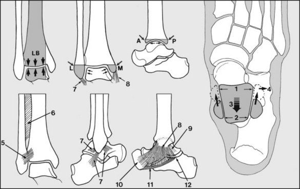
(a) The inferior tibiofibular ligaments (anterior and posterior) (5), which bind the tibia to the fibula. They are assisted by the weak interosseous membrane (6).
(b) The lateral (external) ligament (7) has three parts which arise from the fibula; distally the anterior and posterior fasciculi are attached to the talus, and the central slip is attached to the calcaneus.
(c) The medial ligament (8), immensely strong, is triangular in shape (hence the alternative term deltoid ligament), and is attached proximally to the medial malleolus. Its deep fibres (9) pass to the medial surface of the talus, and its superficial part is attached to the navicular (10), the spring ligament (11) and the calcaneus (12).
Other Common Conditions Seen Around the Ankle
Tenosynovitis
Inflammatory changes in the tendon sheaths behind the malleoli may give rise to pain at the sides of the ankle joint. Tenosynovitis may follow unusual excessive activity or be associated with degenerative changes, flat foot or rheumatoid arthritis. There is puffy swelling in the line of the tendons, with tenderness extending often for several centimetres along their length. Tibialis posterior and peroneus longus are most frequently involved, and stretching of these structures during inversion and eversion of the foot gives rise to pain. Spontaneous rupture is not uncommon. Symptoms generally respond to immobilisation for short periods in a below-knee walking plaster or an orthosis.


