Fig. 48.1
(a, b) ABC in the upper metaphysis of a humerus. Lytic, rapidly expansible lesion
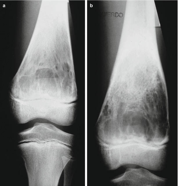
Fig. 48.2
(a, b) Two months follow-up shows a rapid ballooned expansion of the lower femoral metaphysis affected by an ABC
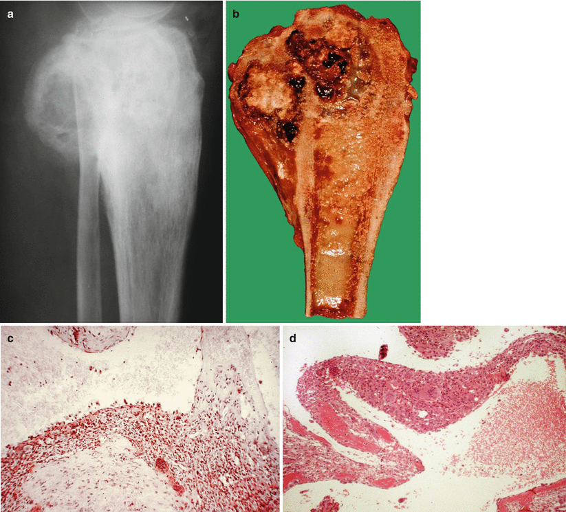
Fig. 48.3
Secondary ABC may affect other lesions. Example: (a–d) chondroblastoma of long evolution with soft tissue involvement and minimal secondary ABC
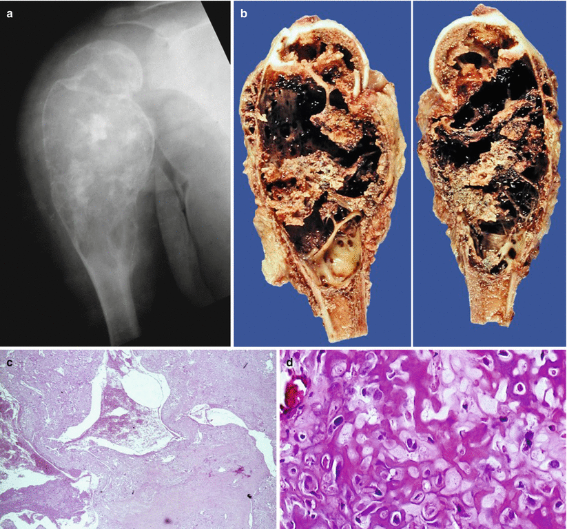
Fig. 48.4
(a–d) Secondary ABC in conventional central osteosarcoma in the upper metaphysis of a humerus
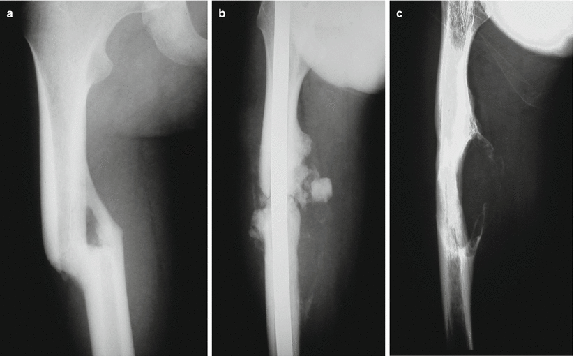
Fig. 48.5
(a–c) Secondary ABC developed after surgical treatment of a femoral shaft fracture
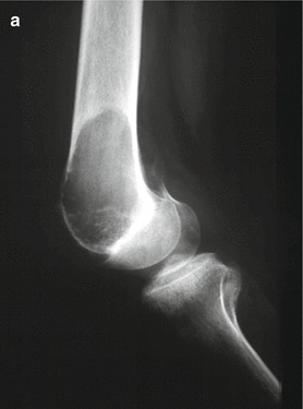
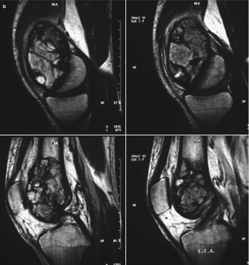
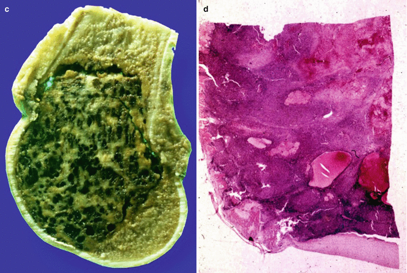
Fig. 48.6
(a–d) Secondary ABC in a pre-existent GCT, with minor cystic component arising in lower epiphysis of femur
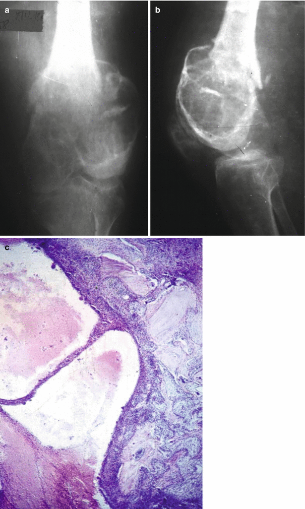
Fig. 48.7
(a–c) Secondary ABC, predominantly cystic, arising in giant cell tumor located in the lower epiphysis of the femur
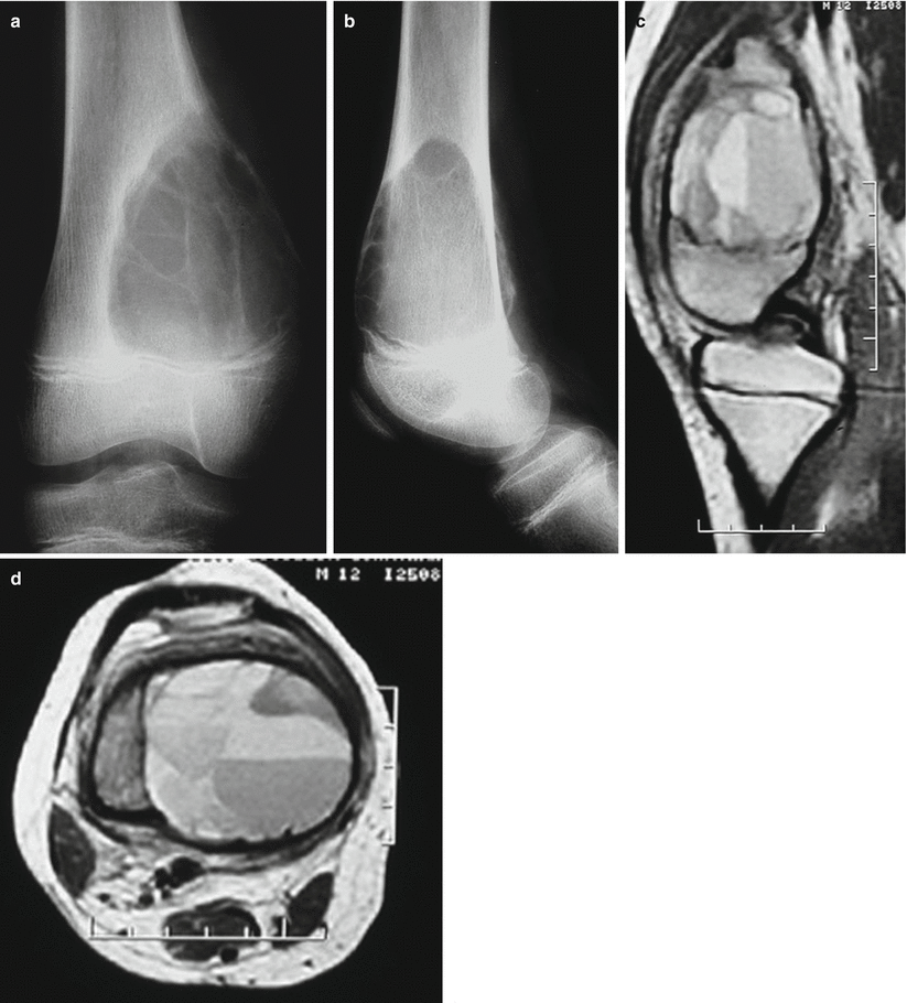
Fig. 48.8
(a, b) Anteroposterior and lateral roentgenographic lytic image of ABC in the lower metaphysis of the femur. Most ABCs are completely lytic, but a few contain traces of mineral. (c) MRI shows the internal septation with multiple fluid-fluid levels
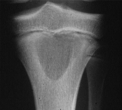
Fig. 48.9
Less commonly, the lesion is centrically situated
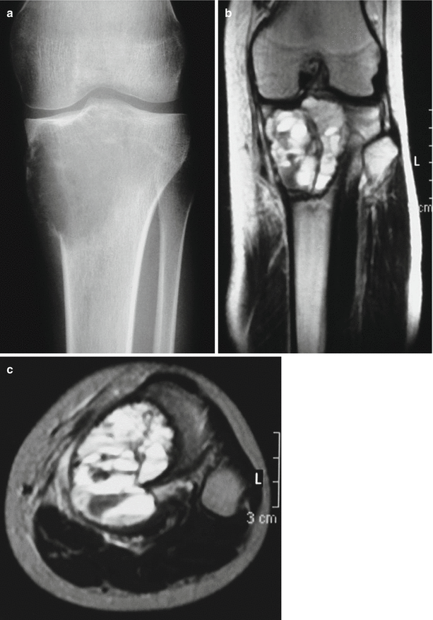
Fig. 48.10
(a) ABC in epiphyseal location. (b, c) Coronal and axial MRI showing the typical fluid-fluid levels
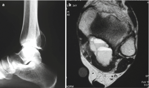
Fig. 48.11
(a) Radiograph shows an expansible lesion on lower epiphysis of the tibia corresponding to ABC. (b) Axial MRI shows the multicameral feature of the lesion with fluid-fluid levels
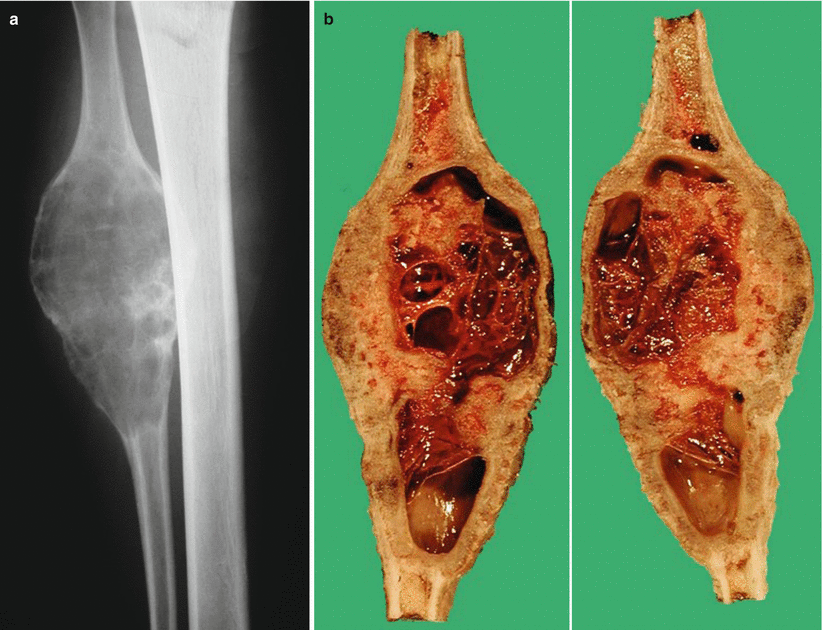
Fig. 48.12
(a) ABC in a fibula diaphysis. (b) Gross section of the surgical specimen with multiple unclotted blood-filled spaces separated by septae of different thicknesses
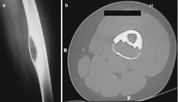
Fig. 48.13
(a) Intracortical diaphyseal ABC of a femur. X-ray shows an intracortical lucency with a shell of cortical bone at the external contour of the lesion. (b) Axial CT scan shows a cystic radiolucent lesion arising in the cortex of the femur
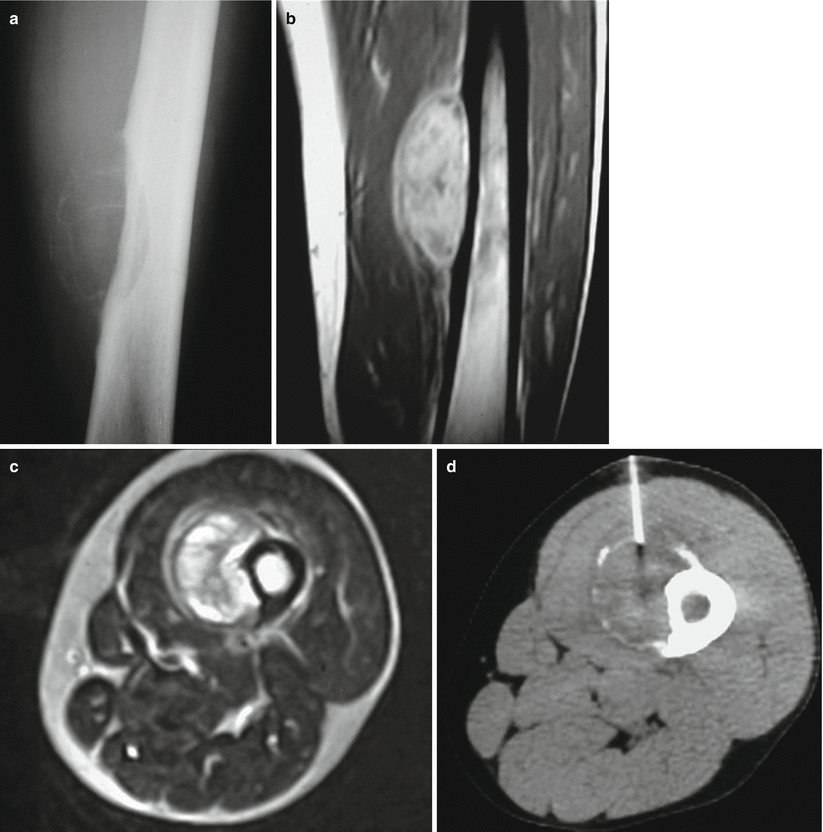
Fig. 48.14
(a) Subperiosteal location of ABC in the shaft of a femur. (b, c) Coronal and axial MRI views show the precise location of the lesion and the typical fluid-fluid levels. (d) Core needle biopsy guided by CT
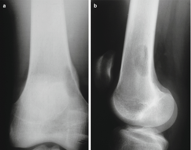
Fig. 48.15
(a, b) Anteroposterior radiograph showing early ABC situated eccentrically in the medullary cavity on the lower metaphysis of the femur
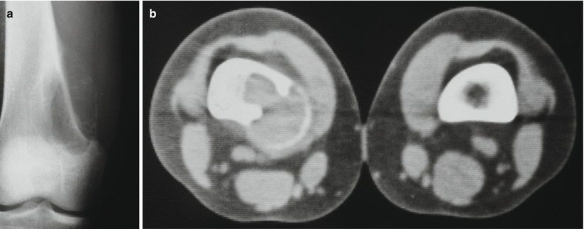
Fig. 48.16
(a) Late ballooning or expansion of the affected bone. The lesion tends to involve the cortex and may destroy it completely and bulge out into the soft tissue where it usually forms a thin sclerotic rim of calcification and periosteal new bone formation. (b) CT scan highlights the calcified rim at the periphery
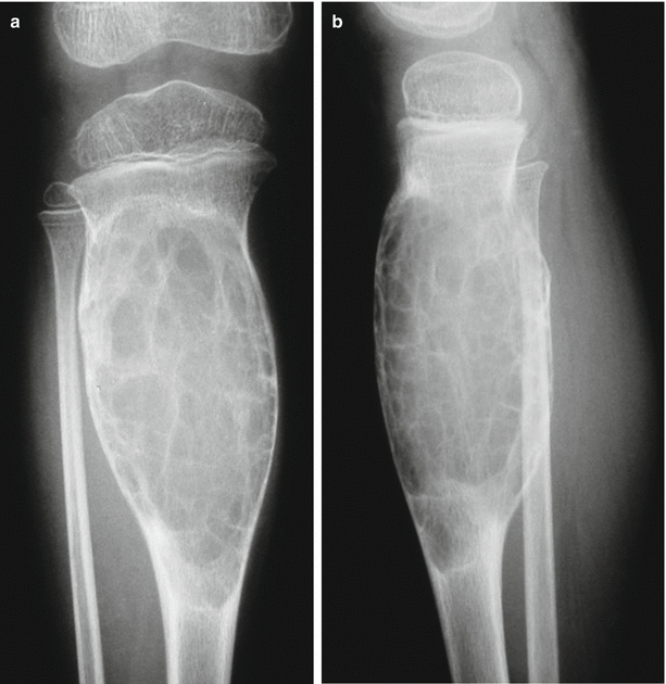
Fig. 48.17
(a, b) Anteroposterior X-ray showing a growing lesion that involves the meta-diaphysis of the tibia. The lesion presents a multiloculated appearance
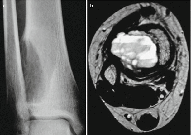
Fig. 48.18
(a) Roentgenogram showing an ABC in distal metaphysis of the tibia. (b) MRI shows the so-called hematocrit effect
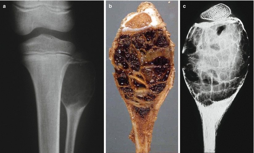
Fig. 48.19
(a) ABC affecting the upper metaphysis of the fibula. (b) Typical gross appearance of multicameral hemorrhagic cyst. (c) X-ray of slab of the gross specimen
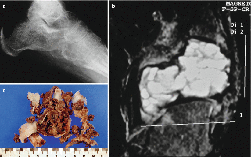
Fig. 48.20
(a) ABC located in the tarsal region. (b) Typical multicameral cystic lesion in MRI. (c) Gross appearance of the surgical specimen with blood-filled cystic areas and septae of different sizes
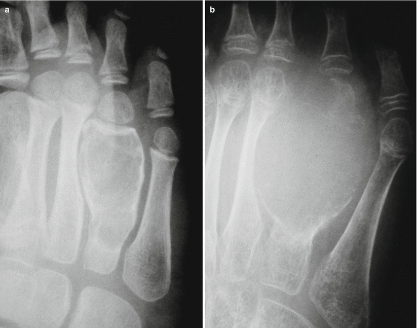
Fig. 48.21
(a) ABC in the fourth metatarsal bone. (b) Evolution: 4 months later
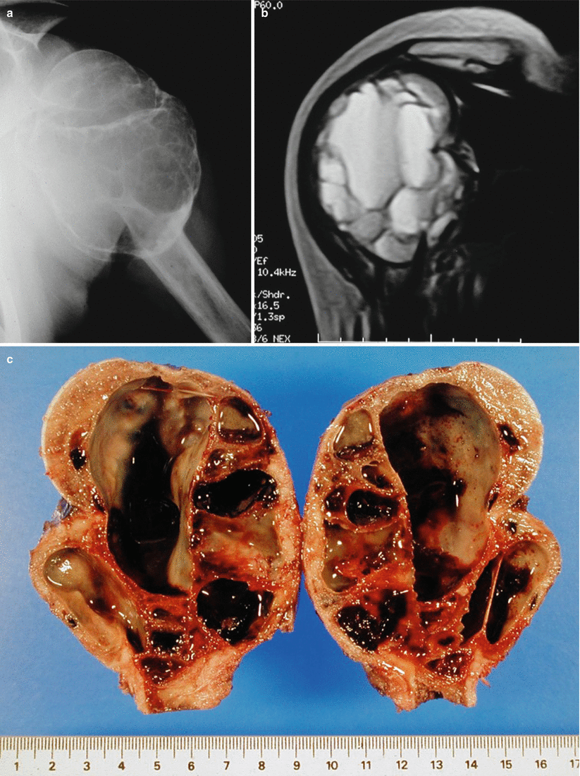
Fig. 48.22
(a) ABC in the upper end of the humerus with a great expansion of the cortex. (b) MRI shows a typical multicameral appearance of the lesion. (c) Gross specimen showing complete septae. Some cavities are filled with nonclotted blood
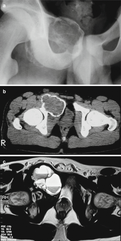
Fig. 48.23


(a) Ballooned destructive lesion in the iliopubic area of the right pelvis. (b) CT shows a well-delimitated lesion surrounded by a thin periosteal bone shell. (c) MRI showing fluid-fluid levels
Related posts:

Stay updated, free articles. Join our Telegram channel
Full access? Get Clinical Tree
