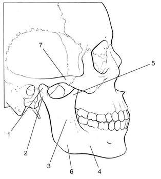
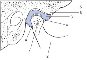
The mandible has a horizontal part (the body) and a vertical part (the ramus). They meet at the mandibular angle. The cranial end of the ramus has two processes: anteriorly the coronoid process and posteriorly the condylar process, which has a head on top and a distinct neck below. The squamous portion of the temporal bone contains the articular surface with a concave articular fossa posteriorly and a convex articular tubercle anteriorly. The articular surface is about three times as large as that of the mandibular head and is covered with fibrocartilage which continues anteriorly into the articular tubercle, the posterior aspect of which is the most important part of the joint. From the temporal bone develops a zygomatic process which, together with the temporal process of the zygomatic bone, forms the zygomatic arch (Fig. 1). The joint capsule is wide and loose on the upper aspect around the mandibular fossa. Distally, it diminishes in a funnel shaped manner to become attached to the mandibular neck (Fig. 2). Its laxity prevents rupture even after dislocation.
Applied anatomy of the temporomandibular joint

Bones
Joint capsule and ligaments
![]()
Stay updated, free articles. Join our Telegram channel

Full access? Get Clinical Tree


Musculoskeletal Key
Fastest Musculoskeletal Insight Engine