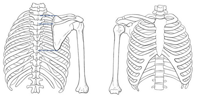
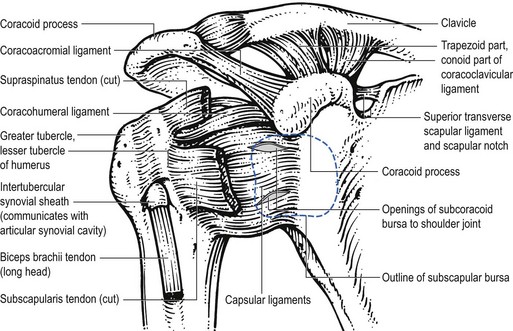
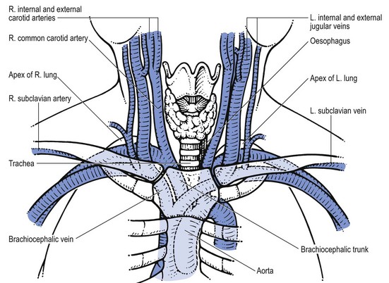
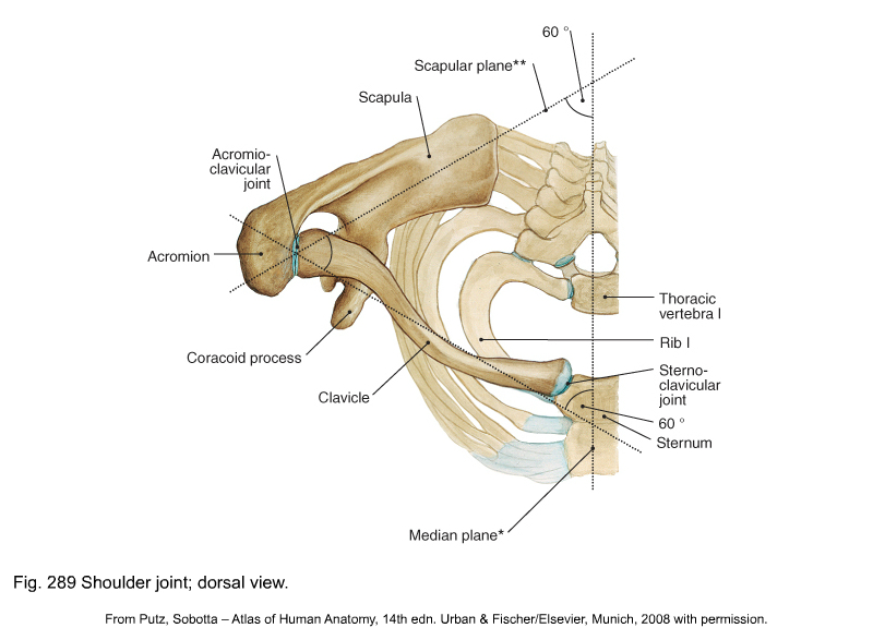
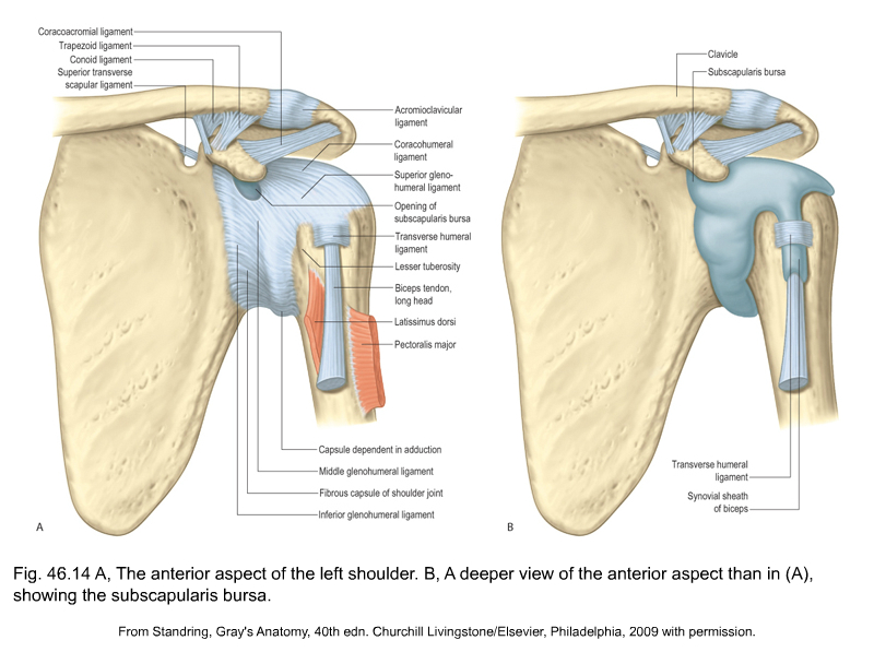
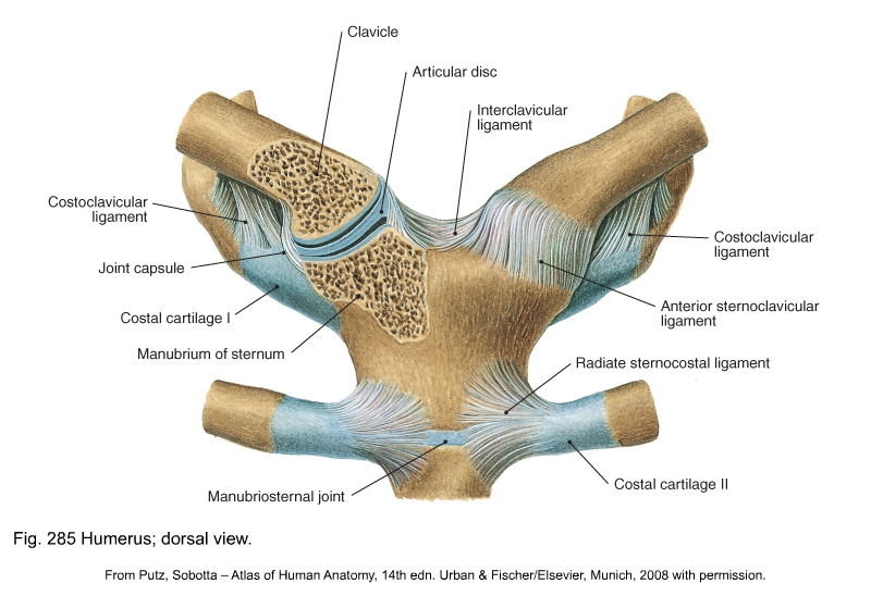
The shoulder girdle forms the connection between the spine, the thorax and the upper limb. It contains three primary articulations, all directly related to the scapula: the acromioclavicular joint, the sternoclavicular joint and the scapulothoracic gliding surface (see Putz, Fig. 289). The shoulder girdle acts as a unit: it cannot be functionally separated from the secondary articulations, i.e. the lower cervical spine, the cervicothoracic junction and the upper thoracic spine, to which it is connected via the costovertebral joints. The clavicle articulates at the medial aspect with the sternum – the sternoclavicular joint – and at the lateral aspect with the acromion to form the acromioclavicular joint. In this way it connects the scapula to the trunk (Fig. 1). The rather flat articular surface at the lateral end of the clavicle articulates with the flat articular surface at the medial border of the acromion. The joint has a capsule which is reinforced by ligaments: cranially the superior acromioclavicular ligament and caudally the inferior one. The joint often contains an intra-articular disc, which is sometimes incomplete (meniscoid) and is subject to early degeneration. The joint line runs obliquely, from craniolateral to caudomedial (Fig. 2). Extra-articular ligaments are important for the stability of the joint and to keep the movements of the lateral end of the clavicle within a certain range. Together they form the roof of the shoulder joint (see Standring, Fig. 46.14). They are the coracoacromial ligament – between the lateral border of the coracoid process and the acromion – and the coracoclavicular ligament. The latter consists of: • The trapezoid ligament which runs from the medial border of the coracoid process to the trapezoid line at the inferior part of the lateral end of the clavicle. • The conoid ligament which is spanned between the base of the coracoid process and the conoid tubercle just medial to the trapezoid line. Movements in the acromioclavicular joint are directly related to those in the sternoclavicular joint and those of the scapula. This joint is also discussed inthe online chapter Applied anatomy of the shoulder. The sternoclavicular joint is more complex. It is formed by the articular surface at the medial aspect of the clavicle and the articular surface at the superolateral corner of the sternal manubrium. The two joint surfaces are discongruent and this is resolved by the presence of an intra-articular disc, which divides the joint into two cavities. The rather loose joint capsule is reinforced by anterior and posterior sternoclavicular ligaments. There are two extracapsular ligaments: the interclavicular ligament, which interconnects both clavicles and covers the jugular notch, and the costoclavicular ligament, between the sternal end of the first rib and the medial part of the clavicle (see Putz, Fig. 285). Close behind the joint lie some vital structures: some important blood vessels (aorta, brachiocephalic trunk, brachiocephalic vein, subclavian artery, subclavian vein, jugular vein and carotid artery), the trachea, the oesophagus, the lung and pleura (Fig. 3). Movements in the sternoclavicular joint are possible around three axes: • Clavicular elevation and depression occur around an anteroposterior axis • Clavicular protraction – the lateral end of the clavicle moves forwards, and retraction – the lateral end of the clavicle moves backwards, around a vertical axis • Backwards and forwards rotation of the clavicle around its longitudinal axis.
Applied anatomy of the shoulder girdle

Osteoligamentous structures
Acromioclavicular joint
Sternoclavicular joint
![]()
Stay updated, free articles. Join our Telegram channel

Full access? Get Clinical Tree


anatomy of the shoulder girdle

