Fig. 12.1
Cervical spine: head in forward projection. Narrowing of intervertebral spaces with considerable subchondral sclerotisation of articular surfaces and non-homogeneous mild opacity of intervertebral spaces, uncovertebral arthrosis and arthrosis of intervertebral joints grade II in the whole part, more accentuated on the left side and in distal half of C-spine
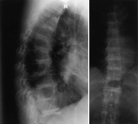
Fig. 12.2
 Genetics of Alkaptonuria
Detection of Homogentisic Acid in Plasma and Urine
Coincidence of Alkaptonuric Ochronosis with Other Diseases
Therapy of Alkaptonuria
Analysis of X-Ray Symptomatology of Ochronotic Arthropathy in the Area of Peripheral Joints
Clinical Manifestation of Ochronotic Arthropathy in Spine
Genetics of Alkaptonuria
Detection of Homogentisic Acid in Plasma and Urine
Coincidence of Alkaptonuric Ochronosis with Other Diseases
Therapy of Alkaptonuria
Analysis of X-Ray Symptomatology of Ochronotic Arthropathy in the Area of Peripheral Joints
Clinical Manifestation of Ochronotic Arthropathy in Spine


Thoracic spine: considerable subchondral sclerotisation of articular surfaces of vertebral bodies deformed by frontal as well as thicker lateral beak-like osteophytes creating the picture of ankylosing hyperostosis; slightly reduced intervertebral space filled with central lamellar inhomogeneous calcifications in the whole part, even synostosis of vertebral bodies at Th 10–11 segment
Related posts:
Genetics of Alkaptonuria
Detection of Homogentisic Acid in Plasma and Urine
Coincidence of Alkaptonuric Ochronosis with Other Diseases
Therapy of Alkaptonuria
Analysis of X-Ray Symptomatology of Ochronotic Arthropathy in the Area of Peripheral Joints
Clinical Manifestation of Ochronotic Arthropathy in Spine
Stay updated, free articles. Join our Telegram channel
Full access? Get Clinical Tree
