Fig. 12.1
Cervical spine: head in forward projection. Narrowing of intervertebral spaces with considerable subchondral sclerotisation of articular surfaces and non-homogeneous mild opacity of intervertebral spaces, uncovertebral arthrosis and arthrosis of intervertebral joints grade II in the whole part, more accentuated on the left side and in distal half of C-spine
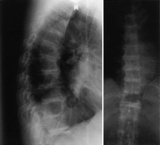
Fig. 12.2


Thoracic spine: considerable subchondral sclerotisation of articular surfaces of vertebral bodies deformed by frontal as well as thicker lateral beak-like osteophytes creating the picture of ankylosing hyperostosis; slightly reduced intervertebral space filled with central lamellar inhomogeneous calcifications in the whole part, even synostosis of vertebral bodies at Th 10–11 segment
Related posts:


Stay updated, free articles. Join our Telegram channel
Full access? Get Clinical Tree
