Fig. 53.1
SPECT/CT of a patient identified a lateral sided patellofemoral overloading and progression of OA
53.1 Anatomy of the Knee
The size and the shape of the patella show significant interindividual difference. The thickness at the central ridge seems to range from 17 to 33 mm and the mediolateral width between 25 and 64 mm [10, 11]. The patella is larger in size in males than in females. There is a remarkable variation in terms of size of the medial and lateral patellar facet, as described by Wiberg [12].
The trochlea as the anatomical counterpart shows a wide variation in shape, depth, and angulation and one may question whether the currently available implants in TKR take the individual anatomy of patients into consideration [13] (see Chaps. 8 and 9).
The relevance of primary patellar resurfacing is not clear given the fact that postoperative anterior knee pain is similar whether or not resurfacing has been done. However, if resurfacing is done, it is recommended to use the onlay technique rather than the inlay technique, because painful nerve endings have been found at the patellar margins that are left unchanged in inlay patellar designs. Resurfacing aims for full coverage without overhang, leaving a symmetric patellar bone stock between 12-15mm. Thus, the component usually covers the whole bone proximal to distal but leaves bone uncovered mediolaterally. The component should be placed as medial possible to optimize tracking, and the excessive lateral patellar facet should be removed to decompress the lateral retinaculum [14–16].
53.2 Component Design
It has been shown that patellofemoral pressure increases up to 1.5–2.5 times after TKR in comparison with the natural knee [17]. Studies have also reported on the impact of the component design on patellofemoral pressure [18].
The design of TKR may also play an important role in development of postoperative patellofemoral complications. For example, the original Insall-Burstein posterior-stabilized prosthesis design had a high incidence of patellar clunk, up to 21 % [19]. Other TKR implants have a significantly thicker anterior flange and a considerable degree of flexion built into the femoral component. These design features might be associated with patellofemoral overloading. Hence, in recent years, great effort was undertaken to optimize the TKR design in terms of improved patellofemoral tracking. Design modifications such as introduction of side-specific components, anatomically oriented trochlear grooves, and longer and deeper trochlear grooves aim for a better patellofemoral performance. In addition, more sizes of patellar and femoral components have the potential to reduce the incidence of patellofemoral complications [19].
The “patella friendly” femoral component design in TKR has an extended anterior flange and a deeper and wider trochlea groove with anatomical orientation of 4-5° valgus to the mechanical axis. The patella engages earlier during flexion, which improves stabilization and increases the patellofemoral contact area. A significantly decrease of complications has been reported when using a “patella friendly” design [20, 21]. However, the analysis of the literature by van Jonbergen showed that the “patella friendly” design did not reduce the rate of anterior knee pain, which brings into question the relevance of the differences in the currently used femoral knee designs. In addition, a recent metaanalysis found no differences in clinical outcome of patellar pain related to different femoral designs grouped patellar friendly or unfriendly [22].
Surgical errors and implant design contribute heavily to patellofemoral maltracking and patellar dislocation [23]. The depth of the femoral artificial trochlea can be too deep, which may cause excessive stress to the patella with subsequent pain, implant wear, or even patellar fracture. On the other hand, a shallow groove may predispose to instability and subluxation. In addition, the orientation of the groove may play an important role. The ideal path would be the direction in line with the pull of the quadriceps muscle. However, with a standard joint line perpendicular to the mechanical axis, the tracking of the patella would be between 3° and 5° directed laterally to adjust for a compromise of the natural valgus pull of the quadriceps muscle, which is attached to the femur and the pelvis. Modern TKR designs are adjusted to these biomechanical considerations.
Older knee implant designs caused patellar maltracking and pain. Contemporary designs adjust for these known disadvantages, making the choice of implant less important in the analysis of anterior knee pain following TKR.
53.3 TKR Component Placement
Proper placement and sizing of the femoral and tibial component are essential for patellofemoral symptoms. Oversizing of the femoral component may cause excessive stress to the extensor mechanism and the patellar joint surface causing synovitis, scarring, repetitive hematoma, wear, and decreased range of motion.
Femoral and tibial component malposition may occur in all three planes of the knee:
1.
Axial plane: internal and external rotation
2.
Coronal plane: varus and valgus position, medial and lateral shift and joint line elevation
3.
Sagittal plane: anterior and posterior shift, flexion and extension
One of the most common reasons for patella maltracking seems to be femoral and tibial component malrotation. Berger et al. found that patellofemoral complications were associated with combined (tibial and femoral) internal rotation, and the greater the rotational abnormality, the worse the anterior knee pain [24]. Rotation of the femoral component influences the tension of the medial and lateral retinaculum. Excessive internal rotation causes stress to the lateral retinaculum with subsequent maltracking of the patella and significant pain at the lateral patella border.
Recently, Vanbiervliet et al. performed an in vitro study to address the influence of malrotation on patellofemoral wear during gait [25]. They found that patellar maltracking due to an internally rotated femoral component led to a significant increase in patellar wear [25].
As instrumentation of TKR offers a standard of 3° of external rotation referencing the posterior femoral condyles, this technical error might be the most frequent cause of patellofemoral pain. Surgeons should be familiar with the limitations of such standard reference and adjust it to the individual knee. Femoral component placement parallel to the epicondylar axis has been shown to be more reliable and reduces patella subluxation and lateral patellar pain [24, 26].
Excessive internal rotation of the femoral component causes stress to the lateral retinaculum with subsequent lateral tracking of the patella and significant pain at the lateral patellar border.
Excessive internal rotation of the tibial component results in a lateralized position of the tibial tubercle. In this setting, the patellar tendon and the quadriceps tendon (Q-angle) no longer run in a similar axis and, with contraction of the quadriceps muscle, the patella might dislocate laterally until the patella and quadriceps tendon are in one line again. The lower leg and foot will be more externally rotated (Figs. 53.2, 53.3). The same problem occurs if the whole TKR is set in excessive valgus. Finally, the joint line of the implanted TKR in relation to the patella should reconstruct a Caton-Deschamps Index of about one. If the tibial bone is not cut adequately, the new joint line will be elevated, causing early contact of the anterior tibial plateau with the patella. Newer inlay designs adjust for this problem by molding the polyethylene at the anterior part, allowing higher degrees of flexion without patellar conflict.


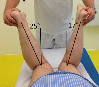
Fig. 53.2
Femoral component: patient prone, thighs parallel, knees flexed 90°. The examiner rotates the hips maximally inwards. The position of the femoral component can be estimated compared to the normal side: The patient’s left side has a TKR. Angle at the left side is 17° compared to 25° on the unreplaced side, indicating that the femoral component of the left knee is placed in excessive external rotation compared to the patient’s normal femoral rotation. A difference of more than 10 to 20° indicates the need for CT-scan to quantify the amount of malrotation

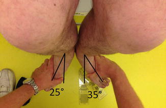
Fig. 53.3
Tibial component: patient sitting with thighs parallel, knee flexed 90°. The examiner rotates the lower leg maximally outwards. The tibial component can be estimated compared to the normal side: the patient’s left side had TKR. Angle at the left side is 35° compared to 25° on the unreplaced side, indicating that the tibial component is placed in excessive internal rotation compared to the patient’s normal tibial rotation. A difference of more than 10 to 20° indicates the need for CT-scan to quantify the amount of malrotation
The medial or lateral shift of the femoral component shows a direct effect on patella tracking. Medialization of the femoral component in the frontal plain causes less coverage of the lateral femoral bone. This may lead to lateral patella subluxation due to the maltracking between the medialized trochlea groove and the patella. Excessive stress occurs to the lateral retinaculum causing pain when the patella is forced to follow the medialized trochlear groove.
A slight lateral shift of the femoral component moves the trochlear groove under the patella and may improve patella tracking. However, there is a minimal risk of causing medial patella subluxation.
Excessive medialization of the femoral component causes not only uncovering of the lateral femur but can also lead to subluxation of the patella.
Patella maltracking has been reported to be the most frequent complication after TKR. However, dislocation caused by ligamentous insufficiency is relatively rare [27–29]. Whiteside and Ohl recommended tibial tubercle transfer for patella dislocation and reported stable function without tibial tubercle nonunion in their series. Rand et al. reported high rates of complications and nonunion using tubercle transfer in TKR. Because the primary restraint against lateral patellar dislocation is the medial patellofemoral ligament (MPFL), we recommend reconstruction of the MPFL (Figs. 53.4, 53.5, and 53.6). This can be performed either with autologous semitendinosus, quadriceps tendon, or synthetic band (Fig. 53.5). We used a synthetic band successfully in four patients, even in the presence of mild component malrotation when component exchange was not advisable because of medical reasons (i.e., cemented long stem TKR in elderly patients with various comorbidities).


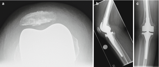
Fig. 53.4
Preoperative X-ray series (a) skyline, (b) lateral, (c) anteroposterior (ap)-view of a 78-year-old female suffering from repetitive patellar dislocations. Status post primary TKR 12 years ago and revision TKR to a hinged design 2 years ago. Infection was ruled out, and CT scan revealed an internally rotated femoral component of 4° in respect to the epicondylar axis. The tibial component was normally rotated. On examination, she demonstrated an excessive patellar motion and a positive apprehension test indicative for patella instability caused by an insufficient medial patellofemoral ligament. On X-rays, there was a mild overall valgus alignment of the knee, a normal patella height but clearly dislocated patella at the ap- and skyline views

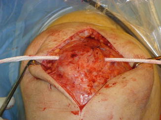
Fig. 53.5
Intraoperative pictures showing a LARS band inserted through a drill hole at the superior pole of the patella and fixed with a interference screw at the medial epicondyle and laterally to the iliotibial band

Fig. 53.6
Two years postoperatively, the patient was satisfied. No patellar dislocation has occurred. Radiographs showed (a) a stable and well centered patella. (b) Lateral, (c) ap-view postoperatively showed a correct patella position
In conclusion, implant design and placement have to be analyzed in detail. Radiographic imaging can be used to evaluate most of these factors. Rotation should be measured on 3D-CT. The femoral component should be parallel to the epicondylar line, and the tibial component should be 18° internally rotated to the tibial tubercle according to the CT evaluation published by Berger et al. [24]. Excessive combined malrotation of the femoral and tibial component may lead to implant removal in patellofemoral pain syndrome after TKR.
These patients may require revision surgery and the components need to be repositioned. However, one should be aware that revision surgery might be difficult when changing the femoral rotation due to the significant bone loss on the lateral femoral condyle when the component was placed in internal rotation. In general, wedges are required on the lateral side for the femoral component.
53.4 Soft Tissue Envelope
The medial and lateral collateral ligament acts almost isometrically throughout the range of knee motion, showing a maximal strain of less than 2 % [30]. The mean length of the medial patellofemoral ligament increases up to 30° of knee flexion and shortens continuously until full flexion with approximations of changing its insertion site of 0.25 mm/10° of knee flexion. When we are unable to restore the minor changes in soft tissue strain, the performance of the total knee prosthesis is compromised.
Stay updated, free articles. Join our Telegram channel

Full access? Get Clinical Tree


