Fig. 70.1
Plain radiograph of the wrist showing an expansile mass of amyloid in the distal ulna
Epidemiology
The average age of these patients is 57 years.
Both men and women are affected.
Radiographic Features
Radiologically, amyloidoma of the bone is an expansile lytic lesion. However, stippled radiodensity reminiscent of cartilage calcification may be present (Fig. 70.2).
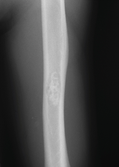
Fig. 70.2
Plain radiograph of a lytic lesion in amyloidoma of bone of the humerus showing focal radiodensities within the lytic area
Image Differential Diagnosis
Metastatic Carcinoma
Patients usually have a history of a primary neoplasia.
Myeloma
Since patients have a monoclonal gammopathy, the lesions may be interpreted as foci of myeloma.
Pathology
Histologic Features
Waxy eosinophilic deposits, usually with a foreign body giant cell reaction, are present in the marrow space (Fig. 70.3).
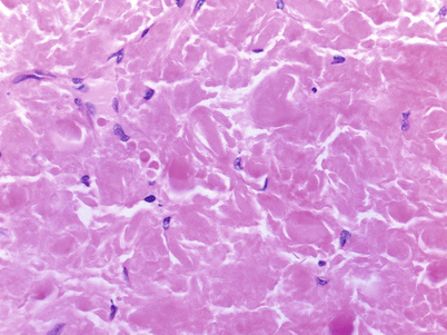
Fig. 70.3
Photomicrograph of amyloid showing eosinophilic waxy amorphous material
The eosinophilic material is identifiable as AL amyloid by permanganate-resistant Congo red birefringence.Related posts:

Stay updated, free articles. Join our Telegram channel

Full access? Get Clinical Tree


