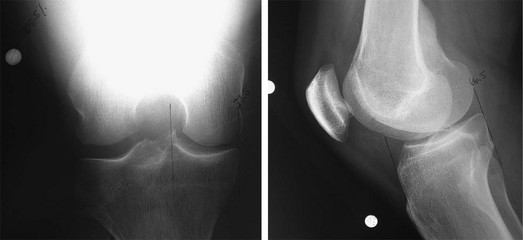
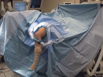
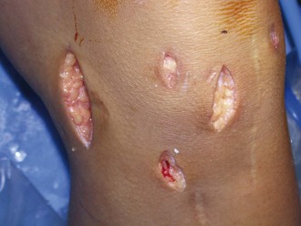
Chapter 56 It is generally believed that any significant meniscectomy alters the biomechanical and biologic environment of the normal knee, eventually resulting in pain, recurrent swelling, and effusions. Overt secondary osteoarthritis is often the endpoint.1,2 Recognition of these consequences has led to a strong commitment within the orthopedic community to meniscus-sparing interventions. However, there are cases in which meniscal preservation is not possible. In carefully selected patients, meniscal allografts can restore nearly normal knee anatomy and biomechanics, providing excellent pain relief and improved function.9 Several techniques exist for allograft meniscus transplantation, including bone plugs, a keyhole technique, and a dovetail technique. We prefer the bridge-in-slot technique4 because of its simplicity and secure bone fixation, the ability to more easily perform concomitant procedures such as osteotomy and ligament reconstruction, and the advantages of maintaining the relationship of the native anterior and posterior horns of the meniscus. • Magnetic resonance imaging with or without the intra-articular administration of contrast material is performed to assess extent of meniscectomy, degree of articular cartilage damage, and presence of subchondral edema in the involved compartment. • Technetium bone scanning may indicate stress overload in the involved compartment or overt osteoarthritis. Meniscal allografts are size and compartment specific. Preoperative measurements are obtained from anteroposterior and lateral radiographs with magnification markers placed on the skin at the level of the joint line. After radiographic magnification is accounted for, meniscal width is measured on the anteroposterior radiograph from the edge of the ipsilateral tibial spine to the edge of the tibial plateau. Meniscal length is calculated by multiplying the depth of the tibial plateau (as measured on lateral radiographs) by 0.8 for the medial meniscus and 0.7 for the lateral meniscus (Fig. 56-1). On the basis of the surgeon’s, anesthesiologist’s, and patient’s preferences, the procedure can be performed with general, spinal, or regional anesthesia or a combination thereof. The patient is positioned supine on a standard operating room table, with a thigh tourniquet, and the extremity is placed in a standard leg holder allowing full knee flexion or hyperflexion (Fig. 56-2). The posteromedial or posterolateral corner must be freely accessible for inside-out meniscus suturing to be performed.
Allograft Meniscus Transplantation
Bridge-in-Slot Technique
Preoperative Considerations
Imaging
Other Modalities
Surgical Planning
Allograft Sizing
Surgical Technique
Surgical Landmarks, Incisions, and Portals
Allograft Meniscus Transplantation: Bridge-in-Slot Technique


