Aging and Activity Tolerance
Implications for Orthotic and Prosthetic Rehabilitation
Kevin K. Chui and Michelle M. Lusardi
Learning Objectives
On completion of this chapter, the reader will be able to do the following:
Many individuals who rely on orthotic or prosthetic devices in order to walk or to accomplish functional tasks have impairments of the musculoskeletal or neuromuscular systems that limit the efficiency of their movement and increase the energy cost of their daily and leisure activities. The separate and interactive effects of aging, inactivity, and cardiac or pulmonary disease can also compromise the capacity for muscular “work,” tolerance of activity, and ability to function.
Consider this example: a 79-year-old woman with insulin-controlled type 2 diabetes has been referred for physical therapy evaluation after transfemoral amputation following a failed femoral-popliteal bypass. She has been on bed rest for several weeks because of her multiple surgeries. The physical effort required by rehabilitation and prosthetic training may initially feel overwhelming to this woman. In her deconditioned state, preprosthetic ambulation with a walker is likely to increase her heart rate (HR) close to the upper limits of a safe target HR for aerobic training. What, then, is her prognosis for functional use of a prosthesis? What are the most important issues to address in her plan of care? What intensity of intervention is most appropriate given her deconditioned state? In what setting and for how long will care be provided? These are questions without simple answers.
The physical therapist, orthotist, and prosthetist must recognize factors that can be successfully modified to enhance performance and activity tolerance when making decisions about prescription and intervention strategies. Aerobic fitness should be a key component of the rehabilitation program for those who will be using a prosthesis or orthosis for the first time. It is vitally important that rehabilitation professionals recognize and respond to the warning signs of significant cardiopulmonary or cardiovascular dysfunction during treatment and training sessions.
Although the anatomical and physiological changes in the aging cardiopulmonary system are important to our discussion, our focus is on the contribution of cellular and tissue-level changes to performance of the cardiopulmonary and cardiovascular systems and, subsequently, on the individual’s ability to function. This view provides a conceptual framework for answering four essential questions:
• Is this individual capable of physical work?
• If so, what is the energy cost of doing this work?
• Is it possible for this individual to become more efficient or more able to do physical work?
Oxygen transport system
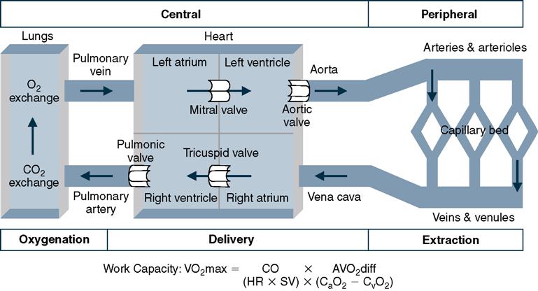
The foundation for the functional view of the cardiopulmonary system is the equation for the oxygen transport system (Figure 2-1). Aerobic capacity (VO2max) is the body’s ability to deliver and use oxygen (maximum rate of oxygen consumption) to support the energy needs of demanding physical activity. VO2max is influenced by three factors: the efficiency of ventilation and oxygenation in the lungs, how much oxygen-rich blood can be delivered from the heart (cardiac output, or CO) to active peripheral tissues, and how well oxygen is extracted from the blood to support muscle contraction and other peripheral tissues during activity (arterial-venous oxygen difference, or AVO2diff).1–3 Aerobic capacity can be represented by the following formula:

The energy cost of doing work is based on the amount of oxygen consumed for the activity, regardless of whether the activity is supported by aerobic (with oxygen) or anaerobic (without oxygen) metabolic mechanisms for producing energy. VO2max provides an indication of the maximum amount of work that can be supported.1–3
CO is the product of two elements. The first is the HR, the number of times that the heart contracts, or beats, per minute. The second is stroke volume (SV), the amount of blood pumped from the left ventricle with each beat (measured in milliliters or liters). Cardiac output is expressed by the following formula (measured in milliliters or liters per minute):

As a product of HR and SV, CO is influenced by four factors: (1) the amount of blood returned from the periphery through the vena cava, (2) the ability of the heart to match its rate of contraction to physiological demand, (3) the efficiency or forcefulness of the heart’s contraction, and (4) the ability of the aorta to deliver blood to peripheral vessels. The delivery of oxygen to the body tissues to be used to produce energy for work is, ultimately, a function of the central components of the cardiopulmonary system.1–3
The second determinant of aerobic capacity, the AVO2diff, reflects the extraction of oxygen from the capillary by the surrounding tissues. The AVO2diff is determined by subtracting the oxygen concentration on the venous (postextraction) side of the capillary bed (CvO2) from that of the arteriole (preextraction) side of the capillary bed (CaO2), according to the formula:

The smaller vessels and capillaries of the cardiovascular system are involved in the process of extraction of oxygen from the blood by the active tissues. Extraction of oxygen from the blood to be used to produce energy for the work of the active tissues is a function of the peripheral components of the cardiopulmonary system.1–3
During exercise or a physically demanding activity, CO must increase to meet the need for additional oxygen in the more active peripheral tissues. This increased CO is the result of a more rapid HR and a greater SV: As the return of blood to the heart increases, the heart contracts more forcefully and a larger volume of blood is pumped into the aorta by the left ventricle. Chemical and hormonal changes that accompany exercise enhance peripheral shunting of blood to the active muscles, and oxygen depletion in muscle assists transfer of oxygen from the capillary blood to the tissue at work.3,4
The efficiency of central components, primarily of CO, accounts for as much as 75% of VO2max. Peripheral oxygen extraction (AVO2diff) contributes the remaining 25% to the process of making oxygen available to support tissue work.5 In healthy adults, under most conditions, more oxygen is delivered to active tissues (muscle mass) than is necessary.3,5 For those who are significantly deconditioned or who have cardiopulmonary or cardiovascular disease, the ability to deliver oxygen efficiently to the periphery as physical activity increases may be compromised. With normal aging, there are age-related physiological changes in the heart itself that limit maximum attainable HR. Because of these changes, it is important to assess whether and to what degree SV can be increased effectively if rehabilitation interventions are to be successful.
The aging heart
The ability to plan an appropriate intervention to address cardiovascular endurance and conditioning in older adults who may need to use a prosthesis or orthosis is founded on an understanding of “typical” age-related changes in cardiovascular structure and physiology, as well as on the functional consequences of these changes.
Cardiovascular Structure
Age-related structural changes in the cardiovascular system occur in five areas: myocardium, cardiac valves, coronary arteries, conduction system, and coronary vasculature (i.e., arteries)6–9 (Table 2-1). Despite these cellular and tissue level changes, a healthy older heart can typically meet energy demands of usual daily activity. Cardiovascular disease, quite prevalent in later life, and a habitually sedentary lifestyle can, however, significantly compromise activity tolerance.10
Table 2-1
Age-Related Changes in the Cardiovascular System
| Structure | Change | Functional Consequences |
| Heart | Deposition of lipids, lipofuscin, and amyloid within cardiac smooth muscle Increased connective tissue and fibrocity Hypertrophy of left ventricle Increased diameter of atria Stiffening and calcification of valves Fewer pacemaker cells in sinoatrial and atrioventricular nodes Fewer conduction fibers in bundle of His and branches Less sensitivity to extrinsic (autonomic) innervation Slower rate of tension development during contraction | Less excitability Diminished cardiac output Diminished venous return Susceptibility to dysrhythmia Reduction in maximal attainable heart rate Less efficient dilation of cardiac arteries during activity Less efficient left ventricular filling in early diastole, leading to reduced stroke volume Increased afterload, leading to weakening of heart muscle |
| Blood vessels | Altered ratio of smooth muscle to connective tissue and elastin in vessel walls Decreased baroreceptor responsiveness Susceptibility to plaque formation within vessel Rigidity and calcification of large arteries, especially aorta Dilation and increased tortuosity of veins | Less efficient delivery of oxygenated blood to muscle and organs Diminished cardiac output Less efficient venous return Susceptibility to venous thrombosis Susceptibility to orthostatic hypotension |
Myocardium
With advanced age, cells of the myocardium show microscopical signs of degeneration including increases in myocardial fat content (i.e., storage of triglyceride droplets within cardiomyocytes); however, the relationship between the quantity of fat and disease severity remains unclear.11 Unlike aging skeletal muscle cells, there is minimal atrophy of cardiac smooth muscle cells. More typically, there is hypertrophy of the left ventricular myocardium, increasing the diameter of the left atrium.12–14 These changes have been attributed to cardiac tissue responses to an increased systolic blood pressure (SBP) and to reduced compliance of the left ventricle and are associated with an increase in weight and size of the heart.13–17
Valves
The four valves of the aged heart often become fibrous and thickened at their margins, as well as somewhat calcified.18 Calcification of the aorta at the base of the cusps of the aortic valve (aortic stenosis) is clinically associated with the slowed exit of blood from the left ventricle into the aorta.19 Such aortic stenosis contributes to a functional reduction in CO. A baroreflex-mediated increase in SBP attempts to compensate for this reduced CO.20,21 Over time, the larger residual of blood in the left ventricle after each beat (increased end systolic volume, or ESV) begins to weaken the left ventricular muscle.22 The left ventricular muscle must work harder to pump the blood out of the ventricle into a more resistant peripheral vascular system.23,24
Calcification of the annulus of the mitral valve can restrict blood flow from the left atrium into the left ventricle during diastole. As a result, end diastolic volume (EDV) of blood in the left ventricle is decreased because the left atrium does not completely empty. Over time, this residual blood in the left atrium elongates the muscle of the atrial walls and increases the diameter of the atrium of the heart.23–25
Coronary Arteries
Age-related changes of the coronary arteries are similar to those in any aged arterial vessel: an increase in thickness of vessel walls and tortuosity of its path.26 These changes tend to occur earlier in the left coronary artery than in the right.27 When coupled with atherosclerosis, these changes may compromise the muscular contraction and pumping efficiency and effectiveness of the left ventricle during exercise or activity of high physiological demand.2,3,28
Conduction System
Age-related changes in the conduction system of the heart can have a substantial impact on cardiac function. The typical 75-year-old has less than 10% of the original number of pacemaker cells of the sinoatrial node.29,30 Fibrous tissue builds within the internodal tracts as well as within the atrioventricular node, including the bundle of His and its main bundle branches.29,30 As a consequence, the ability of the heart to coordinate the actions of all four of its chambers may be compromised.29 Arrhythmias are pathological conditions that become more common in later life; they are managed pharmacologically or with implantation of a pacemaker/defibrillator.31 Rehabilitation professionals must be aware of the impact of medications or pacemaker settings on an individual’s ability to physiologically respond to exercise and to adapt to the intervention, whether it be a conditioning program or early mobility after a medical/surgical event, accordingly.32
Arterial Vascular Tree
Age-related changes in the arterial vascular tree, demonstrated most notably by the thoracic aorta and eventually the more distal vessels, can disrupt the smooth or streamline flow (i.e., laminar flow) of blood from the heart toward the periphery.31,33 Altered alignment of endothelial cells of the intima creates rough or turbulence flow (i.e., nonlaminar flow), which increases the likelihood of deposition of collagen and lipid.34 Fragmentation of elastic fibers in the intima and media of larger arterioles and arteries further compromises the functionally important “rebound” characteristic of arterial vessels.35 Rebound normally assists directional blood flow through the system, preventing the backward reflection of fluid pressure waves of blood. This loss of elasticity increases vulnerability of the aorta, which, distended and stiffened, cannot effectively resist the tensile force of left ventricular ejection. Not surprisingly, the incidence of abdominal aortic aneurysms rises sharply among older adults, and stiffness (distensibility) of the ascending aorta is associated with severity of coronary artery disease.36,37
Cardiovascular Physiology
Although the physiological changes in the cardiovascular system are few, their impact on performance of the older adult can be substantial. The nondiseased aging heart continues to be an effective pump, maintaining its ability to develop enough myocardial contraction to support daily activity. The response of cardiac muscle to calcium (Ca++) is preserved, and its force-generating capacity maintained.38 Two aspects of myocardial contractility do, however, change with aging: the rate of tension development in the myocardium slows and the duration of contraction and relaxation is prolonged.39,40
Sensitivity Beta-Adrenergic
One of the most marked age-related changes in cardiovascular function is the reduced sensitivity of the heart to sympathetic stimulation, specifically to the stimulation of beta-adrenergic receptors.40,41 Age-related reduction in beta-adrenergic sensitivity includes a decreased response to norepinephrine and epinephrine released from sympathetic nerve endings in the heart, as well as a decreased sensitivity to any of these catecholamines circulating in the blood.41,42 Normally, norepinephrine and epinephrine are potent stimulators of ventricular contraction.
An important functional consequence of the change in receptor sensitivity is less efficient cardioacceleratory response, which leads to a lower HR at submaximal and maximal levels of exercise or activity.43 The time for HR rise to the peak rate is prolonged, so more time is necessary to reach the appropriate HR level for physically demanding activities. A further consequence of this reduced beta-adrenergic sensitivity is less than optimal vasodilation of the coronary arteries with increasing activity.41,44 In peripheral arterial vessels, beta-adrenergic receptors do not appear to play a primary role in mediating vasodilation in the working muscles.45
Baroreceptor Reflex
Age-related change in the cardiovascular baroreceptor reflex also contributes to prolongation of cardiovascular response time in the face of an increase in activity (physiological demand).40 The baroreceptors in the proximal aorta appear to become less sensitive to changes in blood volume (pressure) within the vessel. Normally, any drop in proximal aortic pressure triggers the hypothalamus to begin a sequence of events that leads to increased sympathetic stimulation of the heart. Decreased baroreceptor responsiveness may increase an older individual’s susceptibility to orthostatic (postural) and postprandial (after eating) hypotension, or compromise their tolerance of the physiological stress of a Valsalva maneuver associated with breath holding during strenuous activity.46,47 Clinically, this is evidenced by lightheadedness when rising from lying or sitting, especially after a meal, or if one tends to hold one’s breath during effortful activity.
The consequences of age-related physiological changes on the cardiovascular system can often be managed effectively by routinely using simple lower extremity warmup exercises before position changes. Several repetitions of ankle and knee exercises before standing up, especially after a prolonged time sitting (including for meals) or lying down (after a night’s rest), help maximize blood return to the heart (preload), assisting cardiovascular function for the impending demand. In addition, taking a bit more time in initiating and progressing difficulty of activities may help the slowed cardiovascular response time reach an effective level of performance. Scheduling physical therapy or physical activity remote from mealtimes might also be beneficial for patients who are particularly vulnerable to postprandial hypotension.
Functional Consequences of Cardiovascular Aging
What are the functional consequences of cardiovascular aging for older adults participating in exercise or rehabilitation activities? This question can best be answered by focusing on what happens to the CO (Figure 2-2). The age-related structural and physiological changes in the cardiovascular system give rise to two loading conditions that influence CO: cardiac filling (preload) and vascular impedance (afterload).3,20
Preload
Cardiac filling/preload determines the volume of blood in the left ventricle at the end of diastole. The most effective ventricular filling occurs when pressure is low within the heart and relaxation of the muscular walls of the ventricle is maximal.1,5 Mitral valve calcification, decreased compliance of the left ventricle, and the prolonged relaxation of myocardial contraction can contribute to a less effective filling of the left ventricle in early diastole.48 Doppler studies of the flow of blood into the left ventricle in aging adults demonstrate decreased rates of early filling, an increased rate of late atrial filling, and an overall decrease in the peak filling rate.5,22,48 When compared with healthy 45- to 50-year-old adults, the early diastolic filling of a healthy 65- to 80-year-old is 50% less.5,22,49 This reduced volume of blood in the ventricle at the end of diastole does not effectively stretch the ventricular muscle of the heart, compromising the Frank-Starling mechanism and the myocontractility of the left ventricle.50 The functional outcome of decreased early diastolic filling and the reduced EDV is a proportional decrease in SV, one of the determinants of CO and, subsequently, work capacity (VO2max).5,21,40
Afterload
High vascular impedance and increased afterload disrupts flow of blood as it leaves the heart toward the peripheral vasculature. Increased afterload is, in part, a function of age-related stiffness of the proximal aorta, an increase in systemic vascular resistance (elevation of SBP, hypertension), or a combination of both factors.40,51 Ventricular contraction that forces blood flow into a resistant peripheral vascular system produces pressure waves in the blood. These pressure waves reflect back toward the heart, unrestricted by the stiffened walls of the proximal aorta. The reflected pressure waves, aortic stiffness, and increased systemic vascular resistance collectively contribute to an increased afterload in the aging heart.39,51 Increased afterload is thought to be a major factor in the age-associated decrease in maximum SV, hypertrophy of the left ventricle, and prolongation of myocardial relaxation (e.g., slowed relaxation in the presence of a persisting load on the heart).6,7,9
An unfortunate long-term consequence of increased afterload is weakening of the heart muscle itself, particularly of the left ventricle. Restricted blood flow out of the heart results in a large residual volume (RV) of blood in the heart at the end of systole when ventricular contraction is complete. Large ESVs gradually increase the resting length of ventricular cardiac muscle, effectively weakening the force of contraction.2,6,7,9,22,52
Left Ventricular Ejection Fraction
Left ventricular ejection fraction (LVEF) is the proportion of blood pumped out of the heart with each contraction of the left ventricle, which is expressed by the following equation:

At rest the LVEF does not appear to be reduced in older adults. Under conditions of maximum exercise, however, the rise in LVEF is much less than in younger adults.21,53,54 This reduced rise in the LVEF with maximal exercise clearly illustrates the impact that preload and afterload functional cardiovascular age-related changes have on performance.
A substantial reduction in EDV, an expansion of ESV, or a more modest change in both components may account for the decreased LVEF of the exercising older adult:

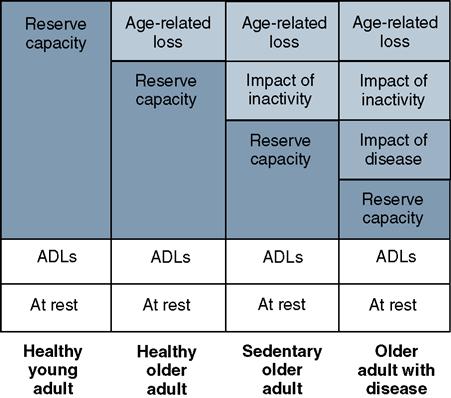
When going from resting to maximal exercise conditions, the amount of blood pumped with each beat for young healthy adults increases 20% to 30% from a resting LVEF of 55% to an exercise LVEF of 80%. For a healthy older adult, in contrast, LVEF typically increases less than 5% from rest to maximal exercise.53,55 The LVEF may actually decrease in adults who are 60 years of age and older.53,56 As LVEF and CO decrease with aging, so does the ability to work over prolonged periods (functional cardiopulmonary reserve capacity) because the volume of blood delivered to active tissue decreases (Figure 2-3). Functional reserve capacity is further compromised by the long-term effects of inactivity and by cardiopulmonary pathology.21,28,57,58 The contribution of habitual exercise to achieving effective maximum exercise LVEF is not well understood but the decline may not be as substantial for highly fit older adults.21
Pulmonary function in later life
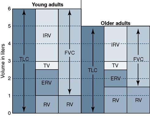
Several important age-related structural changes of the lungs and of the musculoskeletal system have a significant impact on pulmonary function.59 These include change in the tissues and structures making up the lungs and airways, alteration in lung volume, reduced efficiency of gas exchange, and a mechanically less efficient ventilatory pump related to changes in alignment and posture60 (Table 2-2). Although a healthy adult at midlife uses only 10% of the respiratory system capacity at rest, aging of the pulmonary system, especially when accompanied by chronic illness or acute disease, negatively affects the ability of the lungs to respond to increasing demands of physical activity61 (Figure 2-4). Age-related changes in the pulmonary and musculoskeletal systems also contribute to an increase in the physiological work of breathing.
Table 2-2
Summary of Age-Related Changes in the Cardiopulmonary System and Functional Consequences
| Anatomical Changes | Physiological Changes | Consequences | Change in Lung Function Tests |
| Rearrangement and fragmentation of elastin fibers Stiffened cartilage in compliant articulation of ribs and vertebrae Increasing stiffness and compression of annulus fibrosis in intervertebral disks Reduction of strength and endurance of respiratory musculature | Less elastic recoil for expiration Greater compliance of lung Decreased vital capacity, forced More rigid thoracic cage Decreased volume of maximum voluntary ventilation and maximum sustained ventilatory capacity Greater mismatch between ventilation and perfusion within lung | Greater airspace within alveoli, less surface area for O2/CO2 exchange thoracic cage Increased work of breathing Less force during inspiration Less efficient cough Diminished exercise tolerance Reduced resting PaO2 | Increased functional residual capacity and residual volume tissue Shorter, less vital capacity, and forced expiratory volume in 1 second (FEV1) Decreased maximum inspiratory pressure, maximum expiratory pressure, and maximum voluntary ventilation |

Changes within the Lung and Airway
The production of elastin, which is the major protein component of the structure of the lungs, decreases markedly in late life. The elastic fibers of the lung become fragmented, and, functionally, the passive elastic recoil or rebound important for expiration becomes much less efficient. The elastic fibers that maintain the structure of the walls of the alveoli also decrease in number. This loss of elastin means loss of alveoli and consequently less surface area for the exchange of oxygen, as well as an increase in RV associated with more “dead space” within the lung where air exchange cannot occur.60,61 There may be as much as a 15% decrease in the total number of alveoli per unit of lung volume by the age of 70 years.62
With aging, there is also an increase in diameter of major bronchi and large bronchioles, as well as a decreased diameter of smaller bronchioles, often leading to a slight increase in resistance to air flow during respiration.62 This contributes to greater physical work to breathe as age advances.
Starting at midlife and continuing into later life, there tends to be a growing mismatch between lung area ventilated with each breath and lung area perfused by pulmonary arterioles and capillaries, attributed to alteration in alveolar surface, vascular structures, and posture.63 This mismatch compromises the efficiency of diffusion of oxygen across the alveoli into the capillary bed (i.e., decreasing arterial oxygen tension) within the lung becomes less efficient from midlife into later life.60,63
Changes in the Musculoskeletal System
The decreasing elastic recoil and alveolar surface area for oxygen exchange may be further compounded by increased stiffness (loss of flexibility), “barreling” of the thoracic rib cage that houses the lungs, and a decrease in height as intervertebral disks narrow and stiffen.64 Much of the stiffness is attributed to changes in the articulation between rib and vertebrae, as well as decreased elasticity of intercostal muscle and soft tissue.65 Although the stiffened rib cage may be as much a consequence of a sedentary lifestyle as of advancing age, lack of flexibility compromises inspiration and also decreases elastic recoil of expiration.66 In addition, the forward head and slight kyphosis that tend to develop with aging alter rib and diaphragm position, decreasing mechanical efficiency of inspiration.61,64,66 The net effect of a stiffer thoracic cage is an increase in the work of taking a breath since muscles of respiration must work harder during inspiration to counteract the stiffness.61
The striated muscles of respiration are composed of a combination of type I (slow twitch, fatigue resistant, for endurance) and type II (fast twitch, for power) fibers and are susceptible to the same age-related changes in strength and endurance that have been observed in muscles of the extremities.67 Normally, type I muscle fibers are active during quiet breathing while recruitment of type II fibers is triggered by increasing physiological demand as activity increases. Age-related decrement in the strength and efficiency of the diaphragm, intercostals, abdominal muscles, and other accessory muscles of respiration affects the effectiveness and work of breathing.61,68 Altered posture and higher RV within the lung also contribute to an increased work of breathing; when the diaphragm rests in less than optimal position and configuration for contraction, accessory muscles become active sooner, as physiological demand increases. Oxygen consumption in respiratory muscles, as in all striated muscle, decreases linearly with age, making older muscle more vulnerable to the effects of fatigue in situations of high physical demand, especially in the presence of lung disease or injury.60
Control of Ventilation
The rate of breathing (breaths per minute) is matched to physiological demand by input from peripheral mechanoreceptors in the chest wall, lungs, and thoracic joints, as well as centers in the brainstem of central nervous systems (CNS) and peripheral aortic and carotid bodies that are sensitive to concentration of CO2, O2, and hydrogen ions (pH) in the blood.69 With aging, stiffness of the thorax tends to reduce efficiency of mechanoreceptors, and the CNS and peripheral nervous system (PNS) centers that monitor CO2, O2, and pH to detect hypoxia during activity slowly begin to decline.
Gradual loss of descending motor neurons within the CNS also occurs, with less efficient activation of neurons innervating muscles of respiration via the phrenic nerve to the diaphragm for inspiration and of spinal nerves to intercostals for expiration.63 These three factors combine to compromise the individual’s ability to quickly and accurately respond to increasing physiological demand and increase the likelihood of dyspnea during activity.
Functional Consequences of Pulmonary Aging
With less recoil for expiration and reduced flexibility for inspiration, the ability to work is compromised in two ways (see Figure 2-4). First, vital capacity (VC), the maximum amount of air that can be voluntarily moved in and out of the lungs with a breath, is decreased by 25% to 40%. Second, RV, the air remaining in the lungs after a forced expiration, is increased by 25% to 40%.60 This combination of reduced movement of air with each breath and increased air remaining in the lung between breaths leads to higher lung-air carbon dioxide content and, eventually, lower oxygen saturation of the blood after air exchange. The increase in RV also affects the muscles of inspiration: the dome of the diaphragm flattens and the accessory respiratory muscles are elongated. As a result of these length changes, the respiratory muscles work in a mechanically disadvantageous range of the length-tension curve, and the energy cost of the muscular work of breathing rises.61
Functionally, the amount of air inhaled per minute (minute ventilation) is a product of the frequency of breathing times the tidal volume (volume of air moving into and out of the lungs with each usual breath). In healthy individuals, the increased ventilatory needs of low-intensity activities are usually met by an increased depth of breathing (i.e., increased tidal volume).70 Frequency of breathing increases when increased depth alone cannot meet the demands of activity, typically when tidal volume reaches 50% to 60% of the VC.70 For the older adult with reduced VC who is involved in physical activity, tidal volume can quickly exceed this level, so that frequency of breathing increases much earlier than would be demonstrated by a young adult at the same intensity of exercise.71 Because the energy cost of breathing rises sharply with the greater respiratory muscle work associated with an increased respiratory rate, an important consequence of increased frequency of breathing is fatigue.72 This early reliance on an increased frequency of breathing, combined with a large RV and its higher carbon dioxide concentration in lung air, results in a physiological cycle that further drives the need to breathe more frequently. Overworked respiratory muscles are forced to rely on anaerobic metabolism to supply their energy need, resulting in a buildup of lactic acid. Because lactic acid lowers the pH of the tissues (acidosis), it is also a potent physiological stimulus for increased frequency of breathing.72–74 The older person can be easily forced into a condition of rapid, shallow breathing (shortness of breath) to meet the ventilatory requirements of seemingly moderate-intensity exercise.
Implications for intervention
Rehabilitation professionals must consider two questions about the implications of age-related changes in the cardiovascular and cardiopulmonary systems on an older person’s ability to do physical work. First, what precautions should be observed to avoid cardiopulmonary and cardiovascular complications? Second, what can be done to optimize cardiopulmonary and cardiovascular function for maximal physical performance?
Precautions
Because of the combined effects of the age-related changes in the cardiovascular and cardiopulmonary systems, the high incidence of cardiac and pulmonary pathologies in later life, and the deconditioning impact of bed rest and inactivity, older patients who require orthotic or prosthetic intervention may be vulnerable if exercise or activity is too physiologically demanding. Although most older adults can tolerate and respond positively to exercise, exercise is not appropriate in a number of circumstances (Table 2-3).
Table 2-3
Signs and Symptoms of Exercise Intolerance
| Category | Cautionary Signs/Symptoms | Contraindications to Exercise |
| Heart rate | < 40 bpm at rest > 130 bpm at rest Little HR increase with activity Excessive HR increase with activity Frequent arrhythmia | Prolonged at maximum activity |
| ECG | Any recent ECG abnormalities | Prolonged arrhythmia or tachycardia Exercise-induced ECG abnormalities Third-degree heart block |
| Blood pressure | Resting SBP >165 mm Hg Resting DBP >110 mm Hg Lack of SBP response to activity Excessive BP response to activity | Resting SBP >200 mm Hg Resting DBP >110 mm Hg Drop in SBP >20 mm Hg in exercise Drop in DBP during exercise |
| Angina | Low threshold for angina | Resting or unstable angina New jaw, shoulder, or left arm pain |
| Respiratory rate | Dyspnea >35 breaths/min | Dyspnea >45 breaths/min |
| Blood gas values | O2 saturation <90% | O2 saturation <86% |
| Other symptoms | Mild to moderate claudication Onset of pallor Facial expression of distress Lightheadedness or mild dizziness Postactivity fatigue >1 hr Slow recovery from activity | Severe, persistent claudication Cyanosis, severe pallor, or cold sweat Facial expression of severe distress Moderate to severe dizziness, syncope Nausea, vomiting Increasing mental confusionOnset of ataxia, incoordination |
| Additional considerations | Fever >100°F Aortic stenosis Recent mental confusion Abnormal electrolytes (potassium) Known left main coronary artery disease Idiopathic hypertrophic subaortic stenosis Compensated heart failure | Any acute illness Digoxin toxicity Overt congestive heart failure Untreated second or third degree heart block Acute pericarditis <4 to 6 weeks after myocardial infarction <2 days after pulmonary embolism Acute thrombophlebitis Acute hypoglycemia |
Modified from Hillegass E (ed), Essentials of Cardiopulmonary Physical Therapy, 3rd ed. St. Louis: Elsevier Saunders, 2011, pp. 307, 564, 586.
Estimating Workload: Heart Rate and Rate Pressure Product
One of the readily measurable consequences of the reduced response of the heart to sympathetic stimulation in later life is a reduction in the maximal attainable HR.39,69,75 This reduction in maximal HR also signals that an older person’s HR reserve, the difference between the rate for any given level of activity and the maximal attainable HR, is limited as well. For older patients involved in rehabilitation programs, the difference between resting and maximal HR is narrowed. One method of estimating maximal (max) attainable HR is the following5:

For healthy individuals, the recommended target HR for aerobic conditioning exercise is between 60% and 80% of maximal attainable HR. For many older adults, especially those who are habitually inactive, resting HR may be close to the recommended range for exercise exertion.76 Consider an 80-year-old individual with a resting HR of 72 beats per minute. His maximal attainable HR is approximately 140 beats per minute (220 − 80 years). A target HR for an aerobic training level of exertion of 60% of maximal HR would be 84 beats per minute. His resting HR is within 12 beats of the HR for aerobic training. Functionally, this means that an activity as routine as rising from a chair or walking a short distance on a level surface may represent physical work of a level of exertion equated with moderate- to high-intensity exercise. Because of the reduction in maximal attainable HR with age, older adults may be working close to their VO2max range even in usual activities of daily living.76,77
Because HR essentially signals the work of the heart, with each beat representing ventricular contraction, increased HR relates closely to increased heart work and increased oxygen consumption by the myocardium.75 Given that afterload on the heart increases with age, the overall work of the heart for each beat is likely greater as well.21,38,39,40 A more representative way to estimate the work of the heart during activity for older adults is the rate pressure product (RPP),78–80 using HR and SBP as follows:

The linear relationship between VO2max and HR for younger adults actually levels off for older adults.81 Because of this, HR alone cannot accurately reflect the physiological work that the older patient experiences; the RPP provides a clearer impression of relative work.80 For older individuals with HR reserve limited by age, adjusting activity to keep the rise in HR within the lower end of the HR reserve is wise, especially for those with known coronary artery compromise.
Blood Pressure as a Warning Sign
An older person’s blood pressure (BP) must also be considered. Hypertension, particularly increased SBP, is common in older adults. SBP also provides a relative indication of the level of afterload on the heart.40,82,83 Resting BP can be used to indicate whether an older person can safely tolerate increased physiological work. Persons with resting BPs of more than 180/95 mm Hg may have difficulty with increased activity. A conservative estimate of the safe range of exercise suggests that exercise should be stopped if and when BP exceeds 220/110 mm Hg, although some consider 220 mm Hg too conservative a limit for older adults.75 SBP should rise with increasing activity or exercise.84
The older adult with limited HR reserve must increase SV to achieve the required CO.21,39,53 SBP rises as SV increases and blood volume in the peripheral vasculature rises.40 If SBP fails to rise or actually decreases during activity, this is a significant concern.75 The drop or lack of change in SBP indicates that the heart is an ineffective pump, unable to contract and force a reasonable volume of blood out of the left ventricle. Continuing exercise or activity in the presence of a dropping SBP returns more blood to a heart that is incapable of pumping it back out to the body. Elevated diastolic blood pressure (DBP) suggests that the left ventricle is maintaining a higher pressure during the filling period.39,40,69 Early diastolic filling during preload will be compromised,40,49 and the heart will be unable to capitalize on the Frank-Starling mechanism to enhance the force of ventricular contraction.21,53
Respiratory Warning Signs
Dyspnea, or shortness of breath, is an important warning sign as well. Age-related changes in the pulmonary system increase the work of breathing, and breathing becomes less efficient as work increases.72 Because an older person is prone to shortness of breath, recovering from shortness of breath during exercise may be difficult. Breathing more deeply requires a disproportionately greater amount of respiratory muscle work, which further increases the cost of ventilation.73,74 The use of supplemental oxygen by nasal cannula for the postoperative or medically ill older adult who is beginning rehabilitation may be quite beneficial.
Oxygen supplementation may prevent or minimize shortness of breath, enabling an older person to tolerate increased activity better and to participate in rehabilitation more fully. During this oxygen-assisted time, any conditioning exercise to improve muscular performance (especially if combined with nutritional support) delivers blood to the working tissues and improves tissue oxygenation, ultimately aiding pulmonary function. Improved muscular conditioning and cardiovascular function may prevent or delay onset of lactic acidemia and the resultant increased desire to breathe that would trigger shortness of breath.72,85
Optimizing Cardiopulmonary Performance
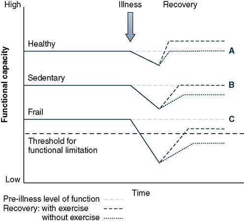
For most older adults, conditioning or training is an effective way to improve function, although some may need a longer training period than younger adults to accomplish their desired level of physical performance.86–90 Physical conditioning, in situations of acute and chronic illness, enables the older person to do more work and better accomplish desired tasks or activities.
Older adults, including those who are quite debilitated, experience improvement in physical performance as a result of conditioning exercises88 (Figure 2-5). For some, significant gains are made as work capacity increases from an initial state below the threshold necessary for function, such that an older person appears to make greater gains than a younger individual in similar circumstances.91,92 In many cases the cardiopulmonary system efficiency gained through conditioning means the difference between independence and dependency, functional recovery and minimal improvement, life without extraordinary means and life support, and for some older individuals life and death.
< div class='tao-gold-member'>
Stay updated, free articles. Join our Telegram channel

Full access? Get Clinical Tree


