Adductor tendinopathy
Athletic pubalgia [5]
Rectus femoris injury
Nerve entrapment (ilioinguinal nerve, iliohypogastric nerve, genitofemoral nerve, obturator nerve) [6]
Hernia (inguinal, femoral, obturator) [7]
Intra-articular hip pathology
• Acetabular labral tear
• Femoral neck stress fracture
• Femoral head osteonecrosis
• Slipped capital femoral epiphysis
Prostatic or urinary tract disease
Intra-abdominal pathology
Avulsion injury
Pelvic fracture
Lumbosacral spine pathology
Iliopsoas pathology [12]
Adductor strains account for a substantial proportion of the groin injuries sustained by athletes. One study found that just over half of groin injuries in soccer players are muscular and/or tendinous in nature [2]. Another prospective investigation of 2,299 professional soccer players found that adductor injuries accounted for 23 % of muscle injuries, and 16 % were re-injuries that led to significantly longer playing time missed [4]. Gibbon found a 23–25 % incidence of groin injuries during a single professional soccer season in England and Europe, with adductor injuries occurring most commonly [13]. Interestingly, in examining all lower extremity muscle injuries, the adductor longus is one of the most prone to strain [14]. Moreover, a recent cadaveric study found decreased vascularity of the adductor longus and adductor brevis tendons near their enthesis [15].
In addition to soccer [1–4, 13, 16], groin injuries are also frequently sustained in American football, ice hockey, and rugby [17–20]. Emery and colleagues found the incidence of groin injury in National Hockey League (NHL) players to be 13–20 per 100 players per year and an average of 6.59 sessions missed following injury [17]. A study of Finnish elite hockey players found the groin to be the most common area of strain [19]. Consistent results were also found in a prospective study of elite Swedish hockey players [20].
Risk factors for groin injuries have also been examined in the soccer players. With regards to ice hockey, one study found a higher incidence during preseason training camp and a higher incidence during games as opposed to during practice [17]. Another investigation associated groin injuries to lower levels of off-season training, previous injury, and veteran status [18]. Peak isometric adductor torque and total abduction flexibility were not found to be associated with groin injury [18]. However, another analysis of professional hockey players found a strong association between adductor weakness and adductor strain with a 17-fold increased risk of adductor strain if preseason adductor strength was less than 80 % of abductor strength [21]. Thus, lack of off-season sport-specific training in hockey appears to be a specific risk factor for in-season groin injuries.
Risk factors for groin injuries have also been examined in soccer players. Engebretsen et al. found that weak adductors and a history of prior groin injury were associated with future groin injury [22]. Another investigation found decreased preseason hip abduction range of motion to be a risk factor for groin strain [23]. Arnason and colleagues performed a prospective study of 306 professional soccer players and found that previous groin injury and decreased hip abduction range were associated with groin strain [24].
Diagnosis
History, Signs, and Symptoms
Adductor tendinopathy is typically the result of noncontact injuries. Its onset is usually atraumatic, although acute groin strains can become recalcitrant to treatment, resulting in symptoms lingering greater than 12 weeks. Symptoms are gradually progressive and are aggravated by sprinting, lunging, and pivoting. Pain is consistently located below the inguinal ligament, over the pubic origin of the adductor longus. Systematic history and review of symptoms must be conducted to rule out other potential causes of groin pain.
Physical Exam
The correct diagnosis of adductor strain relies heavily on an accurate physical exam [3]. Examination of the athlete with an isolated adductor injury reveals full range of motion of the hip. Tenderness is elicited with palpation of the pubis at the pelvic origin of the adductor longus. The athlete will experience pain at the adductor longus musculotendinous junction with resisted adduction. Importantly, both symptoms of pain and findings of tenderness are elicited below the inguinal ligament. These physical examination maneuvers have been shown to have good intra- and inter-observer reliability [25]. Systematic examination must be performed and referral to other specialties may be considered, as there can be several processes occurring concomitantly [26].
Imaging
Plain radiographs that include anteroposterior (AP) pelvis as well as AP and lateral views of the involved hip should be performed to investigate for potential important findings such as femoral acetabular impingement, hip arthrosis, and stress fractures. Radiographs are typically unremarkable; however, changes may be seen in long-standing cases of adductor tendinopathy [27]. Ultrasound is an imaging modality that can be useful [28]; however, it is typically not necessary for diagnosis. Magnetic resonance imaging (MRI) is also not routinely ordered unless there is suspicion of concomitant intra-articular hip pathology (e.g., labral tear) and/or a rectus abdominis injury. MRI has been found by some authors to be useful in diagnosing adductor longus enthesis edema [29]. If MRI is obtained, increased T2 signal may be present within the adductor longus tendon [30–33].
Injection Test
An injection test can be performed using lidocaine in order to confirm the diagnosis of adductor tendinopathy. An athlete with chronic adductor pathology should respond with a decrease in tenderness on palpation and a decrease in pain with resisted adduction. Some authors have used injection of local anesthetic and steroid for both diagnosis and treatment [33, 34]. Schilders et al. found that competitive athletes with adductor tendinopathy in the setting of normal MRI findings commonly have at least 1 year of pain relief following injection [33]. For recreational athletes, these authors found that steroid injection provided less predictable results, and results did not correlate with MRI findings [34]. It is essential to be cognizant that athletes sometimes experience symptoms only after competition, so an injection test may not be reliable in this setting.
Nonoperative Treatment
Adductor injuries are initially treated conservatively with rest, ice, and nonsteroidal anti-inflammatory medications [35]. Once acute pain has decreased, physical therapy focusing on stretching and strengthening is initiated. A recent systematic review has supported the use of physical therapy for adductor injuries [36]. Hölmich et al. have found that active therapy focusing on strengthening the adductor, abdominal, and low back musculature and incorporating coordination and balance exercises is more effective than passive therapy [37]. A follow-up study found that this program produces a durable effect lasting up to 8–12 years [38]. With regards to prevention, one investigation in professional hockey players found that preseason adductor strengthening led to a decrease in the incidence of adductor muscle strain [39].
Natural History
Studies investigating the natural history of chronic adductor tendinopathy in athletes have found variable results in response to nonoperative treatment. Martens and colleagues found that only 38 % of athletes with chronic adductor or rectus abdominis tendinopathy had good or excellent results with nonoperative management [40]. In contrast, Hölmich et al. found that active therapy was successful in 79 % of athletes at 18.5 weeks while passive therapy consisting of stretching and modalities was successful in only 14 % of athletes [37]. It is important to note that there is a significantly longer loss of playing time after re-injury as compared to after initial injury [3].
Surgical Indications
Surgical intervention for chronic adductor tendinopathy can be considered when the athlete experiences pain and tenderness at the origin of the adductor musculature, and there has been lack of symptomatic relief despite at least 12 weeks of conservative management. Inability to return to play should also be taken into account when considering surgical intervention. We have found that performing a simple adductor longus tenotomy often successfully treats football players with isolated adductor longus tendinopathy, who do not respond to nonoperative treatments within 12 weeks.
Surgical Technique
Setup
The patient is placed in the supine position with both hips flexed and maximally abducted (Fig. 15.1). The knees are flexed, and the plantar aspects of the feet are placed in contact with each other. The affected limb is draped free to allow movement as needed during the procedure.
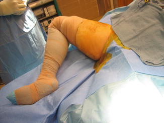

Fig. 15.1
Operative positioning
Procedure
The incision is made at least 1 cm distal to the inguinal crease to prevent postoperative wound maceration from moisture and to allow better healing with less chance of wound breakdown (Fig. 15.2). After dissection through the skin and subcutaneous tissue, the superficial fascia over the adductor longus is incised and elevated as a distinct layer for subsequent closure. A right-angle clamp is placed under the origin of the adductor longus tendon (Fig. 15.3). It is important to watch for and to take care in protecting the obturator nerve during this maneuver.
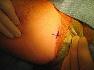
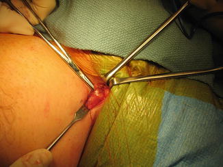

Fig. 15.2
Placement of the incision at least 1 cm distal to the inguinal crease

Fig. 15.3
Placement of a right-angle clamp under the origin of the adductor longus tendon, taking care to protect the obturator nerve
After isolation of the adductor longus tendon, the tendon is fully released as close as possible to its origin on the pubis using electrocautery (Fig. 15.4). Use of electrocautery signals if the release is too close to the obturator nerve. It is essential to note that some adductor longus muscle fibers attach directly to the pubis, and these fibers must be addressed to perform a complete release. Finally, it is important to digitally displace the tendon distally into the thigh to prevent reattachment (Fig. 15.5).
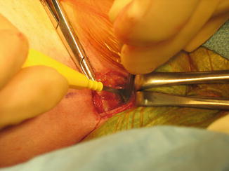
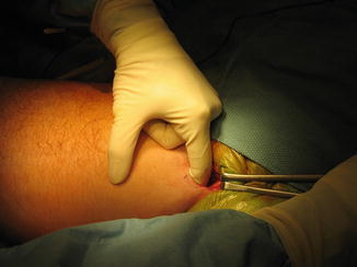

Fig. 15.4
Release of the adductor longus tendon using electrocautery

Fig. 15.5
Displacement of the incised tendon distally into the thigh to prevent reattachment
The wound is irrigated and meticulous hemostasis is ensured to prevent hematoma formation and wound drainage. A multilayer water-tight closure is performed to prevent seroma formation and drainage.
Postoperative Protocol
Following adductor release, an abduction pillow is utilized. Patients are discharged home on the day of the procedure and are allowed to weight bearing as tolerated with crutches.
Role of Herniorrhaphy
The treatment of sports hernias will be addressed in more detail elsewhere in this book. However, it is important to note that sports hernia frequently coexists with adductor tendinopathy [5, 41, 42]. Examination is notable for tenderness along the inguinal canal, tenderness over the distal rectus abdominis attachment, and pain with resisted sit-up [41].
The procedure is performed after induction of general anesthesia. We prefer the technique developed by Dr. David Berger. An incision is made and carried down through Scarpa’s fascia to the external oblique. The external oblique is incised to the external ring. The spermatic cord is mobilized. A direct inguinal hernia is typically visualized, and the hernia is dissected free. The aponeurosis of the internal oblique and transversus abdominis muscle is relaxed by performing a short fasciotomy to the muscle fascia. A prolene hernia system (Ethicon, Bridgewater, NJ) is placed through the defect and sutured in place with interrupted zero Ethibond sutures (Ethicon, Bridgewater, NJ). Hemostasis is ensured, and a layered closure is performed. Interrupted 3–0 silk is used to close the external oblique, a running 3–0 chromic is utilized to close Scarpa’s fascia, and a running 4–0 Monocryl (Ethicon, Bridgewater, NJ) is used to close skin in subcuticular fashion.
Postoperative Rehabilitation
Physical therapy plays an important role in the patient’s postoperative recovery following adductor release. It is generally divided into three phases. Phase I is performed during the first week after surgery. The goals are to enable wound healing and to prevent reattachment of the adductor longus tendon. The athlete’s leg(s) are kept abducted as much as possible for the first 3 days. Repeated abduction stretching is performed throughout the day.
Phase II occurs during postop weeks 2–5. The goals of this phase are to keep the adductor muscles stretched, prevent excess scarring, and improve flexibility. Stretching exercises consist of (1) progressively abducting the legs while standing, (2) shifting weight to one side over a flexed knee while keeping the opposite knee straight, (3) placing one foot on a table with the knee in extension and reaching toward the toes, and (4) lying face down and grabbing one ankle at a time and trying to pull the heel to the buttocks. Strengthening is performed by standing single-leg abduction exercises, supine single-leg leg lifts, standing single-leg circumduction maneuvers, and straight leg sit-ups.
Phase III begins at 6 weeks post-op. It entails a gradual return to running and sport-specific training. Return to sport is only allowed when wound healing is complete, the athlete has full hip range of motion, there is painless adductor muscle function, and preoperative groin pain has resolved.
Patients often feel a “pop” or “tear” at around 8 weeks after they return to running. This is typically due to tearing of adhesions and, in fact, advances recovery and alleviates any residual postoperative soreness or symptoms.
Results
Isolated Adductor Release
The senior author’s personal experience in treating athletes with isolated release of the adductor longus has been presented previously [48]. The study group included 15 patients with a minimum 5-year follow-up. At average follow-up of 7.2 years, 14 patients (93.3 %) had significant improvement in their symptoms, and 12 patients (80 %) were asymptomatic. There were no complications. Fourteen patients (93.3 %) returned to their respective sport, and 13 patients (86.7 %) returned to their previous level of competition and ability. The average length of time to return to sport was 14.3 weeks. It took an average of 26.2 weeks for athletes to be symptom free during competition. Eleven athletes reported that they (73.3 %) would undergo surgery again [48].
Results of adductor release in the literature encompass of a variety of series and generally have had successful outcomes. The first published series of adductor releases by Neuhaus in 1983 found good to excellent results in 19 of 23 tenotomies [49]. In 1992, Akermark reported on 16 patients with isolated release of the adductor longus [50]. Mean preoperative symptom duration was 18.3 months, and 12 of 16 athletes were forced to stop sports participation due to their symptoms. Following surgery, 15 of 16 were able to return to sports, and all subjects were improved or free of symptoms at 35 months. There was decreased muscle strength observed, but this did not influence athletic activity [50].
Stay updated, free articles. Join our Telegram channel

Full access? Get Clinical Tree


