Fig. 7.1
Graphic representation of vascular anatomy of the metaphysis of a tubular bone. Bacteria enter the nutrient artery and lodge into the venous sinusoids
Terminal arterioles lead to venous sinusoids where blood flow is slow and there is paucity of fixed phagocytes in the walls of the vessels. This creates an ideal environment for bacterial lodgments and proliferation. The bacteria and their released toxins initiate an inflammatory response, chemotactic recruitment of leukocytes, edema, ischemia, and eventually bone necrosis (Fig. 7.2).
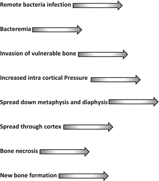
Fig. 7.2
The sequence of events from bacteremia to bone necrosis
Although bones infected with AHO are predominately large tubular bones, no bone is exempt. The femur and tibia are in the lead at 36 % and 33 % respectively as sites for AHO.
By comparison, the calcaneus accounts for 4 %, the metatarsals 2 %, the talus 1 %, and the combined bones of the pelvis 3 % [1, 2].
Where the capsule of a joint encompasses part of the metaphysis such as in the hip, ankle, elbow, and shoulder, extension through the boney cortex and periosteum may lead to septic arthritis. Additionally, in children less than 18 months of age, there may be a patent transphyseal vessel running from the metaphysis, through the physis (growth plate), through the epiphysis, and into the joint space creating continuity between the metaphysis and the adjacent joint. The appearance of septic arthritis in a young child should therefore arouse suspicion of osteomyelitis in an adjacent long bone (Fig. 7.3).
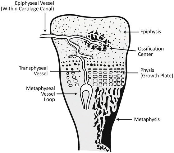
Fig. 7.3
Graphic representation of the metaphysis of a tubular bone of an infant or young child showing the transphyseal vessel
Bacteremia is not an uncommon event and, alone, does not account for the development of AHO. Simultaneous disruption of bone microarchitecture from minor, often not recalled, trauma and bacteremia from a distant focus set the stage for bacterial lodgment and proliferation.
This is often referred to as the “bone bruise theory” of skeletal infection.
Making the Diagnosis
Signs and Symptoms
Sustained pain with palpation and pressure over the end of a long bone or directly over a cuboid bone or flat bone along with systemic signs of illness such as fever, malaise, and anorexia justify consideration of osteomyelitis. Additional symptoms may include abnormal gait, with reluctance to bear weight or use an extremity. Other localizing features include localized swelling, warmth, redness, and decreased range of motion. A very late sign occurs when the inflammatory process has penetrated the boney cortex, the periosteum, and soft tissue and forms a fistulous tract to the surface of the skin resulting in purulent drainage.
Laboratory Aides
AHO is primarily a clinical diagnosis. Laboratory investigations may help confirm or refute clinical suspicions, but are not absolutes.
Non-specific tests are looking for evidence of inflammatory response. The complete blood count (CBC) usually, but not always, shows a leukocytosis with a predominance of neutrophils (granulocytes). Commonly the total white cell count will be over 12,000 with 60 % or more neutrophils. The c-reactive protein (CRP) is quick to rise with inflammation and quick to fall once appropriate surgical and/or medical treatments are initiated. The erythrocyte sedimentation rate (ESR) rises slowly and may still be rising even after appropriate therapy is initiated. These inflammatory markers should be followed during the course of treatment to assure successful eradication of the infection. These test results are variable depending on the pathogenicity of the organism and the inflammatory response of the host.
Microbiology
The organism responsible for AHO can, more often than not, be found in culture if the cultures are obtained and treated appropriately. Specimens of blood (two independent cultures), joint fluid (if applicable), infected bone and subperiosteal fluid should be obtained. Purulent fluid consists of white blood cells and engulfed bacteria. Optimal cultures are from the bone tissue surrounding the infected fluid.
Scrapings of 1 g of bone tissue (about the size of a pea) and rapid transport to the microbiology laboratory in a transport medium will keep organism alive until agar plates and broth have been inoculated. Generally, aerobic and anarobic cultures are sufficient. Fungal and mycobacteria (TB) cultures may be added in unusual circumstances such as with immunodeficiency and infection not responding to conventional therapy. Successful identification of infecting bacteria should occur in greater than 90 % of cases of AHO. Lack of growth from a culture may occur when antibiotics are given before cultures are obtained, when there are delays in processing the tissue for culture, and when inadequate specimens are submitted to the microbiologist. Direct visualization of the specimen by gram stain may give immediate information of the nature of the infectant such as gram positive cocci or gram negative rod. Once the organism is identified subsequent susceptibility testing will identify optimal antibiotic choices.
At all ages, Staphylococcus aureus (coagulase positive staph) is the most prelevent organism. Patients with staph aureus osteomyelitis commonly have this organism as part of their own flora, colonizing anterior nares and skin. Other notable bacteria causing AHO are group B streptococcus in infants, coagulase negative staphylococcus (staph epidermidis), gram negative bacteria such as Kingella kingae, and enteric bacteria including Pseudomonas aeruginosa. Gonoccal joint infection should be considered in sexually active teens and young adults.
Imaging
Conventional radiography is of limited value in the early detection of AHO because boney changes may take 10–14 days to appear. X-rays are, however, useful to rule out other conditions such as fractures, cysts, or malignancy. The first signs to appear are soft tissue swelling adjacent to the infected metaphysis. The swelling may be accompanied by the displacement of radiolucent muscle and fat planes. The bone cortex is porous. With increasing edema within the metaphysis, infected tissue spreads down the shaft of the metaphysis and diaphysis and outward through the cortex, elevating the periosteum. The periosteum is a tough fibrous sheet that has its own blood supply and remains viable despite the infection all round it. Periosteal reaction and boney rarefaction in the metaphysis are the first osseous changes reflecting bone necrosis and resorption. Dead bone, the sequestrum, becomes surrounded with periosteal new bone, the involucrum. This can be seen on plain X-rays at 2–3 weeks of the onset of symptoms (Fig. 7.4).
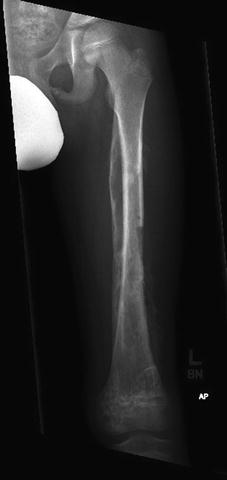
Fig. 7.4
X-ray of femur with advanced osteomyelitis. This depicts necrotic bone distally, lytic lesions of the diaphysis and metaphysis, periosteal reaction and beginning new bone Formation
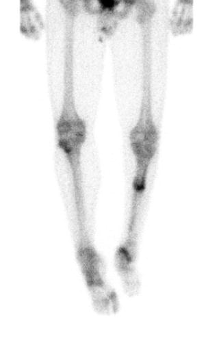
Skeletal scintigraphy, or bone scanning, can detect boney changes of osteomyelitis in the first 24–36 h of symptomatic infection [5]. A radioactive marker, technetium methylene diphosphate, is injected intravenously and flows to all tissues. It will be more densely concentrated in areas of hyperemia. The compound is taken up by osseous tissue and attaches to the boney matrix. The body is scanned at intervals. The marker will remain longer in bone and especially so in inflamed bone where there is greater osseous blood flow. The resulting scanned image, if positive, will show a defined increased uptake or “hot spot” (Figs. 7.5 and 7.6). When a bone scan is performed late in the course of infection, perhaps 10–15 days into the disease, there may be a “cold spot” indicating that the bone is necrotic and without blood supply. All hot spots are not osteomyelitis. Similar appearing scans may be encountered with tumors, such as Ewings sarcoma, or a skeletal presentation of acute lymphoblastic leukemia.