Fig. 10.1
The Matles test or the knee flexion test: the foot on the affected side falls into neutral or dorsiflexion
Sometimes diagnostic imaging may be required to verify a clinical suspicion or for chronic injuries. Plain lateral radiographs may reveal an irregular configuration of the fat-filled triangular space anterior to the Achilles tendon and between the posterior aspect of the tibia and the superior aspect of the calcaneus. It is also helpful to exclude bone injuries in case of acute trauma. Ultrasound (US) and magnetic resonance imaging (MRI) are widely used, even if there is no clear evidence that they improve the rate of correct diagnosis. According to the AAOS guidelines for acute AT rupture, there is not enough evidence to recommend for or against the routine use of US and MRI to confirm the diagnosis [16]. A recent study showed that physical examination, including an abnormal calf squeeze test, a palpable defect, and decreased resting tension, is more sensitive in diagnosing an acute complete AT rupture than MRI (sensitivity 100 % vs 90.9 %) [17]. Moreover MRI is time consuming and expensive and can lead to a delay in treatment (mean time to surgery 5.6 days vs 12.4 day in MRI group). The authors concluded recommending careful evaluation and judicious use of advanced imaging as needed.
10.4 Treatment Strategy
The consensus for athletes is surgery [2], as it provides early functional treatment, less calf atrophy, and the best functional performance with a lower re-rupture rate. Open, percutaneous, or minimally invasive procedures have been successfully used. Open surgery provides good strength to the repair, low re-rupture rates, and reliably good endurance and power to the gastrocnemius-Achilles tendon complex. However, open surgical approaches resulted in high risk of infection and morbidity. Review articles and meta-analysis showed high costs and a 20-fold higher rate of complications in open procedures than conservative treatment [13]. Therefore, minimally invasive procedures have been successfully used to avoid these complications. Minimally invasive AT repair provides many advantages. Major advantages are less iatrogenic damage to normal tissues, lower postoperative pain, accurate opposition of the tendon ends minimizing surgical incisions, and improved cosmetics. A recent systematic review reported a rate of superficial infections of 0.5 and 4.3 % after minimally invasive and open surgery, respectively [18]. Shorter hospitalization time and average time to return to working activities was also showed. Functional outcomes were not significantly different between minimally invasive and open surgery. Although sural nerve injury has been reported as a potential complication of this kind of surgery, new techniques have minimized the risk of sural nerve damage [18].
10.5 Percutaneous Achilles Tendon Repair: Surgical Technique
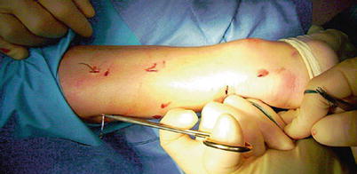
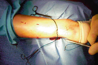
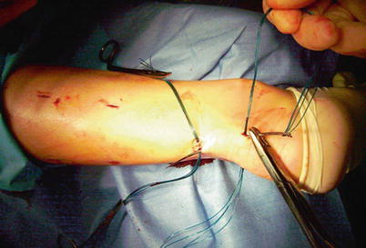
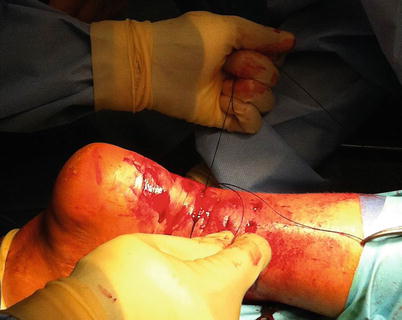
A 1 cm transverse incision is made over the defect using a size 11 blade. Four longitudinal stab incisions are made lateral and medial to the tendon 6 cm proximal to the palpable defect. Two further longitudinal incisions on either side of the tendon are made 4–6 cm distal to the palpable defect. Forceps are then used to mobilize the tendon from beneath the subcutaneous tissues. A 9 cm Mayo needle is threaded with two double loops of Number 1 Maxon, and this is passed transversely between the proximal stab incisions through the bulk of the tendon (Fig. 10.2). The bulk of the tendon is surprisingly superficial. The loose ends are held with a clip. In turn, each of the ends is then passed distally from just proximal to the transverse Maxon passage through the bulk of the tendon to pass out of the diagonally opposing stab incision. A subsequent diagonal pass is then made to the transverse incision over the ruptured tendon. To prevent entanglement, both ends of the Maxon are held in separate clips. This suture is then tested for security by pulling with both ends of the Maxon distally. Another double loop of Maxon is then passed between the distal stab incisions through the tendon (Fig. 10.3) and in turn through the tendon and out of the transverse incision starting distal to the transverse passage (Fig. 10.4). The ankle is held in full plantar flexion, and in turn the opposing ends of the Maxon thread are tied together with a double-throw knot, and then three further throws before being buried using the forceps (Fig. 10.5). A clip is used to hold the first throw of the lateral side to maintain the tension of the suture. We use 3-0 Vicryl suture to close the transverse incision and Steri-Strips close the stab incisions. A nonadherent dressing is applied. A full plaster cast is applied in the operating room with the ankle in physiologic equinus. The cast is split on both medial and lateral sides to allow for swelling. The patient is discharged on the same day of the operation.


Fig. 10.2
A 9 cm Mayo is threaded with two double loops of Number 1 Maxon, and this is passed transversely between the proximal stab incisions through the bulk of the tendon

Fig. 10.3
Another double loop of Maxon is then passed between the distal stab incisions through the tendon

Fig. 10.4
The double loop of Maxon is passed in turn through the tendon and out of the transverse incision starting distal to the transverse passage

Fig. 10.5
The two tendon stumps are sutured together with the ankle in full plantar flexion
10.6 Rehabilitation and Return to Play
Following percutaneous repair, patients are encouraged to bear weight on the operated limb as soon as possible as tolerated. The cast is removed at 2 weeks postoperatively, and a boot with the ankle in a plantigrade position is used. Removal of the boot under supervision of a physiotherapist allows the ankle to be plantar flexed fully but not dorsiflexed. These exercises are performed against manual resistance. At 6 weeks postoperatively, the boot is removed, and the patient referred to physiotherapy for active mobilization. At 12 weeks postoperatively, patients are assessed as to whether they were able to undertake more vigorous physiotherapy and are encouraged to gradually return to their normal activities. Progressive activities are incorporated as strength allows, with the aim to return to unrestricted activities by 6 months following surgery. Patients are reviewed at 3-month intervals and discharged at 9 or 12 months after the operation once they are able to perform at least five toe raises unaided on the operated leg and after they returned to their normal activities.
10.7 Discussion
AT ruptures are common in athletes. Surgical repair provides good results in young and active people. Open, percutaneous, or minimally invasive procedures have been successfully used. Open surgery provides good strength to the repair, low re-rupture rates, and reliably good endurance and power to the gastrocnemius-Achilles tendon complex. However, open surgical repair may result in high risk of infection and morbidity. Review articles and meta-analysis showed high costs and a 20-fold higher rate of complications in open procedures than conservative treatment [6]. Therefore, minimally invasive procedures have been successfully used to avoid these complications. Minimally invasive Achilles tendon repair provides many advantages. Major advantages are less iatrogenic damage to normal tissues, lower postoperative pain, accurate opposition of the tendon ends minimizing surgical incisions, shorter hospitalization time, lower rate of postsurgical infections, and improved cosmesis [18]. Excellent results have been reported in 17 elite athletes after percutaneous surgical repair of Achilles tendon ruptures [19]. All patients returned to high-level competition, with an average time to return to full sport participation of 4.8 months (range 3.2–6.5).
Rehabilitation of the Achilles tendon is complex and often nonstandardized. Detailed postoperative physical therapy programs for the AT often vary. Return to activity (RTA) can be defined as the time in which patients initiate their desired activity or sport that was limiting them. Evidence-based study of physical therapy regimens with regard to the foot and ankle is very limited. Modalities that have been rigorously studied have shown little benefit, including ultrasound, massage, and injections [20, 21]. Both eccentric exercises and extracorporeal shockwave therapy (ESWT) have been studied with a wide range of results. When patients can return to sports reducing the risk of further injuries is a big question, in particular for athletes because physicians are often faced with the pressing requirements of the athlete himself, the coach, and the team. Recurrence of AT tendinopathy and reinjury risk has been reported to be higher after short recovery periods [22]. Saxena et al. reported that simple parameters such as single-legged concentric strength, range of motion, and muscle girth can predict the ability to RTA [21]. If patients meet all 3 of these criteria, they are allowed to return to sport (Table 10.1), and the mean time to RTA after AT surgical repair was 21.8 ± 4.0 weeks. Females were more likely to have a delay in RTA.
Table 10.1


Parameters used in assessing the time frame of patients undergoing Achilles tendon surgery return to activity (RTA)
Stay updated, free articles. Join our Telegram channel

Full access? Get Clinical Tree


